Abstract
Bisphenol-A (BPA) is a ubiquitous endocrine-disrupting chemical. Recently, many issues have arisen surrounding the disease pathogenesis of BPA. Therefore, several studies have been conducted to investigate the proteomic biomarkers of BPA that are associated with disease processes. However, studies on identifying highly sensitive biological cell model systems in determining BPA health risk are lacking. Here, we determined suitable cell model systems and potential biomarkers for predicting BPA-mediated disease using the bioinformatics tool Pathway Studio. We compiled known BPA-mediated diseases in humans, which were categorized into five major types. Subsequently, we investigated the differentially expressed proteins following BPA exposure in several cell types, and analyzed the efficacy of altered proteins to investigate their associations with BPA-mediated diseases. Our results demonstrated that colon cancer cells (SW480), mammary gland, and Sertoli cells were highly sensitive biological model systems, because of the efficacy of predicting the majority of BPA-mediated diseases. We selected glucose-6-phosphate dehydrogenase (G6PD), cytochrome b-c1 complex subunit 1 (UQCRC1), and voltage-dependent anion-selective channel protein 2 (VDAC2) as highly sensitive biomarkers to predict BPA-mediated diseases. Furthermore, we summarized proteomic studies in spermatozoa following BPA exposure, which have recently been considered as another suitable cell type for predicting BPA-mediated diseases.
Keywords: bisphenol-A (BPA) toxicity, cell model types, biomarker, pathway studio, spermatozoa
1. Introduction
Endocrine disrupting chemicals (EDCs) are exogenous chemicals that can interrupt the endocrine system, from the chemical’s biosynthesis to its elimination, thus producing adverse effects on endocrine homeostasis in the body. Exposure to EDCs is associated with abnormal developmental, neurological, immune, and reproductive functions in both wild animals and humans [1,2]. Among the EDCs, bisphenol-A (BPA) is one of the most ubiquitous and extensively used, which has raised concerns about its prevalence in humans. According to the U.S. Centers for Disease Control and Prevention (CDC), perceptible levels of BPA are detected in urine for over 90% of the population in the U.S. [3]. Recent studies have revealed that BPA is closely associated with several diseases, such as cancer, diabetes, cardiovascular disease, thyroid dysfunction, developmental disorders, miscarriages, and reproductive disorders [4]. Since BPA is regarded as a harmful EDC, which may influence the endocrine system via binding with several physiological receptors preventing the binding of natural hormones, a large number of studies have been conducted, to investigate the effect of BPA and its underlying mechanism for the pathogenesis of diseases using both in vivo and in vitro trials [5,6].
It has been suggested that “omics” disciplines have opened a new avenue for identifying high quality biomarkers, which are reliable for the prediction of various diseases [7]. Most importantly, in the past few years, researchers have successfully engaged in comprehensive proteomic studies of various diseases following exposure to BPA both in vivo and in vitro [8,9,10,11,12]. In addition, bioinformatics has also improved following the development of proteomics, allowing us to make correlations between proteome profiling and interacting proteins, cellular regulation, and associated diseases. Therefore, these comprehensive studies may be considered as accurate and sensitive tools for predicting diseases and hazards to human health. Agarwal et al. (2015) demonstrated that BPA-induced hippocampal neurodegeneration in BPA-treated rats [13]. Moreover, Ge et al. (2014) found 36 proteins were significantly differentially expressed between control and BPA-treated groups in Sertoli cells [12]. Other research groups have also shown that prepubertal exposure to BPA altered mammary gland proteome profiling [14]. A direct comparison of proteome profiling between control and treatment groups may, therefore, be helpful to predict the human health hazards that follow BPA exposure.
BPA has two proposed mechanisms of action: a genomic, and a non-genomic pathway [9,15,16]. In the genomic pathway (nuclear), BPA binds endocrine receptors (ERs) and induces the dimerization of ERs. Subsequently, ER dimers attach to DNA, either directly or indirectly, by binding other transcription factors, including specificity protein 1 and activator protein 1 [15,16]. Alternatively, BPA can affect cell functions through the non-genomic pathway by binding membrane-bound receptors, leading to the activation of kinase signaling pathways [15,17]. During these signaling cascades, the membrane-bound receptor acts with G-protein-coupled receptors (GPCRs), and can cause fast estrogenic signaling by activation of the mitogen-activated protein kinase (MAPK) and phosphatidylinositol 3-kinase (PI3K). Calcium fluctuation, as well as cyclic adenosine monophosphate (cAMP) synthesis [18,19,20,21], may also occur.
Early studies demonstrated that spermatozoa include GPCRs and non-genomic ERs, and thus, spermatozoa have been considered as an alternative model system to investigate the proteome profiling following exposure to BPA, together with the investigation of BPA effects. There is ample experimental data on reproductive disease following exposure to BPA. Higher urinary BPA is significantly correlated with implantation failure, severe endometriosis, low sperm count, increased morphologically abnormal spermatozoa, and DNA damage [22,23,24]. Several studies have examined proteome profiling after BPA exposure, which may also identify precise biomarkers for the prediction of several diseases in this model system.
Therefore, the goal of this study was to identify a more sensitive model system for studying the effects of BPA exposure. Simultaneously, by comparing predicted proteomic biomarkers of BPA exposure in different cell types, we aimed to evaluate more sensitive biomarkers, capable of predicting BPA-mediated disease conditions using a bioinformatics application. In addition, we summarized recently applied proteomics studies in spermatozoa, which could be considered a highly sensitive and effective alternative model for BPA-mediated disease pathogenesis, most importantly, reproductive abnormalities.
2. Results and Discussion
2.1. Bisphenol-A (BPA) Induces a Wide Range of Diseases in Humans
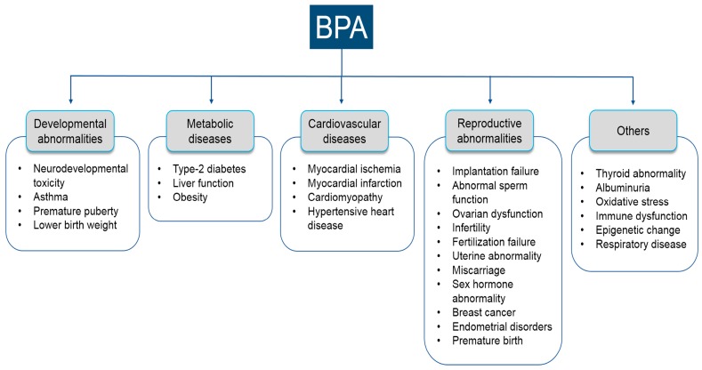
The effects of BPA on several diseases have been extensively studied, using both animal and human models [4,5,6]. Our search revealed several BPA-mediated diseases, which were broadly characterized into five major categories: developmental, metabolic, reproductive, cardiovascular, and other diseases (Figure 1). In the developmental category, the common diseases included neurodevelopmental toxicity [25,26,27,28,29,30], asthma [31], premature puberty [32,33], and lower birth weight [34,35]. Common diseases in the metabolic category were type-2 diabetes [36,37,38], liver function [39,40], and obesity [41,42,43]. Main diseases in the cardiovascular category included myocardial ischemia [44,45,46], myocardial infarction [39,45], cardiomyopathy [47], and hypertensive heart disease [48]. Major diseases in the reproductive subcategories were implantation failure [23], sperm function [22,49,50], infertility [51,52], ovarian dysfunction [53,54,55], fertilization [56,57], uterine abnormality [58], miscarriages [59,60], abnormal homeostasis of sex hormones [61,62], breast cancer [63,64], endometrial disorders [65,66], and premature birth [65]. In addition, we did not characterize some diseases into specific subcategories, which are also predisposed following BPA exposure, such as thyroid abnormality [67,68], albuminuria [69,70], oxidative stress [71,72], immune dysfunction [73], epigenetic changes [74], and respiratory disease [75]. From these findings, it is clear that exposure to BPA predisposes humans to a wide variety of disease conditions. Several recent studies considered laboratory animal models in vivo, or in vitro cell systems, to help us elucidate BPA-mediated disease conditions. However, more studies should be conducted to better elucidate the underlying molecular mechanisms of such disease pathogenesis. In addition, this study will provide vital information for future development of a theranostic approach to BPA toxicity in clinical conditions.
Figure 1.
Summary of bisphenol-A-mediated diseases broadly characterized into five major categories.
2.2. Compilation of Proteomics Studies Screening BPA Toxicity
Several efforts have been undertaken to evaluate potential adverse health effects of BPA exposure. The recent development of proteomic approaches has provided researchers with useful tools for identifying protein markers of toxic chemical exposure. Proteomics investigations have been conducted in several cell systems to investigate potential biomarkers of BPA exposure, most importantly, in Sertoli cells [12,76], thyroid [77], serum [8], mammary gland [14,78], zebrafish brain [79], rat hippocampus [13], mouse prefrontal cortex [28], colon cancer cells (SW480) [80], and Leydig cells [81].
In this study, we examined the predicted proteomic biomarkers from different cells using the Pathway Studio program. This program is capable of unraveling protein–protein interactions associated with disease biology. In addition, it is capable of providing exclusive statistical application for normalization of data, as well as exact identification of certain genes or proteins in health and diseases. Therefore, it has been considered as a comprehensive biology-based investigational framework, ideal for biomarker analysis and its correspondence relationship with disease modeling.
Our preliminary analysis showed that altered proteins from the majority of the cell types (BPA-exposed) represented an association with BPA-mediated diseases (Tables S1–S9). However, SW480, mammary gland, and Sertoli cells were selected as the most suitable cells for predicting the majority of the diseases caused by BPA (capable of predicting all diseases types; Tables S1–S9). Furthermore, we investigated the altered spermatozoa proteome [9,10] following BPA exposure in detail, which could be another model system for predicting BPA-mediated reproductive diseases.
2.3. Efficacy of the BPA-Induced Differential Proteome in SW480 Cells in Predicting BPA-Induced Diseases
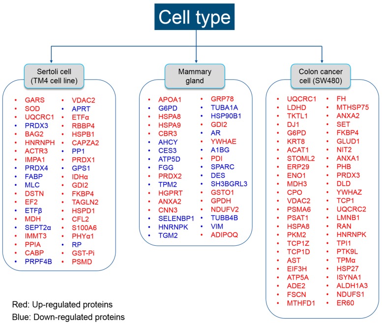
The SW480 cell line is derived from human colon cells, and is widely used for studying colon cancer [82,83,84,85]. Chen et al. (2015) showed that exposure to BPA (10−8 and 10−5 M) for 48 h, in vitro, is capable of promoting metastasis [80], and modifying the protein profile of colorectal cancer. They found 56 differentially expressed proteins following exposure to BPA by MALDI–TOF–MS/MS analysis (see Figure 2).
Figure 2.
Differentially expressed proteins following bisphenol-A exposure in three cell types. Differentially expressed proteins in the Sertoli cell were summarized according to [12,76]. Differentially expressed proteins in mammary gland cell were summarized according to [14,78]. Differentially expressed proteins in colon cancer cell were summarized according to [80].
We analyzed all proteins by the Pathway Studio program, to determine whether a predicted protein marker is capable of detecting BPA-associated diseases significantly. Our data revealed that 9 proteins out of 56 (i.e., MTHFD1, ANXA2, G6PD, ACAT1, UQCRC2, UQCRC1, VDAC2, HNRNPK, and HNRNPL) were significantly associated with major disease categories (developmental, metabolic, reproductive, cardiovascular, and other) (Table 1, p < 0.05). Although several other proteins, such as LDLR, PSMA6, ENO1, GLUD1, YWHAZ, SET, and PKM, also represented an association to some BPA-mediated diseases, the relationship was not significant (p > 0.05).
Table 1.
Summary of bisphenol-A-induced differential proteomes in human cancer colon cell (SW480) and their association with bisphenol-A-mediated diseases. “-” = No specific search results were found.
| Major Disease Category | Specific Disease | Overlapping Entitles | p-Value |
|---|---|---|---|
| Reproductive | Implantation failure | MTHFD1, ANXA2 | <0.01 |
| Developmental | Neurodevelopmental toxicity | G6PD, ACAT1, UQCRC2, UQCRC1, VDAC2, HNRNPK, HNRNPL | <0.01 |
| Metabolic | Type-2 diabetes | UQCRC2, UQCRC1, VDAC2 | <0.01 |
| Cardiovascular | - | UQCRC2, UQCRC1, VDAC2, MTHFD1, ANXA2 | <0.01 |
Altered MTHFD1 and ANXA2 were significantly associated with reproductive diseases (implantation failure). A study reported by Rozen’s group has also shown that the lack of MTHFDA synthetase significantly affects embryonic defects in female mice. The study demonstrated that disruption of the maternal MTHFD1 gene damages fetal growth [86]. There are also ample studies that suggest ANXA2 is closely involved with embryo implantation and breast cancer, by acting as an adhesion molecule on the endometrium [87,88,89].
Neurodevelopmental toxicity also can be predicted by G6PD, ACAT1, UQCRC2, UQCRC1, VDAC2, HNRNPK, and HNRNPL proteins, as expressed differentially in the SW480 cell line following BPA exposure. Extensive studies have been conducted to investigate the effects of these proteins and their mechanisms of neurodevelopmental toxicity. Jeng et al. (2013) provided a direct clue that lack of G6PD can cause oxidative DNA damage, which can lead to neurodegenerative disease [90]. ACAT1 is widely known as a critical enzyme, which reversibly converts two units of acetyl-CoA to acetoacetyl-CoA in mitochondria. Therefore, abnormal expression of ACAT1 may affect proper levels of acetyl-CoA, which could cause neurodegeneration [91,92]. On the other hand, alterations of UQCRC1, UQCRC2, and VDAC2 expression, have been reported to be associated with mitochondrial dysfunction, and thus, lead to oxidative stress and reactive oxygen species production [93,94,95]. It is reported that overexpression of UQCRC1 is associated with the development of abnormal behavior, and that UQCRC2 genes and VDAC2 are down-regulated in hypertensive rats, and in the hippocampus, under prenatal stress [96,97,98].
These three proteins are also associated with metabolic disease and cardiovascular diseases. VDAC2 is down-regulated in type-2 diabetic mouse liver [99]. Furthermore, VDAC2 has been studied as a regulating factor for cardiac rhythmicity by uptake of mitochondrial Ca2+ [100,101]. Nicolas et al. (2017) demonstrated that UQCRC2 is significantly reduced in pancreatic islets of diabetic phenotype mice [102]. In addition, the other studies also have shown that UQCRC2 is differentially expressed in rat insulinoma cell lines, and is significantly reduced under hyperglycemia, which is closely related with cardiac metabolic pathways [103,104]. Therefore, it is tempting to hypothesize that SW480 is a highly sensitive model to screen BPA toxicity, considering its efficacy to predict BPA-mediated disease by differentially expressed protein markers.
2.4. Efficacy of BPA-Induced Differential Proteome in Mammary Gland Cells to Predict BPA-Induced Diseases
Betancourt et al. (2012) have reported that prepubertal exposures to BPA-induced altered carcinogenesis in mammary gland cells of rats, which were subsequently predisposed to differential protein expression. They found 18 differentially expressed proteins between BPA-exposed and control rats by 2-dimensional gel electrophoresis followed by MS analysis [14]. In another study, the same research group also investigated differential protein expression in rat mammary glands following exposure to BPA in utero [78]. In the latter study, the differentially expressed proteome was explored by both MALDI-TOF-TOF and LC-MS/MS analysis. Here, we compiled all differentially expressed proteins from both studies. A total of 39 proteins were found to be expressed differentially in rat mammary glands following BPA exposure (see Figure 2). Pathway Studio analysis of these proteins revealed that 9 of 39 proteins (FGG, GTM2, HPRT1, HSPA5, G6PD, ADIPOQ, DES, ANXA2, and HPRT1) represent a significant association with all major disease categories (Table 2, p < 0.05).
Table 2.
Summary of bisphenol-A-induced differential proteome in mammary gland cells and their association with bisphenol-A-mediated diseases.
| Major Disease Category | Specific Disease | Overlapping Entitles | p-Value |
|---|---|---|---|
| Reproductive | Implantation failure | FGG | <0.05 |
| Developmental | Neurodevelopmental toxicity | TGM2, HPRT1, G6PD | <0.05 |
| Metabolic | Type-2 diabetes | ADIPOQ, HSPA5 | <0.05 |
| Cardiovascular | Cardiomyopathy | FGG, DES, ANXA2 | <0.05 |
We found that FGG expression was significantly correlated with reproductive diseases. Lin et al. (2012) have reported that FGG was differentially expressed in the plasma of a uterine leiomyoma patient [105]. Subsequently, FGG was overexpressed in plasma prior to preeclampsia, which is a risky pregnancy syndrome. FGG is widely known as a blood coagulation protein, and thus, might also predict cardiovascular diseases [106].
Three proteins, TGM2, HPRT1, and G6PD, were significantly correlated with neurodevelopmental toxicity. It is reported that TGM2 plays a role in cellular apoptosis, thus, it is involved in various diseases, such as diabetes and neurodegeneration [107,108]. Kang et al. (2011) demonstrated that a lack of HPRT1 is associated with uncontrolled neurodevelopmental pathways [109]. In addition, a study reported by Cristini and colleagues has described that HPRT1 has a critical role in human fetal neurodevelopment [110].
Altered ADIPOQ and HSPA5 expression were significantly correlated with type-2 diabetes. HSPA5 is known to be upregulated in human type-2 diabetes and high-fat-diet-induced diabetes in mice [111,112]. Several researchers have demonstrated that ADIPOQ is closely associated with type-2 diabetes [113,114,115]. Furthermore, although our Pathway Studio program did not show a significant association, these two proteins are also closely linked with cardiovascular diseases. ADIPOQ has been studied as a vital factor in the cardiac metabolic pathway that may be helpful in treating diabetes and cardiovascular disease [116,117]. It is reported that HSPA5 is significantly increased in the hypertensive heart, which may lead to heart failure [118,119]. Altered FGG is found in men with myocardial infarction, arterial disease, and heart failure through blood coagulation [120,121]. DES is also considered as a potential biomarker for the prediction of cardiovascular diseases. It is reported that aberrant DES aggregation can lead to cardiac diseases [122].
2.5. Efficacy of BPA-Induced Differential Proteome in Sertoli Cell to Predict BPA-Induced Diseases
Sertoli cells are the “nurse” cells of the testis that help in spermatogenesis and regulate uninterrupted production of spermatozoa. Because of their important role in spermatogenesis, Sertoli cells are the foremost target of toxicant-induced reproductive abnormalities. Ge et al. (2014) have analyzed the effects of BPA on Sertoli cell (TM4 cell line) proliferation and altered proteome by MALDI-TOF-MS/MS analysis [12]. Others have also described the altered proteome in Sertoli cells (TM4 cell line) treated with BPA by MALDI-TOF-MS/MS analysis [76]. Here, we collected all differentially expressed proteins from both studies (Figure 2). A total of 42 differentially expressed proteins were analyzed by the Pathway Studio program to investigate the interactions between proteins and diseases. Five different proteins (e. g., HSPB1, SOD1, UQCRC1, VDAC2, and HSPB1) were found to be associated with various BPA-associated diseases such as reproductive, developmental, metabolic, and cardiovascular, following exposure to BPA (Table 3, p < 0.05).
Table 3.
Summary of bisphenol-A-induced differential proteome in Sertoli cells (mouse TM4 cells) and association with bisphenol-A-mediated diseases.
| Major Disease Category | Specific Disease | Overlapping Entities | p-Value |
|---|---|---|---|
| Reproductive | Implantation failure | HSPB1 | <0.05 |
| Developmental | Neurodevelopmental toxicity | SOD1, UQCRC1, VDAC2 | <0.05 |
| Metabolic | Type-2 diabetes | UQCRC1, VDAC2, SOD1 | <0.05 |
| Cardiovascular | Myocardial ischemia | UQCRC1, VDAC2, SOD1 | <0.05 |
Our Pathway Studio results revealed that HSPB1 is significantly associated with reproductive disease (implantation failure). It is reported that HSPB1 protein is overexpressed in uterine distension during pregnancy, as well as inducing the distension of the myometrium, owing to the growing fetus [123,124].
In the Sertoli cell system, we also found that type-2 diabetes and cardiovascular diseases could be predicted by three different proteins (UQCRC1, VDAC2, and SOD1). Neves et al. (2012) described the vital role of SOD1 in preventing cardiovascular diseases by countering to oxidative stress caused by the type-2 diabetes [125]. Consistent with this finding, other research groups have demonstrated that the distribution of SOD1 is different between diabetes patients and healthy controls [126]. Moreover, it is reported that SOD1 deficiency has critical implications for the pathogenesis of cardiac diseases [127]. Therefore, considering the efficacy of the three cell types (SW480, mammary gland, and Sertoli cells) in predicting BPA-exposure-related disease, it is tempting to conclude that all cells provide considerable efficacy to predict BPA toxicity. Similar studies should be conducted, considering both in vitro and in vivo experimental design, to increase the specificity and efficacy of the identified biomarkers to predict BPA-associated diseases.
2.6. Commonly Expressed Proteins in Three Major Cell Types Following BPA Exposure
Among the various protein biomarkers, we noticed that G6PD, UQCRC1, and VDAC2 were associated with the same disease categories in more than one of the three cell types. G6PD is associated with neurodevelopmental diseases in both mammary gland and SW480 cell lines. UQCRC1 and VDAC2 were associated with neurodevelopmental disease, type-2 diabetes, and cardiovascular disease in both Sertoli cell and SW 480 cell lines (Table 4).
Table 4.
Commonly expressed proteins in mammary gland, Sertoli, and human SW480 cells following exposure to BPA and their efficacy to predict BPA-mediated diseases. “-” = No specific search results were found.
| Major Disease Category |
Specific Disease | Cell Type | p-Value | ||
|---|---|---|---|---|---|
| Mammary Gland | Sertoli Cell | SW480 | |||
| Reproductive | - | - | - | - | - |
| Developmental | Neurodevelopmental disease | G6PD | UQCRC1, VDAC2 |
G6PD, UQCRC1, VDAC2 | <0.05 |
| Metabolic | Type-2 diabetes | - | UQCRC1, VDAC2 |
UQCRC1, VDAC2 | <0.05 |
| Cardiovascular disease | Myocardial ischemia | - | UQCRC1, VDAC2 |
UQCRC1, VDAC2 | <0.05 |
A large number of studies have been conducted to investigate the effect of G6PD and its underlying mechanisms. It is reported that G6PD is a catalytic enzyme in the pentose phosphate pathway to provide the energy to cells by maintaining NADPH [128]. G6PD deficiency is a common disease worldwide known commonly as favism, which can lead to hemolysis and other conditions, such as hemolytic anemia, diabetes, and neonatal jaundice [129]. It is worth noting that G6PD deficiency is caused by an X-chromosome abnormality, which may be damaged by BPA exposure. Many studies have reported that BPA has an impact on G6PD [129,130,131,132]. As aforementioned, UQCRC1 and VDAC2 play a vital role in the mitochondrial respiratory chain, resulting in decreased ATP synthesis, increased oxidative stress, and reactive oxygen species production [93,94,95]. BPA is reported to be significantly related to the generation of ATP, oxidative stress, and especially mitochondrial dysfunction, in a manner similar to the biological functions of these proteins [9,10,133,134,135,136]. Based on these previous studies, we can anticipate biological functions or associated diseases of specific proteins. G6PD, UQCRC1, and VDAC2 are well-studied proteins in both signaling pathways and diseases; therefore, these three proteins could become highly utilized biomarkers to predict diseases that are closely associated with BPA exposure. However, further studies are required to confirm the safety and efficacy of these biomarkers for possible clinical applications. Interestingly, we did not find any single protein common in all cell types. This may be because the exposure scheme of BPA, and the doses of exposure in each study, were different.
2.7. Efficacy of BPA-Induced Differential Proteome in Spermatozoa to Predict BPA-Induced Diseases
An intensive search in literature revealed that mature spermatozoa contain both GPCRs and non-genomic ERs; thus, this is an excellent model to screen the molecular mechanism of EDC action [130,137,138]. Although there are numerous studies where spermatozoa were used to investigate the action of EDCs including BPA, few studies have been conducted to demonstrate the proteomic change of spermatozoa following BPA exposure. Recently, we introduced spermatozoa as a model cell to investigate protein biomarkers following both in vitro and in vivo exposure to BPA. This model is highly sensitive because mature mammalian spermatozoa are mostly incapable of protein synthesis; thus, predicted protein biomarkers in spermatozoa provide considerable constancy for use in clinical conditions [139,140,141].
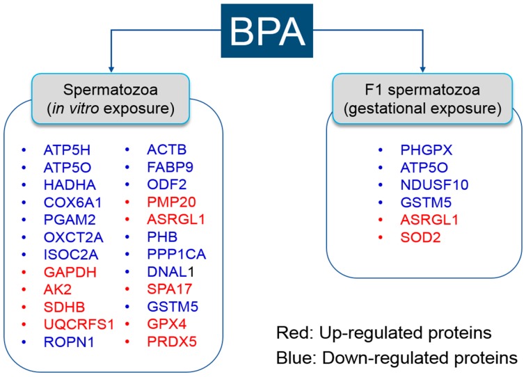
Our research group demonstrated that BPA affects sperm function, fertility, and proteome profiles in mice in vitro [10,135]. Subsequently, we have also reported that sperm function, fertility, and proteome were influenced in F1 spermatozoa following gestational exposure to BPA [9]. Here, we compiled all differentially expressed proteins from both in vitro and in vivo studies (Figure 3).
Figure 3.
List of differentially expressed proteins following bisphenol-A exposure in spermatozoa. Differentially expressed proteins in the spermatozoa in vitro were summarized according to [10,135]. Differentially expressed proteins in F1 spermatozoa following gestational exposure to bisphenol-A were summarized according to [9].
We then analyzed them to investigate the linkage between altered protein profiles and BPA-mediated diseases by the Pathway Studio program. Three altered proteins (e.g., UQCRFS1, SDHB, and ATP5O) out of 30 were matched with BPA-mediated diseases, as shown in Table 5.
Table 5.
Summary of bisphenol-A-induced differential proteomes in spermatozoa and their association with bisphenol-A-mediated diseases. “-” = No specific search results were found.
| Major Disease Category | Specific Disease | Overlapping Entities | p-Value |
|---|---|---|---|
| Reproductive | - | - | - |
| Developmental | Neurodevelopmental toxicity | UQCRFS1, SDHB, ATP5O | <0.01 |
| Metabolic | Type-2 diabetes | UQCRFS1, SDHB, ATP5O | <0.01 |
| Cardiovascular | Myocardial ischemia | UQCRFS1, ATP5O | <0.01 |
UQCRFS1, SDHB, and ATP5O were significantly associated with both neurodevelopmental toxicity and type-2 diabetes (p < 0.05). In addition, both UQCRFS1 and ATP5O presented a statistically significant association with cardiovascular diseases (p < 0.05). However, contrary to our expectation, there was no significant association between the differentially expressed proteins and BPA-mediated reproductive diseases. The statistical value output, given by Pathway Studio, is compiled based on a curated pathway, which is derived from millions of full-text articles, 25 million abstracts, and more than 164,000 clinical trials. Therefore, this may be because the exact pathways of specific reproductive diseases are not yet well established compared to other diseases. Despite many studies already reporting the association between specific proteins and fertility, relatively few studies are performed on the underlying mechanism of specific reproductive diseases. For instance, several studies have extensively reported that PRDXs plays a vital role in fertility through preventing oxidative stress; however, the underlying mechanisms are not fully understood [142,143,144,145]. Therefore, we decided to link the differentially expressed proteins to BPA-mediated reproductive diseases manually, using the Pathway Studio program without statistical analysis.
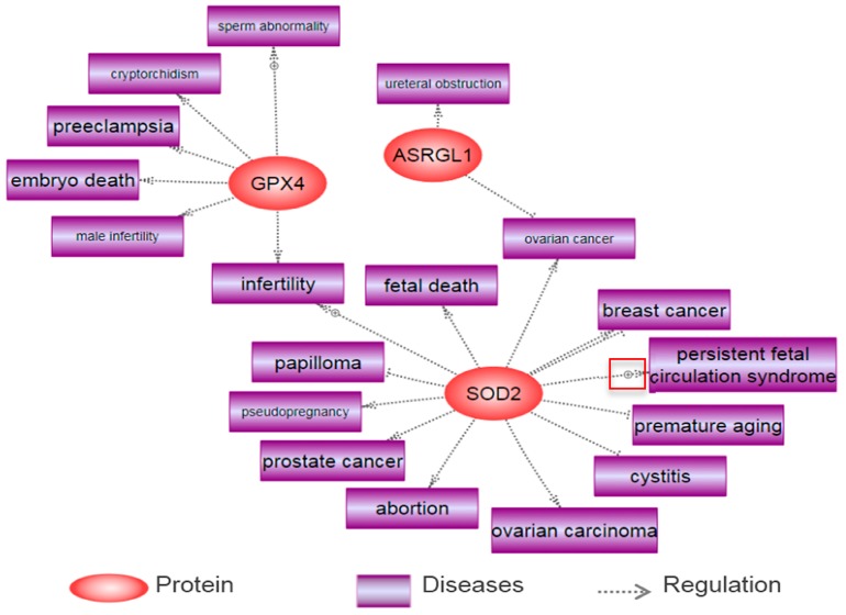
We found three proteins (GPX4, ASRGL1, and SOD2) out of six differentially expressed proteins in F1 spermatozoa following BPA exposure were related to several reproductive diseases using the Pathway Studio program. The major diseases include sperm abnormality, cryptorchidism, preeclampsia, male infertility, embryo death, ureteral obstruction, ovarian cancer, cystitis, breast cancer, prostate cancer, abortion, pseudo pregnancy, ovarian carcinoma, papilloma, fetal death, and infertility. These have been summarized in Figure 4.
Figure 4.
Reproductive diseases associated with several proteins following in vitro exposure to bisphenol-A in spermatozoa.
Imai et al. (2009) reported that abnormalities of spermatozoa and seminiferous tubules are found in spermatocyte-specific GPX4 knockout mice [146]. Consistently, it is reported that the deficiency of GPX4 in these mice resulted in embryonic death through oxidative stress, which may cause male infertility [147,148]. SOD2 is a widely known effective antioxidant enzyme, which can prevent oxidative stress. Several studies have revealed that this protein has an impact on reproductive diseases. Uriu-Adams et al. (2005) reported that disruption of SOD leads to fetal death in mice, and other research also revealed that a genetic polymorphism in SOD determines the risk of infertility in patients [149,150]. In addition, it is reported that ASRGL causes ureteral obstruction through inflammatory complication of acute pancreatitis [151]. Few studies have been conducted to demonstrate the underlying mechanisms of action of specific proteins on reproductive diseases, although the relationship between these proteins and disease was insignificant.
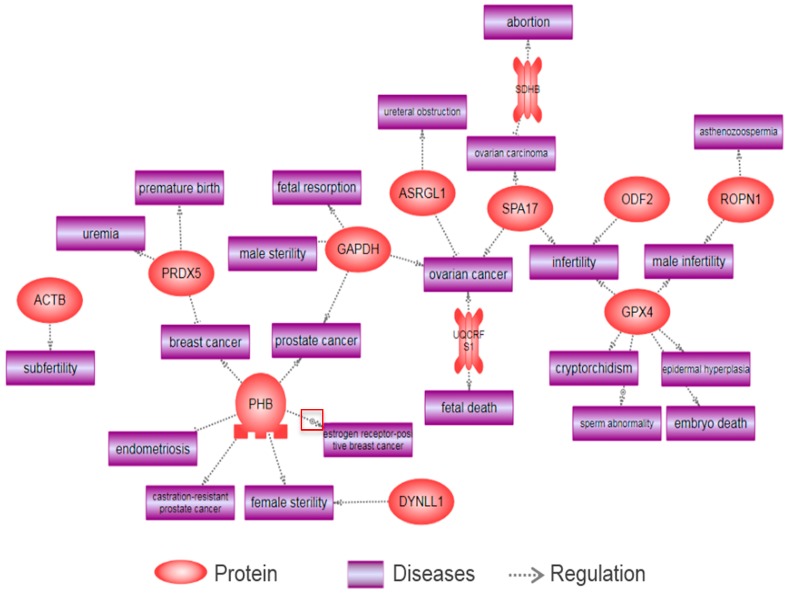
Using an in vitro approach, we found that BPA is capable of altering a total of 24 proteins in spermatozoa. Subsequently, 13 differentially expressed proteins (e.g., ACTB, DYNLL1, ASRGL1, PHB, SPA17, ATP5O, GAPDH, PRDX5, GPX4, UQCRFS1, ROPN1, ODF2, and SDHB) were associated with various reproductive diseases, such as ureteral obstruction, endometriosis, subfertility, prostate cancer, breast cancer, ovarian cancer, male sterility, fetal resorption, abortion, ovarian carcinoma, female sterility, infertility, asthenozoospermia, fetal death, male infertility, sperm abnormality, cryptorchidism, embryo death, uremia, and premature birth, as shown by Pathway Studio (Figure 5).
Figure 5.
Reproductive diseases associated with several proteins following gestational exposure to bisphenol-A in F1 spermatozoa.
Consistent with the differentially expressed proteins of F1 spermatozoa, both GPX4 and ASRGL1 were altered by BPA exposure. SPA17 has been reported to induce tissue-specific malignancy in human epithelial ovarian cells [152]. Furthermore, GAPDH, an important catalytic enzyme for glycolysis, has been studied in relation to reproductive diseases. Riley has described that inhibition of GAPDH affects fetal resorption [153]. In another study, the same research group reported that inactive GAPDH causes the apoptosis of blastocysts, and thus results in fetal resorption [154]. Furthermore, it is reported that GAPDH has impacts on male sterility, prostate cancer, and ovarian cancer [155,156,157]. PHB is also associated with several reproductive diseases. He et al. (2015) showed that modification of the PHB gene caused abnormal development and estrogen insensitivity in mice uteri, and resulted in sterility [158]. In addition, another study demonstrated that PHB plays a vital role in the initiation of prostate cancer and early androgen-independent tumors [159]. It is reported that ROPN1 deficiency affects sperm motility by PKA-dependent signaling processes, resulting in male infertility [160]. DYNLL1 has been reported to have an impact on female sterility [161]. Subsequently, it has been reported that PRDX5 has a positive effect on breast cancer by regulating oxidative stress [162]. León et al. (2007) described that SDHB is downregulated in pollen abortions in Arabidopsis [163]. Moreover, other proteins (ODF2, UQCRFS1, and ACTB) are involved in infertility, ovarian cancer, fetal death, and subfertility [164,165,166,167].
In summary, although we could not demonstrate a statistical association between BPA-mediated reproductive diseases and altered protein expression following BPA exposure in spermatozoa, we found that several differentially expressed proteins are correlated with reproductive diseases, according to our bioinformatic data. Therefore, we speculate that spermatozoa could be considered as a potential biological cell model system to predict BPA-mediated reproductive diseases more specifically. However, further studies should be conducted for screening the alteration of the proteome within reproductive diseases, and discovering the underlying mechanisms and pathways of various reproductive diseases. On the other hand, the majority of the cells revealed a sex-based difference following exposure to chemicals and microbial stressors [168]. These differences may affect BPA-induced proteomic profiles in male and female cells in a sex-specific manner. Therefore, rigorous preclinical study with the focus on sex or gender must be considered to develop more specific guidelines of a chemical toxicity [169].
3. Materials and Methods
3.1. Identification of BPA-Mediated Diseases
The PubMed search engine was used to thoroughly search the MEDLINE database for literature to identify BPA-mediated disease/disease conditions. Briefly, we compiled published studies that were conducted as epidemiological studies on human health hazards mediated via either direct or indirect BPA exposure. The various diseases and disorders identified were categorized into five major categories: developmental abnormalities, metabolic disease, cardiovascular disease, reproductive diseases, and others.
3.2. Identification of Model Cell Systems to Identify Proteomic Biomarkers of BPA Exposure
The PubMed search engine was also used to thoroughly identify scientific literature regarding proteomic studies on several cell types, to investigate the differentially expressed proteins following exposure to BPA both in vitro and in vivo.
3.3. Analysis of Disease Pathways by Differentially Expressed Proteins
The differentially expressed proteins following BPA exposure in each cell type were analyzed by the computation bioinformatics program Pathway Studio® 9.0 (Ariadne Genomics, Rockville, MD, USA), to demonstrate whether the proteomics biomarker in different cells could predict BPA-mediated diseases/disease conditions. Briefly, protein names (symbols) were entered into the program to determine significantly matching diseases for each differentially expressed protein, based on the information extracted from the NCBI PubMed database. Related diseases associated with each differentially expressed protein were re-confirmed using a PubMed Medline hyperlink that was embedded in each node. Fisher’s exact test was used to determine whether a disease was statistically correlated with the target protein. p < 0.05 was considered statistically significant.
4. Conclusions
Comprehensive proteomics approach has already been developed as a high-throughput technique, which is helpful to analyze numerous altered proteins in pathological and toxicology studies. A direct comparison of the differentially expressed proteome following exposure to toxic agents/BPA could help identify potential protein biomarkers that predict the abnormality from exposure to specific EDCs. Simultaneously, these protein biomarkers can be linked to extensive biological functions, interacting proteins, and diseases based on these “footprints”. Here, we showed that SW480, mammary gland, and Sertoli cells are the most suitable cell system models to predict BPA-mediated diseases among other examined cell types, such as thyroid, serum, zebrafish brain, rat hippocampus, prefrontal cortex of mice, and Leydig cells. Subsequently, we also demonstrated that G6PD, UQCRC1, and VDAC2 may be the most sensitive and highly utilized protein biomarkers, which can predict all major disease categories following BPA exposure. Furthermore, we showed that various differentially expressed proteins in spermatozoa following BPA exposure are also closely related with many disease categories, including reproductive diseases. Therefore, it seems that spermatozoa might be a good potential biological cell model type to predict BPA-mediated reproductive diseases. However, further studies are needed to evaluate and enhance the specificity and efficacy of potential biomarkers to predict BPA-mediated diseases, and to establish the pathways and mechanisms related to reproductive diseases.
Acknowledgments
This work was supported by Korea Institute of Planning and Evaluation for Technology in Food, Agriculture, Forestry, and Fisheries (IPET) through Agri-Bio Industry Technology Development Program, funded by Ministry of Agriculture, Food, and Rural Affairs (MAFRA) (116172-3). Do-Yeal Ryu was supported by a NRF (National Research Foundation of Korea) grant funded by the Korean Government (NRF-2016-Fostering Core Leaders of the Future Basic Science Program/Global Ph.D. Fellowship Program). Md Saidur Rahman was supported by Korea Research Fellowship (KRF) Program through the National Research Foundation of Korea (NRF) funded by the Ministry of Science and ICT (project no. 2017H1D3A1A02013844).
Abbreviations
| BPA | Bisphenol-A |
| CDC | Centers for Disease Control |
| EDC | Endocrine Disrupting Chemical |
| ER | Endocrine Receptor |
| GPCR | G-protein-Coupled Receptors |
| MAPK | Mitogen-Activated Protein Kinase |
| PI3K | Phosphatidylinositol 3-Kinase |
Supplementary Materials
Supplementary materials can be found at www.mdpi.com/1422-0067/18/9/1909/s1.
Author Contributions
Do-Yeal Ryu and Md Saidur Rahman analyzed the data and created the artwork. Do-Yeal Ryu and Md Saidur Rahman, drafted the manuscript. Myung-Geol Pang supervised the design of the study and critically reviewed the manuscript. All authors critically reviewed the manuscript for intellectual content and gave final approval of the version for publication.
Conflicts of Interest
The authors declare they have no conflict of financial interest. The corresponding author is responsible for submitting a competing financial interest’s statement on behalf of all authors.
References
- 1.Darbre P.D. Endocrine Disruptors and Obesity. Curr. Obes. Rep. 2017;6:18–27. doi: 10.1007/s13679-017-0240-4. [DOI] [PMC free article] [PubMed] [Google Scholar]
- 2.Trasande L., Zoeller R.T., Hass U., Kortenkamp A., Grandjean P., Myers J.P., DiGangi J., Bellanger M., Hauser R., Legler J., et al. Estimating burden and disease costs of exposure to endocrine-disrupting chemicals in the European union. J. Clin. Endocrinol. Metab. 2015;100:1245–1255. doi: 10.1210/jc.2014-4324. [DOI] [PMC free article] [PubMed] [Google Scholar]
- 3.Calafat A.M., Ye X., Wong L.Y., Reidy J.A., Needham L.L. Exposure of the U.S. population to bisphenol A and 4-tertiary-octylphenol: 2003–2004. Environ. Health Perspect. 2008;116:39–44. doi: 10.1289/ehp.10753. [DOI] [PMC free article] [PubMed] [Google Scholar]
- 4.Rochester J.R. Bisphenol A and human health: A review of the literature. Reprod. Toxicol. 2013;42:132–155. doi: 10.1016/j.reprotox.2013.08.008. [DOI] [PubMed] [Google Scholar]
- 5.Peretz J., Vrooman L., Ricke W.A., Hunt P.A., Ehrlich S., Hauser R., Padmanabhan V., Taylor H.S., Swan S.H., VandeVoort C.A., et al. Bisphenol a and reproductive health: Update of experimental and human evidence, 2007–2013. Environ. Health Perspect. 2014;122:775–786. doi: 10.1289/ehp.1307728. [DOI] [PMC free article] [PubMed] [Google Scholar]
- 6.Richter C.A., Birnbaum L.S., Farabollini F., Newbold R.R., Rubin B.S., Talsness C.E., Vandenbergh J.G., Walser-Kuntz D.R., vom Saal F.S. In Vivo effects of bisphenol A in laboratory rodent studies. Reprod. Toxicol. 2007;24:199–224. doi: 10.1016/j.reprotox.2007.06.004. [DOI] [PMC free article] [PubMed] [Google Scholar]
- 7.Benninghoff A. Toxicoproteomics—The next step in the evolution of environmental biomarkers. Toxicol. Sci. 2007;95:1–4. doi: 10.1093/toxsci/kfl157. [DOI] [PubMed] [Google Scholar]
- 8.Betancourt A., Mobley J.A., Wang J., Jenkins S., Chen D., Kojima K., Russo J., Lamartiniere C.A. Alterations in the rat serum proteome induced by prepubertal exposure to bisphenol a and genistein. J. Proteome Res. 2014;13:1502–1514. doi: 10.1021/pr401027q. [DOI] [PMC free article] [PubMed] [Google Scholar]
- 9.Rahman M.S., Kwon W.S., Karmakar P.C., Yoon S.J., Ryu B.Y., Pang M.G. Gestational Exposure to Bisphenol A Affects the Function and Proteome Profile of F1 Spermatozoa in Adult Mice. Environ. Health Perspect. 2017;125:238–245. doi: 10.1289/EHP378. [DOI] [PMC free article] [PubMed] [Google Scholar]
- 10.Rahman M.S., Kwon W.S., Yoon S.J., Park Y.J., Ryu B.Y., Pang M.G. A novel approach to assessing bisphenol-A hazards using an in vitro model system. BMC Genom. 2016 doi: 10.1186/s12864-016-2979-5. [DOI] [PMC free article] [PubMed] [Google Scholar]
- 11.Yang M., Lee H.S., Pyo M.Y. Proteomic biomarkers for prenatal bisphenol A-exposure in mouse immune organs. Environ. Mol. Mutagen. 2008;49:368–373. doi: 10.1002/em.20394. [DOI] [PubMed] [Google Scholar]
- 12.Ge L.C., Chen Z.J., Liu H., Zhang K.S., Su Q., Ma X.Y., Huang H.B., Zhao Z.D., Wang Y.Y., Giesy J.P., et al. Signaling related with biphasic effects of bisphenol A (BPA) on Sertoli cell proliferation: A comparative proteomic analysis. Biochim. Biophys. Acta. 2014;1840:2663–2673. doi: 10.1016/j.bbagen.2014.05.018. [DOI] [PubMed] [Google Scholar]
- 13.Agarwal S., Tiwari S.K., Seth B., Yadav A., Singh A., Mudawal A., Chauhan L.K., Gupta S.K., Choubey V., Tripathi A., et al. Activation of Autophagic Flux against Xenoestrogen Bisphenol-A-induced Hippocampal Neurodegeneration via AMP kinase (AMPK)/Mammalian Target of Rapamycin (mTOR) Pathways. J. Biol. Chem. 2015;290:21163–21184. doi: 10.1074/jbc.M115.648998. [DOI] [PMC free article] [PubMed] [Google Scholar] [Retracted]
- 14.Betancourt A.M., Wang J., Jenkins S., Mobley J., Russo J., Lamartiniere C.A. Altered carcinogenesis and proteome in mammary glands of rats after prepubertal exposures to the hormonally active chemicals bisphenol a and genistein. J. Nutr. 2012;142:1382S–1388S. doi: 10.3945/jn.111.152058. [DOI] [PMC free article] [PubMed] [Google Scholar]
- 15.Shanle E.K., Xu W. Endocrine disrupting chemicals targeting estrogen receptor signaling: Identification and mechanisms of action. Chem. Res. Toxicol. 2011;24:6–19. doi: 10.1021/tx100231n. [DOI] [PMC free article] [PubMed] [Google Scholar]
- 16.Heldring N., Pike A., Andersson S., Matthews J., Cheng G., Hartman J., Tujague M., Strom A., Treuter E., Warner M., et al. Estrogen receptors: How do they signal and what are their targets. Physiol. Rev. 2007;87:905–931. doi: 10.1152/physrev.00026.2006. [DOI] [PubMed] [Google Scholar]
- 17.Nadal A., Ropero A.B., Laribi O., Maillet M., Fuentes E., Soria B. Nongenomic actions of estrogens and xenoestrogens by binding at a plasma membrane receptor unrelated to estrogen receptor α and estrogen receptor β. Proc. Natl. Acad. Sci. USA. 2000;97:11603–11608. doi: 10.1073/pnas.97.21.11603. [DOI] [PMC free article] [PubMed] [Google Scholar]
- 18.Aronica S.M., Kraus W.L., Katzenellenbogen B.S. Estrogen action via the cAMP signaling pathway: Stimulation of adenylate cyclase and cAMP-regulated gene transcription. Proc. Natl. Acad. Sci. USA. 1994;91:8517–8521. doi: 10.1073/pnas.91.18.8517. [DOI] [PMC free article] [PubMed] [Google Scholar]
- 19.Chen Z., Yuhanna I.S., Galcheva-Gargova Z., Karas R.H., Mendelsohn M.E., Shaul P.W. Estrogen receptor α mediates the nongenomic activation of endothelial nitric oxide synthase by estrogen. J. Clin. Investig. 1999;103:401–406. doi: 10.1172/JCI5347. [DOI] [PMC free article] [PubMed] [Google Scholar]
- 20.Improta-Brears T., Whorton A.R., Codazzi F., York J.D., Meyer T., McDonnell D.P. Estrogen-induced activation of mitogen-activated protein kinase requires mobilization of intracellular calcium. Proc. Natl. Acad. Sci. USA. 1999;96:4686–4691. doi: 10.1073/pnas.96.8.4686. [DOI] [PMC free article] [PubMed] [Google Scholar]
- 21.Migliaccio A., Di Domenico M., Castoria G., de Falco A., Bontempo P., Nola E., Auricchio F. Tyrosine kinase/p21ras/MAP-kinase pathway activation by estradiol-receptor complex in MCF-7 cells. EMBO J. 1996;15:1292–1300. [PMC free article] [PubMed] [Google Scholar]
- 22.Meeker J.D., Ehrlich S., Toth T.L., Wright D.L., Calafat A.M., Trisini A.T., Ye X.Y., Hauser R. Semen quality and sperm DNA damage in relation to urinary bisphenol A among men from an infertility clinic. Reprod. Toxicol. 2010;30:532–539. doi: 10.1016/j.reprotox.2010.07.005. [DOI] [PMC free article] [PubMed] [Google Scholar]
- 23.Ehrlich S., Williams P.L., Missmer S.A., Flaws J.A., Berry K.F., Calafat A.M., Ye X., Petrozza J.C., Wright D., Hauser R. Urinary bisphenol A concentrations and implantation failure among women undergoing in vitro fertilization. Environ. Health Perspect. 2012;120:978–983. doi: 10.1289/ehp.1104307. [DOI] [PMC free article] [PubMed] [Google Scholar]
- 24.Itoh H., Iwasaki M., Hanaoka T., Sasaki H., Tanaka T., Tsugane S. Urinary bisphenol-A concentration in infertile Japanese women and its association with endometriosis: A cross-sectional study. Environ. Health Prev. Med. 2007;12:258–264. doi: 10.1007/BF02898033. [DOI] [PMC free article] [PubMed] [Google Scholar]
- 25.Bellinger D.C., Trachtenberg F., Zhang A., Tavares M., Daniel D., McKinlay S. Dental amalgam and psychosocial status: The New England Children's Amalgam Trial. J. Dent. Res. 2008;87:470–474. doi: 10.1177/154405910808700504. [DOI] [PMC free article] [PubMed] [Google Scholar]
- 26.Bellinger D.C., Daniel D., Trachtenberg F., Tavares M., McKinlay S. Dental amalgam restorations and children’s neuropsychological function: The New England Children’s Amalgam Trial. Environ. Health Perspect. 2007;115:440–446. doi: 10.1289/ehp.9497. [DOI] [PMC free article] [PubMed] [Google Scholar]
- 27.Maserejian N.N., Trachtenberg F.L., Hauser R., McKinlay S., Shrader P., Tavares M., Bellinger D.C. Dental composite restorations and psychosocial function in children. Pediatrics. 2012;130:e328–338. doi: 10.1542/peds.2011-3374. [DOI] [PMC free article] [PubMed] [Google Scholar]
- 28.Luo G., Wei R., Wang S., Wang J. Paternal bisphenol a diet changes prefrontal cortex proteome and provokes behavioral dysfunction in male offspring. Chemosphere. 2017;184:720–729. doi: 10.1016/j.chemosphere.2017.06.050. [DOI] [PubMed] [Google Scholar]
- 29.Maserejian N.N., Trachtenberg F.L., Hauser R., McKinlay S., Shrader P., Bellinger D.C. Dental composite restorations and neuropsychological development in children: Treatment level analysis from a randomized clinical trial. Neurotoxicology. 2012;33:1291–1297. doi: 10.1016/j.neuro.2012.08.001. [DOI] [PMC free article] [PubMed] [Google Scholar]
- 30.Miodovnik A., Engel S.M., Zhu C., Ye X., Soorya L.V., Silva M.J., Calafat A.M., Wolff M.S. Endocrine disruptors and childhood social impairment. Neurotoxicology. 2011;32:261–267. doi: 10.1016/j.neuro.2010.12.009. [DOI] [PMC free article] [PubMed] [Google Scholar]
- 31.Donohue K.M., Miller R.L., Perzanowski M.S., Just A.C., Hoepner L.A., Arunajadai S., Canfield S., Resnick D., Calafat A.M., Perera F.P., et al. Prenatal and postnatal bisphenol A exposure and asthma development among inner-city children. J. Allergy Clin. Immunol. 2013;131:736–742. doi: 10.1016/j.jaci.2012.12.1573. [DOI] [PMC free article] [PubMed] [Google Scholar]
- 32.Wolff M.S., Britton J.A., Boguski L., Hochman S., Maloney N., Serra N., Liu Z., Berkowitz G., Larson S., Forman J. Environmental exposures and puberty in inner-city girls. Environ. Res. 2008;107:393–400. doi: 10.1016/j.envres.2008.03.006. [DOI] [PMC free article] [PubMed] [Google Scholar]
- 33.Wolff M.S., Teitelbaum S.L., Pinney S.M., Windham G., Liao L., Biro F., Kushi L.H., Erdmann C., Hiatt R.A., Rybak M.E., et al. Investigation of relationships between urinary biomarkers of phytoestrogens, phthalates, and phenols and pubertal stages in girls. Environ. Health Perspect. 2010;118:1039–1046. doi: 10.1289/ehp.0901690. [DOI] [PMC free article] [PubMed] [Google Scholar]
- 34.Miao M., Yuan W., Zhu G., He X., Li D.K. In utero exposure to bisphenol-A and its effect on birth weight of offspring. Reprod. Toxicol. 2011;32:64–68. doi: 10.1016/j.reprotox.2011.03.002. [DOI] [PubMed] [Google Scholar]
- 35.Wolff M.S., Engel S.M., Berkowitz G.S., Ye X., Silva M.J., Zhu C., Wetmur J., Calafat A.M. Prenatal phenol and phthalate exposures and birth outcomes. Environ. Health Perspect. 2008;116:1092–1097. doi: 10.1289/ehp.11007. [DOI] [PMC free article] [PubMed] [Google Scholar]
- 36.Lang I.A., Galloway T.S., Scarlett A., Henley W.E., Depledge M., Wallace R.B., Melzer D. Association of urinary bisphenol A concentration with medical disorders and laboratory abnormalities in adults. JAMA. 2008;300:1303–1310. doi: 10.1001/jama.300.11.1303. [DOI] [PubMed] [Google Scholar]
- 37.Shankar A., Teppala S. Relationship between urinary bisphenol A levels and diabetes mellitus. J. Clin. Endocrinol. Metab. 2011;96:3822–3826. doi: 10.1210/jc.2011-1682. [DOI] [PMC free article] [PubMed] [Google Scholar]
- 38.Silver M.K., O'Neill M.S., Sowers M.R., Park S.K. Urinary bisphenol A and type-2 diabetes in U.S. adults: Data from NHANES 2003–2008. PLoS ONE. 2011 doi: 10.1371/journal.pone.0026868. [DOI] [PMC free article] [PubMed] [Google Scholar]
- 39.Melzer D., Rice N.E., Lewis C., Henley W.E., Galloway T.S. Association of urinary bisphenol a concentration with heart disease: Evidence from NHANES 2003/06. PLoS ONE. :2010. doi: 10.1371/journal.pone.0008673. [DOI] [PMC free article] [PubMed] [Google Scholar]
- 40.Shankar A., Teppala S. Urinary bisphenol A and hypertension in a multiethnic sample of US adults. J. Environ. Public Health. 2012 doi: 10.1155/2012/481641. [DOI] [PMC free article] [PubMed] [Google Scholar]
- 41.Trasande L., Attina T.M., Blustein J. Association between urinary bisphenol A concentration and obesity prevalence in children and adolescents. JAMA. 2012;308:1113–1121. doi: 10.1001/2012.jama.11461. [DOI] [PubMed] [Google Scholar]
- 42.Harley K.G., Aguilar Schall R., Chevrier J., Tyler K., Aguirre H., Bradman A., Holland N.T., Lustig R.H., Calafat A.M., Eskenazi B. Prenatal and postnatal bisphenol A exposure and body mass index in childhood in the CHAMACOS cohort. Environ. Health Perspect. 2013;121:514–520. doi: 10.1289/ehp.1205548. [DOI] [PMC free article] [PubMed] [Google Scholar]
- 43.Shankar A., Teppala S., Sabanayagam C. Urinary bisphenol a levels and measures of obesity: Results from the national health and nutrition examination survey 2003–2008. ISRN Endocrinol. 2012 doi: 10.5402/2012/965243. [DOI] [PMC free article] [PubMed] [Google Scholar]
- 44.Belcher S.M., Gear R.B., Kendig E.L. Bisphenol A alters autonomic tone and extracellular matrix structure and induces sex-specific effects on cardiovascular function in male and female CD-1 mice. Endocrinology. 2015;156:882–895. doi: 10.1210/en.2014-1847. [DOI] [PMC free article] [PubMed] [Google Scholar]
- 45.Patel B.B., Kasneci A., Bolt A.M., Di Lalla V., Di Iorio M.R., Raad M., Mann K.K., Chalifour L.E. Chronic Exposure to Bisphenol A Reduces Successful Cardiac Remodeling After an Experimental Myocardial Infarction in Male C57bl/6n Mice. Toxicol. Sci. 2015;146:101–115. doi: 10.1093/toxsci/kfv073. [DOI] [PubMed] [Google Scholar]
- 46.Patel B.B., Raad M., Sebag I.A., Chalifour L.E. Lifelong exposure to bisphenol a alters cardiac structure/function, protein expression, and DNA methylation in adult mice. Toxicol. Sci. 2013;133:174–185. doi: 10.1093/toxsci/kft026. [DOI] [PubMed] [Google Scholar]
- 47.Xiong Q., Liu X., Shen Y., Yu P., Chen S., Hu J., Yu J., Li J., Wang H.S., Cheng X., et al. Elevated serum Bisphenol A level in patients with dilated cardiomyopathy. Int. J. Environ. Res. Public Health. 2015;12:5329–5337. doi: 10.3390/ijerph120505329. [DOI] [PMC free article] [PubMed] [Google Scholar]
- 48.Bae S., Kim J.H., Lim Y.H., Park H.Y., Hong Y.C. Associations of bisphenol A exposure with heart rate variability and blood pressure. Hypertension. 2012;60:786–793. doi: 10.1161/HYPERTENSIONAHA.112.197715. [DOI] [PubMed] [Google Scholar]
- 49.Xiao G.B., Wang R.Y., Cai Y.Z., He G.H., Zhou Z.J. Effect of bisphenol A on semen quality of exposed workers: A pilot study. Zhonghua Lao Dong Wei Sheng Zhi Ye Bing Za Zhi. 2009;27:741–743. [PubMed] [Google Scholar]
- 50.Li D.K., Zhou Z., Miao M., He Y., Wang J., Ferber J., Herrinton L.J., Gao E., Yuan W. Urine bisphenol-A (BPA) level in relation to semen quality. Fertil. Steril. 2011;95:625–630. doi: 10.1016/j.fertnstert.2010.09.026. [DOI] [PubMed] [Google Scholar]
- 51.Caserta D., Bordi G., Ciardo F., Marci R., La Rocca C., Tait S., Bergamasco B., Stecca L., Mantovani A., Guerranti C., et al. The influence of endocrine disruptors in a selected population of infertile women. Gynecol. Endocrinol. 2013;29:444–447. doi: 10.3109/09513590.2012.758702. [DOI] [PubMed] [Google Scholar]
- 52.Mok-Lin E., Ehrlich S., Williams P.L., Petrozza J., Wright D.L., Calafat A.M., Ye X., Hauser R. Urinary bisphenol A concentrations and ovarian response among women undergoing IVF. Int. J. Androl. 2010;33:385–393. doi: 10.1111/j.1365-2605.2009.01014.x. [DOI] [PMC free article] [PubMed] [Google Scholar]
- 53.Rashidi B.H., Amanlou M., Lak T.B., Ghazizadeh M., Eslami B. A case-control study of bisphenol A and endometrioma among subgroup of Iranian women. J. Res. Med. Sci. 2017 doi: 10.4103/1735-1995.199086. [DOI] [PMC free article] [PubMed] [Google Scholar]
- 54.Fujimoto V.Y., Kim D., vom Saal F.S., Lamb J.D., Taylor J.A., Bloom M.S. Serum unconjugated bisphenol A concentrations in women may adversely influence oocyte quality during in vitro fertilization. Fertil. Steril. 2011;95:1816–1819. doi: 10.1016/j.fertnstert.2010.11.008. [DOI] [PubMed] [Google Scholar]
- 55.Ehrlich S., Williams P.L., Missmer S.A., Flaws J.A., Ye X., Calafat A.M., Petrozza J.C., Wright D., Hauser R. Urinary bisphenol A concentrations and early reproductive health outcomes among women undergoing IVF. Hum. Reprod. 2012;27:3583–3592. doi: 10.1093/humrep/des328. [DOI] [PMC free article] [PubMed] [Google Scholar]
- 56.Bloom M.S., Vom Saal F.S., Kim D., Taylor J.A., Lamb J.D., Fujimoto V.Y. Serum unconjugated bisphenol A concentrations in men may influence embryo quality indicators during in vitro fertilization. Environ. Toxicol. Pharmacol. 2011;32:319–323. doi: 10.1016/j.etap.2011.06.003. [DOI] [PMC free article] [PubMed] [Google Scholar]
- 57.Qiao L., Zheng L., Cai D. Study on the levels of the bisphenol A, octylphenol, 4-nonylphenol in serum of precocious girls. Wei Sheng Yan Jiu. 2010;39:9–12. [PubMed] [Google Scholar]
- 58.Cobellis L., Colacurci N., Trabucco E., Carpentiero C., Grumetto L. Measurement of bisphenol A and bisphenol B levels in human blood sera from healthy and endometriotic women. Biomed. Chromatogr. 2009;23:1186–1190. doi: 10.1002/bmc.1241. [DOI] [PubMed] [Google Scholar]
- 59.Zheng Y.M., Wang Y., Zhao J., Dai Y.H., Luo X.M., Shen Z.J., Chen X., Yuan W., Shen Y.P. Association between serum bisphenol-A and recurrent spontaneous abortion: A 1:2 case-control study, China. Zhonghua Liu Xing Bing Xue Za Zhi. 2012;33:841–845. [PubMed] [Google Scholar]
- 60.Sugiura-Ogasawara M., Ozaki Y., Sonta S., Makino T., Suzumori K. Exposure to bisphenol A is associated with recurrent miscarriage. Hum. Reprod. 2005;20:2325–2329. doi: 10.1093/humrep/deh888. [DOI] [PubMed] [Google Scholar]
- 61.Takeuchi T., Tsutsumi O. Serum bisphenol a concentrations showed gender differences, possibly linked to androgen levels. Biochem. Biophys. Res. Commun. 2002;291:76–78. doi: 10.1006/bbrc.2002.6407. [DOI] [PubMed] [Google Scholar]
- 62.Hanaoka T., Kawamura N., Hara K., Tsugane S. Urinary bisphenol A and plasma hormone concentrations in male workers exposed to bisphenol A diglycidyl ether and mixed organic solvents. Occup. Environ. Med. 2002;59:625–628. doi: 10.1136/oem.59.9.625. [DOI] [PMC free article] [PubMed] [Google Scholar]
- 63.Yang M., Ryu J.H., Jeon R., Kang D., Yoo K.Y. Effects of bisphenol A on breast cancer and its risk factors. Arch. Toxicol. 2009;83:281–285. doi: 10.1007/s00204-008-0364-0. [DOI] [PubMed] [Google Scholar]
- 64.Aschengrau A., Coogan P.F., Quinn M., Cashins L.J. Occupational exposure to estrogenic chemicals and the occurrence of breast cancer: An exploratory analysis. Am. J. Ind. Med. 1998;34:6–14. doi: 10.1002/(SICI)1097-0274(199807)34:1<6::AID-AJIM2>3.0.CO;2-X. [DOI] [PubMed] [Google Scholar]
- 65.Cantonwine D., Meeker J.D., Hu H., Sanchez B.N., Lamadrid-Figueroa H., Mercado-Garcia A., Fortenberry G.Z., Calafat A.M., Tellez-Rojo M.M. Bisphenol a exposure in Mexico City and risk of prematurity: A pilot nested case control study. Environ. Health. 2010 doi: 10.1186/1476-069X-9-62. [DOI] [PMC free article] [PubMed] [Google Scholar]
- 66.Hiroi H., Tsutsumi O., Takeuchi T., Momoeda M., Ikezuki Y., Okamura A., Yokota H., Taketani Y. Differences in serum bisphenol a concentrations in premenopausal normal women and women with endometrial hyperplasia. Endocr. J. 2004;51:595–600. doi: 10.1507/endocrj.51.595. [DOI] [PubMed] [Google Scholar]
- 67.Meeker J.D., Ferguson K.K. Relationship between urinary phthalate and bisphenol A concentrations and serum thyroid measures in U.S. adults and adolescents from the National Health and Nutrition Examination Survey (NHANES) 2007–2008. Environ. Health Perspect. 2011;119:1396–1402. doi: 10.1289/ehp.1103582. [DOI] [PMC free article] [PubMed] [Google Scholar]
- 68.Wang T., Lu J., Xu M., Xu Y., Li M., Liu Y., Tian X., Chen Y., Dai M., Wang W., et al. Urinary bisphenol a concentration and thyroid function in Chinese adults. Epidemiology. 2013;24:295–302. doi: 10.1097/EDE.0b013e318280e02f. [DOI] [PubMed] [Google Scholar]
- 69.Trasande L., Attina T.M., Trachtman H. Bisphenol A exposure is associated with low-grade urinary albumin excretion in children of the United States. Kidney Int. 2013;83:741–748. doi: 10.1038/ki.2012.422. [DOI] [PMC free article] [PubMed] [Google Scholar]
- 70.Li M., Bi Y., Qi L., Wang T., Xu M., Huang Y., Xu Y., Chen Y., Lu J., Wang W., et al. Exposure to bisphenol A is associated with low-grade albuminuria in Chinese adults. Kidney Int. 2012;81:1131–1139. doi: 10.1038/ki.2012.6. [DOI] [PubMed] [Google Scholar]
- 71.Yang Y.J., Hong Y.C., Oh S.Y., Park M.S., Kim H., Leem J.H., Ha E.H. Bisphenol A exposure is associated with oxidative stress and inflammation in postmenopausal women. Environ. Res. 2009;109:797–801. doi: 10.1016/j.envres.2009.04.014. [DOI] [PubMed] [Google Scholar]
- 72.Yi B., Kasai H., Lee H.S., Kang Y., Park J.Y., Yang M. Inhibition by wheat sprout (Triticum aestivum) juice of bisphenol A-induced oxidative stress in young women. Mutat. Res. 2011;724:64–68. doi: 10.1016/j.mrgentox.2011.06.007. [DOI] [PubMed] [Google Scholar]
- 73.Clayton E.M., Todd M., Dowd J.B., Aiello A.E. The impact of bisphenol A and triclosan on immune parameters in the U.S. population, NHANES 2003–2006. Environ. Health Perspect. 2011;119:390–396. doi: 10.1289/ehp.1002883. [DOI] [PMC free article] [PubMed] [Google Scholar]
- 74.Hanna C.W., Bloom M.S., Robinson W.P., Kim D., Parsons P.J., vom Saal F.S., Taylor J.A., Steuerwald A.J., Fujimoto V.Y. DNA methylation changes in whole blood is associated with exposure to the environmental contaminants, mercury, lead, cadmium and bisphenol A, in women undergoing ovarian stimulation for IVF. Hum. Reprod. 2012;27:1401–1410. doi: 10.1093/humrep/des038. [DOI] [PMC free article] [PubMed] [Google Scholar]
- 75.Gascon M., Casas M., Morales E., Valvi D., Ballesteros-Gomez A., Luque N., Rubio S., Monfort N., Ventura R., Martinez D., et al. Prenatal exposure to bisphenol A and phthalates and childhood respiratory tract infections and allergy. J. Allergy Clin. Immunol. 2015;135:370–378. doi: 10.1016/j.jaci.2014.09.030. [DOI] [PubMed] [Google Scholar]
- 76.Lee D.Y., Lee S.S., Joo W.A., Lee E.J., Kim C.W. Analysis of differentially regulated proteins in TM4 cells treated with bisphenol A. Biosci. Biotechnol. Biochem. 2004;68:1201–1208. doi: 10.1271/bbb.68.1201. [DOI] [PubMed] [Google Scholar]
- 77.Lee H.S., Kang Y., Tae K., Bae G.U., Park J.Y., Cho Y.H., Yang M. Proteomic Biomarkers for Bisphenol A-Early Exposure and Women's Thyroid Cancer. Cancer Res. Treat. 2017 doi: 10.4143/crt.2017.001. [DOI] [PMC free article] [PubMed] [Google Scholar]
- 78.Betancourt A.M., Mobley J.A., Russo J., Lamartiniere C.A. Proteomic analysis in mammary glands of rat offspring exposed in utero to bisphenol A. J. Proteom. 2010;73:1241–1253. doi: 10.1016/j.jprot.2010.02.020. [DOI] [PubMed] [Google Scholar]
- 79.Wu X.Y., Lu C.W., Dong X., Zhang Z., Yang M., Xu H. Proteomics analysis of zebrafish brain following chronically exposed to bisphenol A. Toxicol. Environ. Chem. 2017;99:469–481. doi: 10.1080/02772248.2016.1180694. [DOI] [Google Scholar]
- 80.Chen Z.J., Yang X.L., Liu H., Wei W., Zhang K.S., Huang H.B., Giesy J.P., Liu H.L., Du J., Wang H.S. Bisphenol A modulates colorectal cancer protein profile and promotes the metastasis via induction of epithelial to mesenchymal transitions. Arch. Toxicol. 2015;89:1371–1381. doi: 10.1007/s00204-014-1301-z. [DOI] [PubMed] [Google Scholar]
- 81.Chen Z.J., Zhang K.S., Ge L.C., Liu H., Chen L.K., Du J., Wang H.S. Signals involved in the effects of bisphenol A (BPA) on proliferation and motility of Leydig cells: A comparative proteomic analysis. Toxicol. Res. UK. 2016;5:1573–1584. doi: 10.1039/C6TX00258G. [DOI] [PMC free article] [PubMed] [Google Scholar]
- 82.Ahmed D., Eide P.W., Eilertsen I.A., Danielsen S.A., Eknaes M., Hektoen M., Lind G.E., Lothe R.A. Epigenetic and genetic features of 24 colon cancer cell lines. Oncogenesis. :2013. doi: 10.1038/oncsis.2013.35. [DOI] [PMC free article] [PubMed] [Google Scholar]
- 83.Kim D., Mollah M.L., Kim K. Induction of apoptosis of SW480 human colon cancer cells by (−)-epicatechin isolated from Bulnesia sarmienti. Anticancer Res. 2012;32:5353–5361. [PubMed] [Google Scholar]
- 84.Wang X., Fu Z., Zhao Y., Wu X., Shen W. Profile of protein expression of the colon cancer cell line SW480 with survivin/shRNA. Eur. J. Cancer Prev. 2011;20:190–198. doi: 10.1097/CEJ.0b013e3283431c08. [DOI] [PubMed] [Google Scholar]
- 85.Melcher R., Steinlein C., Feichtinger W., Muller C.R., Menzel T., Luhrs H., Scheppach W., Schmid M. Spectral karyotyping of the human colon cancer cell lines SW480 and SW620. Cytogenet. Cell Genet. 2000;88:145–1452. doi: 10.1159/000015508. [DOI] [PubMed] [Google Scholar]
- 86.Christensen K.E., Hou W., Bahous R.H., Deng L., Malysheva O.V., Arning E., Bottiglieri T., Caudill M.A., Jerome-Majewska L.A., Rozen R. Moderate folic acid supplementation and MTHFD1-synthetase deficiency in mice, a model for the R653Q variant, result in embryonic defects and abnormal placental development. Am. J. Clin. Nutr. 2016;104:1459–1469. doi: 10.3945/ajcn.116.139519. [DOI] [PubMed] [Google Scholar]
- 87.Wang B., Ye T.M., Lee K.F., Chiu P.C., Pang R.T., Ng E.H., Yeung W.S. Annexin A2 Acts as an Adhesion Molecule on the Endometrial Epithelium during Implantation in Mice. PLoS ONE. 2015;10 doi: 10.1371/journal.pone.0139506. [DOI] [PMC free article] [PubMed] [Google Scholar]
- 88.Garrido-Gomez T., Dominguez F., Quinonero A., Estella C., Vilella F., Pellicer A., Simon C. Annexin A2 is critical for embryo adhesiveness to the human endometrium by RhoA activation through F-actin regulation. FASEB J. 2012;26:3715–3727. doi: 10.1096/fj.12-204008. [DOI] [PubMed] [Google Scholar]
- 89.Myrvang H.K., Guo X., Li C., Dekker L.V. Protein interactions between surface annexin A2 and S100A10 mediate adhesion of breast cancer cells to microvascular endothelial cells. FEBS Lett. 2013;587:3210–3215. doi: 10.1016/j.febslet.2013.08.012. [DOI] [PubMed] [Google Scholar]
- 90.Jeng W., Loniewska M.M., Wells P.G. Brain glucose-6-phosphate dehydrogenase protects against endogenous oxidative DNA damage and neurodegeneration in aged mice. ACS Chem. Neurosci. 2013;4:1123–1132. doi: 10.1021/cn400079y. [DOI] [PMC free article] [PubMed] [Google Scholar]
- 91.Szutowicz A., Tomaszewicz M., Bielarczyk H. Disturbances of acetyl-CoA, energy and acetylcholine metabolism in some encephalopathies. Acta Neurobiol. Exp. 1996;56:323–339. doi: 10.55782/ane-1996-1137. [DOI] [PubMed] [Google Scholar]
- 92.Choi C.H., Zimon A., Usheva A. Metabolic stress regulates basic transcription through acetyl-coenzyme A. Cell. Mol. Life Sci. 2005;62:625–628. doi: 10.1007/s00018-005-4516-6. [DOI] [PubMed] [Google Scholar]
- 93.Shibanuma M., Inoue A., Ushida K., Uchida T., Ishikawa F., Mori K., Nose K. Importance of mitochondrial dysfunction in oxidative stress response: A comparative study of gene expression profiles. Free Radic. Res. 2011;45:672–680. doi: 10.3109/10715762.2011.564169. [DOI] [PubMed] [Google Scholar]
- 94.Aguilera-Aguirre L., Bacsi A., Saavedra-Molina A., Kurosky A., Sur S., Boldogh I. Mitochondrial dysfunction increases allergic airway inflammation. J. Immunol. 2009;183:5379–5387. doi: 10.4049/jimmunol.0900228. [DOI] [PMC free article] [PubMed] [Google Scholar]
- 95.Pavlov E.V., Priault M., Pietkiewicz D., Cheng E.H., Antonsson B., Manon S., Korsmeyer S.J., Mannella C.A., Kinnally K.W. A novel, high conductance channel of mitochondria linked to apoptosis in mammalian cells and Bax expression in yeast. J. Cell Biol. 2001;155:725–731. doi: 10.1083/jcb.200107057. [DOI] [PMC free article] [PubMed] [Google Scholar]
- 96.Kriaucionis S., Paterson A., Curtis J., Guy J., Macleod N., Bird A. Gene expression analysis exposes mitochondrial abnormalities in a mouse model of Rett syndrome. Mol. Cell. Biol. 2006;26:5033–5042. doi: 10.1128/MCB.01665-05. [DOI] [PMC free article] [PubMed] [Google Scholar]
- 97.Glombik K., Stachowicz A., Slusarczyk J., Trojan E., Budziszewska B., Suski M., Kubera M., Lason W., Wedzony K., Olszanecki R., et al. Maternal stress predicts altered biogenesis and the profile of mitochondrial proteins in the frontal cortex and hippocampus of adult offspring rats. Psychoneuroendocrinology. 2015;60:151–162. doi: 10.1016/j.psyneuen.2015.06.015. [DOI] [PubMed] [Google Scholar]
- 98.Dela Pena I., Kim H.J., Sohn A., Kim B.N., Han D.H., Ryu J.H., Shin C.Y., Noh M., Cheong J.H. Prefrontal cortical and striatal transcriptional responses to the reinforcing effect of repeated methylphenidate treatment in the spontaneously hypertensive rat, animal model of attention-deficit/hyperactivity disorder (ADHD) Behav. Brain Funct. 2014 doi: 10.1186/1744-9081-10-17. [DOI] [PMC free article] [PubMed] [Google Scholar]
- 99.Zhang F., Xu X., Zhang Y., Zhou B., He Z., Zhai Q. Gene expression profile analysis of type 2 diabetic mouse liver. PLoS ONE. 2013 doi: 10.1371/journal.pone.0057766. [DOI] [PMC free article] [PubMed] [Google Scholar]
- 100.Min C.K., Yeom D.R., Lee K.E., Kwon H.K., Kang M., Kim Y.S., Park Z.Y., Jeon H., Kim D.H. Coupling of ryanodine receptor 2 and voltage-dependent anion channel 2 is essential for Ca(2)+ transfer from the sarcoplasmic reticulum to the mitochondria in the heart. Biochem. J. 2012;447:371–379. doi: 10.1042/BJ20120705. [DOI] [PubMed] [Google Scholar]
- 101.Nicholas L.M., Valtat B., Medina A., Andersson L., Abels M., Mollet I.G., Jain D., Eliasson L., Wierup N., Fex M., et al. Mitochondrial transcription factor B2 is essential for mitochondrial and cellular function in pancreatic β-cells. Mol. Metab. 2017;6:651–663. doi: 10.1016/j.molmet.2017.05.005. [DOI] [PMC free article] [PubMed] [Google Scholar]
- 102.Skelin M., Rupnik M., Cencic A. Pancreatic β cell lines and their applications in diabetes mellitus research. ALTEX. 2010;27:105–113. doi: 10.14573/altex.2010.2.105. [DOI] [PubMed] [Google Scholar]
- 103.Gupte A.A., Minze L.J., Reyes M., Ren Y., Wang X., Brunner G., Ghosn M., Cordero-Reyes A.M., Ding K., Pratico D., et al. High-fat feeding-induced hyperinsulinemia increases cardiac glucose uptake and mitochondrial function despite peripheral insulin resistance. Endocrinology. 2013;154:2650–2662. doi: 10.1210/en.2012-2272. [DOI] [PMC free article] [PubMed] [Google Scholar]
- 104.Tapia-Limonchi R., Diaz I., Cahuana G.M., Bautista M., Martin F., Soria B., Tejedo J.R., Bedoya F.J. Impact of exposure to low concentrations of nitric oxide on protein profile in murine and human pancreatic islet cells. Islets. 2014 doi: 10.1080/19382014.2014.995997. [DOI] [PMC free article] [PubMed] [Google Scholar]
- 105.Lin C.P., Chen Y.W., Liu W.H., Chou H.C., Chang Y.P., Lin S.T., Li J.M., Jian S.F., Lee Y.R., Chan H.L. Proteomic identification of plasma biomarkers in uterine leiomyoma. Mol. Biosyst. 2012;8:1136–1145. doi: 10.1039/C2MB05453A. [DOI] [PubMed] [Google Scholar]
- 106.Blumenstein M., McMaster M.T., Black M.A., Wu S., Prakash R., Cooney J., McCowan L.M., Cooper G.J., North R.A. A proteomic approach identifies early pregnancy biomarkers for preeclampsia: Novel linkages between a predisposition to preeclampsia and cardiovascular disease. Proteomics. 2009;9:2929–2945. doi: 10.1002/pmic.200800625. [DOI] [PubMed] [Google Scholar]
- 107.Tatsukawa H., Furutani Y., Hitomi K., Kojima S. Transglutaminase 2 has opposing roles in the regulation of cellular functions as well as cell growth and death. Cell Death Dis. 2016 doi: 10.1038/cddis.2016.150. [DOI] [PMC free article] [PubMed] [Google Scholar]
- 108.Iismaa S.E., Mearns B.M., Lorand L., Graham R.M. Transglutaminases and disease: Lessons from genetically engineered mouse models and inherited disorders. Physiol. Rev. 2009;89:991–1023. doi: 10.1152/physrev.00044.2008. [DOI] [PubMed] [Google Scholar]
- 109.Kang T.H., Guibinga G.H., Jinnah H.A., Friedmann T. HPRT deficiency coordinately dysregulates canonical Wnt and presenilin-1 signaling: A neuro-developmental regulatory role for a housekeeping gene? PLoS ONE. 2011 doi: 10.1371/journal.pone.0016572. [DOI] [PMC free article] [PubMed] [Google Scholar]
- 110.Cristini S., Navone S., Canzi L., Acerbi F., Ciusani E., Hladnik U., de Gemmis P., Alessandri G., Colombo A., Parati E., et al. Human neural stem cells: A model system for the study of Lesch-Nyhan disease neurological aspects. Hum. Mol. Genet. 2010;19:1939–1950. doi: 10.1093/hmg/ddq072. [DOI] [PubMed] [Google Scholar]
- 111.Laybutt D.R., Preston A.M., Akerfeldt M.C., Kench J.G., Busch A.K., Biankin A.V., Biden T.J. Endoplasmic reticulum stress contributes to β cell apoptosis in type 2 diabetes. Diabetologia. 2007;50:752–763. doi: 10.1007/s00125-006-0590-z. [DOI] [PubMed] [Google Scholar]
- 112.Teodoro-Morrison T., Schuiki I., Zhang L., Belsham D.D., Volchuk A. GRP78 overproduction in pancreatic β cells protects against high-fat-diet-induced diabetes in mice. Diabetologia. 2013;56:1057–1067. doi: 10.1007/s00125-013-2855-7. [DOI] [PubMed] [Google Scholar]
- 113.Yao M., Wu Y., Fang Q., Sun L., Li T., Qiao H. Association of ADIPOQ variants with type 2 diabetes mellitus susceptibility in ethnic Han Chinese from northeast China. J. Diabetes Investig. 2016;7:853–859. doi: 10.1111/jdi.12535. [DOI] [PMC free article] [PubMed] [Google Scholar]
- 114.Ramya K., Ayyappa K.A., Ghosh S., Mohan V., Radha V. Genetic association of ADIPOQ gene variants with type 2 diabetes, obesity and serum adiponectin levels in south Indian population. Gene. 2013;532:253–262. doi: 10.1016/j.gene.2013.09.012. [DOI] [PubMed] [Google Scholar]
- 115.Tsai M.K., Wang H.M., Shiang J.C., Chen I.H., Wang C.C., Shiao Y.F., Liu W.S., Lin T.J., Chen T.M., Chen Y.H. Sequence variants of ADIPOQ and association with type 2 diabetes mellitus in Taiwan Chinese Han population. Sci. World J. 2014 doi: 10.1155/2014/650393. [DOI] [PMC free article] [PubMed] [Google Scholar]
- 116.Ikeoka D., Mader J.K., Pieber T.R. Adipose tissue, inflammation and cardiovascular disease. Rev. Assoc. Med. Bras. 2010;56:116–121. doi: 10.1590/S0104-42302010000100026. [DOI] [PubMed] [Google Scholar]
- 117.Han S.H., Sakuma I., Shin E.K., Koh K.K. Antiatherosclerotic and anti-insulin resistance effects of adiponectin: Basic and clinical studies. Prog. Cardiovasc. Dis. 2009;52:126–140. doi: 10.1016/j.pcad.2009.06.004. [DOI] [PubMed] [Google Scholar]
- 118.Bhimji S., Godin D.V., McNeill J.H. Myocardial ultrastructural changes in alloxan-induced diabetes in rabbits. Acta Anat. 1986;125:195–200. doi: 10.1159/000146161. [DOI] [PubMed] [Google Scholar]
- 119.Sun Y., Liu G., Song T., Liu F., Kang W., Zhang Y., Ge Z. Upregulation of GRP78 and caspase-12 in diastolic failing heart. Acta Biochim. Pol. 2008;55:511–516. [PubMed] [Google Scholar]
- 120.Appiah D., Schreiner P.J., MacLehose R.F., Folsom A.R. Association of Plasma γ′ Fibrinogen with Incident Cardiovascular Disease: The Atherosclerosis Risk in Communities (ARIC) Study. Arterioscler. Thromb. Vasc. Biol. 2015;35:2700–2706. doi: 10.1161/ATVBAHA.115.306284. [DOI] [PMC free article] [PubMed] [Google Scholar]
- 121.Fatah K., Silveira A., Tornvall P., Karpe F., Blomback M., Hamsten A. Proneness to formation of tight and rigid fibrin gel structures in men with myocardial infarction at a young age. Thromb. Haemost. 1996;76:535–540. [PubMed] [Google Scholar]
- 122.Wang X., Osinska H., Gerdes A.M., Robbins J. Desmin filaments and cardiac disease: Establishing causality. J. Card Fail. 2002;8:S287–292. doi: 10.1054/jcaf.2002.129279. [DOI] [PubMed] [Google Scholar]
- 123.White B.G., MacPhee D.J. Distension of the uterus induces HspB1 expression in rat uterine smooth muscle. Am. J. Physiol. Regul. Integr. Comp. Physiol. 2011;301:R1418–1426. doi: 10.1152/ajpregu.00272.2011. [DOI] [PubMed] [Google Scholar]
- 124.White B.G., Williams S.J., Highmore K., Macphee D.J. Small heat shock protein 27 (Hsp27) expression is highly induced in rat myometrium during late pregnancy and labour. Reproduction. 2005;129:115–126. doi: 10.1530/rep.1.00426. [DOI] [PubMed] [Google Scholar]
- 125.Neves A.L., Mohammedi K., Emery N., Roussel R., Fumeron F., Marre M., Velho G. Allelic variations in superoxide dismutase-1 (SOD1) gene and renal and cardiovascular morbidity and mortality in type 2 diabetic subjects. Mol. Genet. Metab. 2012;106:359–365. doi: 10.1016/j.ymgme.2012.04.023. [DOI] [PubMed] [Google Scholar]
- 126.Flekac M., Skrha J., Hilgertova J., Lacinova Z., Jarolimkova M. Gene polymorphisms of superoxide dismutases and catalase in diabetes mellitus. BMC Med. Genet. 2008 doi: 10.1186/1471-2350-9-30. [DOI] [PMC free article] [PubMed] [Google Scholar]
- 127.Fukai T., Folz R.J., Landmesser U., Harrison D.G. Extracellular superoxide dismutase and cardiovascular disease. Cardiovasc. Res. 2002;55:239–249. doi: 10.1016/S0008-6363(02)00328-0. [DOI] [PubMed] [Google Scholar]
- 128.Au S.W., Gover S., Lam V.M., Adams M.J. Human glucose-6-phosphate dehydrogenase: The crystal structure reveals a structural NADP(+) molecule and provides insights into enzyme deficiency. Structure. 2000;8:293–303. doi: 10.1016/S0969-2126(00)00104-0. [DOI] [PubMed] [Google Scholar]
- 129.Beutler E. Glucose-6-phosphate dehydrogenase deficiency: A historical perspective. Blood. 2008;111:16–24. doi: 10.1182/blood-2007-04-077412. [DOI] [PubMed] [Google Scholar]
- 130.Choi B.I., Harvey A.J., Green M.P. Bisphenol A affects early bovine embryo development and metabolism that is negated by an oestrogen receptor inhibitor. Sci. Rep. 2016 doi: 10.1038/srep29318. [DOI] [PMC free article] [PubMed] [Google Scholar]
- 131.Kim K.B., Seo K.W., Kim Y.J., Park M., Park C.W., Kim P.Y., Kim J.I., Lee S.H. Estrogenic effects of phenolic compounds on glucose-6-phosphate dehydrogenase in MCF-7 cells and uterine glutathione peroxidase in rats. Chemosphere. 2003;50:1167–1173. doi: 10.1016/S0045-6535(02)00628-8. [DOI] [PubMed] [Google Scholar]
- 132.Chen J.Q., Brown T.R., Russo J. Regulation of energy metabolism pathways by estrogens and estrogenic chemicals and potential implications in obesity associated with increased exposure to endocrine disruptors. Biochim. Biophys. Acta. 2009;1793:1128–1143. doi: 10.1016/j.bbamcr.2009.03.009. [DOI] [PMC free article] [PubMed] [Google Scholar]
- 133.Moon M.K., Kim M.J., Jung I.K., Koo Y.D., Ann H.Y., Lee K.J., Kim S.H., Yoon Y.C., Cho B.J., Park K.S., et al. Bisphenol A impairs mitochondrial function in the liver at doses below the no observed adverse effect level. J. Korean Med. Sci. 2012;27:644–652. doi: 10.3346/jkms.2012.27.6.644. [DOI] [PMC free article] [PubMed] [Google Scholar]
- 134.Kaur K., Chauhan V., Gu F., Chauhan A. Bisphenol A induces oxidative stress and mitochondrial dysfunction in lymphoblasts from children with autism and unaffected siblings. Free Radic. Biol. Med. 2014;76:25–33. doi: 10.1016/j.freeradbiomed.2014.07.030. [DOI] [PubMed] [Google Scholar]
- 135.Rahman M.S., Kwon W.S., Lee J.S., Yoon S.J., Ryu B.Y., Pang M.G. Bisphenol-A affects male fertility via fertility-related proteins in spermatozoa. Sci. Rep. 2015 doi: 10.1038/srep09169. [DOI] [PMC free article] [PubMed] [Google Scholar]
- 136.Lin Y., Sun X., Qiu L., Wei J., Huang Q., Fang C., Ye T., Kang M., Shen H., Dong S. Exposure to bisphenol A induces dysfunction of insulin secretion and apoptosis through the damage of mitochondria in rat insulinoma (INS-1) cells. Cell Death Dis. :2013. doi: 10.1038/cddis.2012.206. [DOI] [PMC free article] [PubMed] [Google Scholar]
- 137.Park Y.J., Mohamed el S.A., Kwon W.S., You Y.A., Ryu B.Y., Pang M.G. Xenoestrogenic chemicals effectively alter sperm functional behavior in mice. Reprod. Toxicol. 2011;32:418–424. doi: 10.1016/j.reprotox.2011.09.011. [DOI] [PubMed] [Google Scholar]
- 138.Mohamed el S.A., Park Y.J., Song W.H., Shin D.H., You Y.A., Ryu B.Y., Pang M.G. Xenoestrogenic compounds promote capacitation and an acrosome reaction in porcine sperm. Theriogenology. 2011;75:1161–1169. doi: 10.1016/j.theriogenology.2010.11.028. [DOI] [PubMed] [Google Scholar]
- 139.Park Y.J., Kwon W.S., Oh S.A., Pang M.G. Fertility-related proteomic profiling bull spermatozoa separated by percoll. J. Proteome Res. 2012;11:4162–4168. doi: 10.1021/pr300248s. [DOI] [PubMed] [Google Scholar]
- 140.Rahman M.S., Kwon W.S., Pang M.G. Prediction of male fertility using capacitation-associated proteins in spermatozoa. Mol. Reprod. Dev. 2017 doi: 10.1002/mrd.22810. [DOI] [PubMed] [Google Scholar]
- 141.Park Y.J., Kim J., You Y.A., Pang M.G. Proteomic revolution to improve tools for evaluating male fertility in animals. J. Proteome Res. 2013;12:4738–4747. doi: 10.1021/pr400639x. [DOI] [PubMed] [Google Scholar]
- 142.Ozkosem B., Feinstein S.I., Fisher A.B., O’Flaherty C. Absence of Peroxiredoxin 6 Amplifies the Effect of Oxidant Stress on Mobility and SCSA/CMA3 Defined Chromatin Quality and Impairs Fertilizing Ability of Mouse Spermatozoa. Biol. Reprod. 2016 doi: 10.1095/biolreprod.115.137646. [DOI] [PMC free article] [PubMed] [Google Scholar]
- 143.Park S.J., Kim J.H., Kim T.S., Lee S.R., Park J.W., Lee S., Kim J.M., Lee D.S. Peroxiredoxin 2 regulates PGF2 α-induced corpus luteum regression in mice by inhibiting ROS-dependent JNK activation. Free Radic. Biol. Med. 2017;108:44–55. doi: 10.1016/j.freeradbiomed.2017.03.013. [DOI] [PubMed] [Google Scholar]
- 144.O'Flaherty C. Peroxiredoxins: Hidden players in the antioxidant defence of human spermatozoa. Basic Clin. Androl. 2014 doi: 10.1186/2051-4190-24-4. [DOI] [PMC free article] [PubMed] [Google Scholar]
- 145.Li L. The Relevance of Mammalian Peroxiredoxins to the Gametogenesis, Embryogenesis, and Pregnancy Outcomes. Reprod. Sci. 2017;24:812–817. doi: 10.1177/1933719116667217. [DOI] [PubMed] [Google Scholar]
- 146.Imai H., Hakkaku N., Iwamoto R., Suzuki J., Suzuki T., Tajima Y., Konishi K., Minami S., Ichinose S., Ishizaka K., et al. Depletion of selenoprotein GPx4 in spermatocytes causes male infertility in mice. J. Biol. Chem. 2009;284:32522–32532. doi: 10.1074/jbc.M109.016139. [DOI] [PMC free article] [PubMed] [Google Scholar]
- 147.Garry M.R., Kavanagh T.J., Faustman E.M., Sidhu J.S., Liao R., Ware C., Vliet P.A., Deeb S.S. Sensitivity of mouse lung fibroblasts heterozygous for GPx4 to oxidative stress. Free Radic. Biol. Med. 2008;44:1075–1087. doi: 10.1016/j.freeradbiomed.2007.12.002. [DOI] [PubMed] [Google Scholar]
- 148.Conrad M. Transgenic mouse models for the vital selenoenzymes cytosolic thioredoxin reductase, mitochondrial thioredoxin reductase and glutathione peroxidase 4. Biochim. Biophys. Acta. 2009;1790:1575–1585. doi: 10.1016/j.bbagen.2009.05.001. [DOI] [PubMed] [Google Scholar]
- 149.Faure C., Leveille P., Dupont C., Julia C., Chavatte-Palmer P., Alifert G., Sutton A., Levy R. Are superoxide dismutase 2 and nitric oxide synthase polymorphisms associated with idiopathic infertility? Antioxid. Redox Signal. 2014;21:565–569. doi: 10.1089/ars.2014.5831. [DOI] [PubMed] [Google Scholar]
- 150.Uriu-Adams J.Y., Keen C.L. Copper, oxidative stress, and human health. Mol. Aspects Med. 2005;26:268–298. doi: 10.1016/j.mam.2005.07.015. [DOI] [PubMed] [Google Scholar]
- 151.Chen C.H., Lu M.Y., Lin K.H., Lin D.T., Peng S.F., Jou S.T. Ureteral obstruction caused by l-asparaginase-induced pancreatitis in a child with acute lymphoblastic leukemia. J. Formos. Med. Assoc. 2004;103:380–384. [PubMed] [Google Scholar]
- 152.Nakazato T., Kanuma T., Tamura T., Faried L.S., Aoki H., Minegishi T. Sperm protein 17 influences the tissue-specific malignancy of clear cell adenocarcinoma in human epithelial ovarian cancer. Int. J. Gynecol. Cancer. 2007;17:426–432. doi: 10.1111/j.1525-1438.2007.00815.x. [DOI] [PubMed] [Google Scholar]
- 153.Riley J.K., Carayannopoulos M.O., Wyman A.H., Chi M., Moley K.H. Phosphatidylinositol 3-kinase activity is critical for glucose metabolism and embryo survival in murine blastocysts. J. Biol. Chem. 2006;281:6010–6019. doi: 10.1074/jbc.M506982200. [DOI] [PubMed] [Google Scholar]
- 154.Riley J.K., Moley K.H. Glucose utilization and the PI3-K pathway: Mechanisms for cell survival in preimplantation embryos. Reproduction. 2006;131:823–835. doi: 10.1530/rep.1.00645. [DOI] [PubMed] [Google Scholar]
- 155.Huang Q., Lan F., Zheng Z., Xie F., Han J., Dong L., Xie Y., Zheng F. Akt2 kinase suppresses glyceraldehyde-3-phosphate dehydrogenase (GAPDH)-mediated apoptosis in ovarian cancer cells via phosphorylating GAPDH at threonine 237 and decreasing its nuclear translocation. J. Biol. Chem. 2011;286:42211–42220. doi: 10.1074/jbc.M111.296905. [DOI] [PMC free article] [PubMed] [Google Scholar]
- 156.Sirover M.A. New insights into an old protein: The functional diversity of mammalian glyceraldehyde-3-phosphate dehydrogenase. Biochim. Biophys. Acta. 1999;1432:159–184. doi: 10.1016/S0167-4838(99)00119-3. [DOI] [PubMed] [Google Scholar]
- 157.Munoz-Bertomeu J., Cascales-Minana B., Irles-Segura A., Mateu I., Nunes-Nesi A., Fernie A.R., Segura J., Ros R. The plastidial glyceraldehyde-3-phosphate dehydrogenase is critical for viable pollen development in Arabidopsis. Plant Physiol. 2010;152:1830–1841. doi: 10.1104/pp.109.150458. [DOI] [PMC free article] [PubMed] [Google Scholar]
- 158.He B., Kim T.H., Kommagani R., Feng Q., Lanz R.B., Jeong J.W., DeMayo F.J., Katzenellenbogen B.S., Lydon J.P., O’Malley B.W. Estrogen-regulated prohibitin is required for mouse uterine development and adult function. Endocrinology. 2011;152:1047–1056. doi: 10.1210/en.2010-0732. [DOI] [PMC free article] [PubMed] [Google Scholar]
- 159.Koushyar S., Jiang W.G., Dart D.A. Unveiling the potential of prohibitin in cancer. Cancer Lett. 2015;369:316–322. doi: 10.1016/j.canlet.2015.09.012. [DOI] [PubMed] [Google Scholar]
- 160.Fiedler S.E., Dudiki T., Vijayaraghavan S., Carr D.W. Loss of R2D2 proteins ROPN1 and ROPN1L causes defects in murine sperm motility, phosphorylation, and fibrous sheath integrity. Biol. Reprod. 2013 doi: 10.1095/biolreprod.112.105262. [DOI] [PMC free article] [PubMed] [Google Scholar]
- 161.Batlevi Y., Martin D.N., Pandey U.B., Simon C.R., Powers C.M., Taylor J.P., Baehrecke E.H. Dynein light chain 1 is required for autophagy, protein clearance, and cell death in Drosophila. Proc. Natl. Acad. Sci. USA. 2010;107:742–747. doi: 10.1073/pnas.0907967107. [DOI] [PMC free article] [PubMed] [Google Scholar]
- 162.Park M.H., Jo M., Kim Y.R., Lee C.K., Hong J.T. Roles of peroxiredoxins in cancer, neurodegenerative diseases and inflammatory diseases. Pharmacol. Ther. 2016;163:1–23. doi: 10.1016/j.pharmthera.2016.03.018. [DOI] [PMC free article] [PubMed] [Google Scholar]
- 163.Leon G., Holuigue L., Jordana X. Mitochondrial complex II Is essential for gametophyte development in Arabidopsis. Plant Physiol. 2007;143:1534–1546. doi: 10.1104/pp.106.095158. [DOI] [PMC free article] [PubMed] [Google Scholar]
- 164.Brockmann C., Huarte J., Dugina V., Challet L., Rey E., Conne B., Swetloff A., Nef S., Chaponnier C., Vassalli J.D. β- and γ-cytoplasmic actins are required for meiosis in mouse oocytes. Biol. Reprod. 2011;85:1025–1039. doi: 10.1095/biolreprod.111.091736. [DOI] [PubMed] [Google Scholar]
- 165.Kaneko S.J., Gerasimova T., Smith S.T., Lloyd K.O., Suzumori K., Young S.R. CA125 and UQCRFS1 FISH studies of ovarian carcinoma. Gynecol. Oncol. 2003;90:29–36. doi: 10.1016/S0090-8258(03)00144-6. [DOI] [PubMed] [Google Scholar]
- 166.Anso E., Weinberg S.E., Diebold L.P., Thompson B.J., Malinge S., Schumacker P.T., Liu X., Zhang Y., Shao Z., Steadman M., et al. The mitochondrial respiratory chain is essential for haematopoietic stem cell function. Nat. Cell Biol. 2017;19:614–625. doi: 10.1038/ncb3529. [DOI] [PMC free article] [PubMed] [Google Scholar]
- 167.Shi Z., Hou J., Guo X., Zhang H., Yang F., Dai J. Testicular phosphoproteome in perfluorododecanoic acid-exposed rats. Toxicol. Lett. 2013;221:91–101. doi: 10.1016/j.toxlet.2013.06.219. [DOI] [PubMed] [Google Scholar]
- 168.Clayton J.A., Collins F.S. Policy: NIH to balance sex in cell and animal studies. Nature. 2014;509:282–283. doi: 10.1038/509282a. [DOI] [PMC free article] [PubMed] [Google Scholar]
- 169.Becker J.B., Arnold A.P., Berkley K.J., Blaustein J.D., Eckel L.A., Hampson E., Herman J.P., Marts S., Sadee W., Steiner M., et al. Strategies and methods for research on sex differences in brain and behavior. Endocrinology. 2005;146:1650–1673. doi: 10.1210/en.2004-1142. [DOI] [PubMed] [Google Scholar]
Associated Data
This section collects any data citations, data availability statements, or supplementary materials included in this article.


