Sir,
Human papillomavirus (HPV) type 7 (HPV 7) is not well known for a long time, it was considered as just the causative virus of butcher's warts, as HPV 7 was first found in butchers, and there is a high prevalence of hand warts in butchers and fish handlers.[1,2,3] Then, it has been detected in a significant proportion of oral papillomas and face warts in patients with human immunodeficiency virus (HIV).[4,5,6,7] In 2010, we[8] found that 17 out of 20 cases were HPV 7 DNA positive in the tissue of warts in toe webs (WTW) within immunocompetent patients which are extremely rare warts localized in the interdigital areas and proposed that WTW can be another characteristic clinical form of HPV 7 infection. In 2015, Miyauchi et al.[9] reported an additional case of HPV7-related WTW that showed quite characteristic multiple lesions. There has no further evidence which can prove HPV 7 played a pathogenic role in WTW yet. Here, we report that HPV 7 messenger RNA (mRNA) was found in two cases with WTW.
This study was conducted at the Affiliated People's Hospital of Jiangsu University and the First People's Hospital of Zhenjiang. These two patients were enrolled from the Outpatient Department of Dermatology between May 2015 and April 2016. The approval of the ethics committee for obtaining and using specimens from the two patients was obtained at the beginning of the study. Case 1: A 58-year-old male patient presented with asymptomatic interdigital toe lesions for 6 months. He was a farmer and had no systemic diseases. Physical examination showed spiky cauliflower-shaped verrucous lesions of 1 cm × 0.3 cm × 0.5 cm in the second interdigital areas of the right feet [Figure 1a]. The whole toe webs were moist and macerated with slight scaling. Dermatophytes were detected positive in the third and fourth interdigital areas by potassium hydroxide test.
Figure 1.
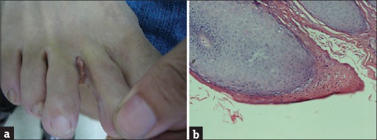
(a) Spiky cauliflower-shaped verrucous lesions in case 1. (b) Parakeratosis and hyperkeratosis in the epidermis, thickened granular layer, and acanthosis. In the granular layer, giant cells with vacuolated cytoplasm (koilocytotic cells) were present, indicating human papillomavirus infection in case 1(haematoxylin and eosin staining; original magnification, ×100)
Case 2: A 72-year-old male patient presented to our hospital with asymptomatic interdigital toe lesions that had first appeared about 3 months earlier. He was a retired worker who liked taking a long walk after dinner. He had well-controlled hypertension. Physical examination showed macerated flat pale patchy verrucous lesions with excoriation in the third interdigital areas of the left feet [Figure 2a]. Dermatophytes were detected positive in the second, third, and fourth interdigital areas.
Figure 2.
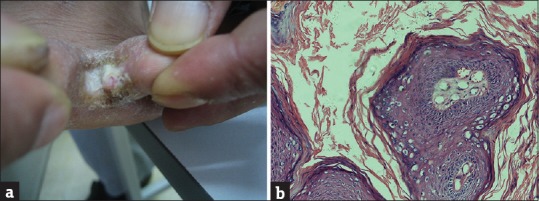
(a) Macerated flat pale patchy verrucous lesions with excoriation in case 2. (b) case 2 (haematoxylin and eosin staining; original magnification, ×100)
There were no other verrucous lesions on the whole body in these two patients. All biochemical examinations showed the results within normal limits, and anti-HIV type 1 and 2 antibodies of the patients’ serum were negative. The pathological features of the lesion were papillomatosis; giant cells with vacuolated cytoplasm (koilocytotic cells) were present in the granular layer, indicating HPV infection [Figures 1b and 2b]. From the clinicopathological findings, the diagnosis of WTW was made. Surgical removal and cauterization by microwave treatment were performed, and topical antifungal agents were applied for 1 month. No recurrence has been observed for the 12 months to date in these two cases.
DNA and mRNA extraction from skin lesions was done by Shanghai Sangon Biological Engineering Technology & Services Co., Ltd., (Shanghai, China). Complementary DNA [cDNA] was synthesized by reverse transcription from mRNA. DNA and cDNA specimens were kept at −20°C for polymerase chain reaction (PCR) amplification and HPV genotyping analysis.
Two degenerate primer systems, MY09/11 and CP65/70, were used to amplify HPV DNA or cDNA templates. In addition, to verify further the PCR product sequencing results, HPV7 type-specific primer sets were designed to amplify HPV7 DNA or cDNA.
The primers were designed according to the literature.[8] House primer (β-actin) was synthesized by ourselves, and the size was 207bp. The PCR reaction volume and conditions were the same as described previously.[8] The purified PCR products were sequenced in both directions by Shanghai Sangon Biological Engineering Technology & Services Co., Ltd., (Shanghai, China). The websites used for sequence blasting are available online at http://blast.ncbi.nlm.nih.gov/Blast.cgi.
The sequences of each primer and the size of the PCR products are shown in Table 1. PCR reaction conditions are shown in Table 2. After PCR amplification, products were subjected to 1.5% agarose gel electrophoresis and analyzed through a gel imaging system.
Table 1.
The primer sequences, and polymerase chain reaction product size
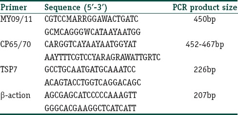
Table 2.
Polymerase chain reaction condition with general, house and type-special primers

The degenerate PCR amplification showed that DNA and cDNA specimens of two toe web tissue specimens were HPV positive. Figure 3 shows electrophoresis of PCR products, DNA sample as templates, and Figure 4 shows electrophoresis of PCR products, cDNA sample as templates. Using either MY09/11 or CP75/60, PCR sequence blasting results consistently revealed that two toe web specimens were positive with HPV 7.
Figure 3.
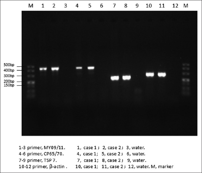
Electrophoresis of polymerase chain reaction products, DNA sample as templates
Figure 4.
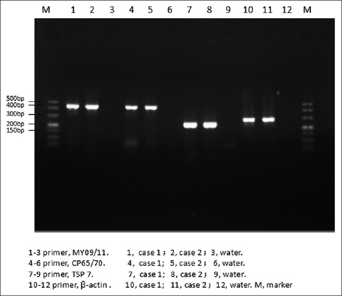
Electrophoresis of polymerase chain reaction products, complementary DNA sample as templates
To the best of our knowledge, only one case series describing similar warts has been reported, in which we summarized the features of 20 cases with WTW associated with HPV 7.[8] And recently, Miyauchi et al. reported an additional case of HPV 7-related WTW in 2015.[9] Our data show other two cases that HPV 7 may lead to interdigital, spiky warts or brownish plaques in immunocompetent patients who have not worked as butchers or fish handlers. Toe webs site as a wet environment and the presence of fungal infections may be the triggering factors.
Furthermore, in these two cases, cDNA samples were both positive for HPV 7. The sequencing results of the purified PCR products from cDNA samples were all positive with HPV 7, and no other HPV types were found. These results indicated that HPV 7 mRNA was detected in lesion tissue, suggesting a further association between the lesion and this virus. HPV7 may play a pathogenic role in WTW.
To the best of our knowledge, there is no further evidence which can prove HPV 7 played a pathogenic role in WTW yet. Here, we report that HPV7 mRNA was found in two cases with WTW. This paper is the first report of an evaluation of the prevalence of HPV7 mRNA in these specific subgroups of patients with warts. Larger samples and further molecular testing of the associations are recommended for future research.
Financial support and sponsorship
This study was financially supported by the opening program of Jiangsu Key Laboratory of Molecular Biology for Skin Diseases and STIs (No. 2015KF05), Nanjing, Jiangsu 210042, People's Republic of China.
Conflicts of interest
There are no conflicts of interest.
References
- 1.Oltersdorf T, Campo MS, Favre M, Dartmann K, Gissmann L. Molecular cloning and characterization of human papillomavirus type 7 DNA. Virology. 1986;149:247–50. doi: 10.1016/0042-6822(86)90126-1. [DOI] [PubMed] [Google Scholar]
- 2.Rüdlinger R, Bunney MH, Grob R, Hunter JA. Warts in fish handlers. Br J Dermatol. 1989;120:375–81. doi: 10.1111/j.1365-2133.1989.tb04163.x. [DOI] [PubMed] [Google Scholar]
- 3.Stehr-Green PA, Hewer P, Meekin GE, Judd LE. The aetiology and risk factors for warts among poultry processing workers. Int J Epidemiol. 1993;22:294–8. doi: 10.1093/ije/22.2.294. [DOI] [PubMed] [Google Scholar]
- 4.Greenspan D, de Villiers EM, Greenspan JS, de Souza YG, zur Hausen H. Unusual HPV types in oral warts in association with HIV infection. J Oral Pathol. 1988;17:482–8. doi: 10.1111/j.1600-0714.1988.tb01321.x. [DOI] [PubMed] [Google Scholar]
- 5.de Villiers EM. Prevalence of HPV 7 papillomas in the oral mucosa and facial skin of patients with human immunodeficiency virus. Arch Dermatol. 1989;125:1590. doi: 10.1001/archderm.125.11.1590. [DOI] [PubMed] [Google Scholar]
- 6.Völter C, He Y, Delius H, Roy-Burman A, Greenspan JS, Greenspan D, et al. Novel HPV types present in oral papillomatous lesions from patients with HIV infection. Int J Cancer. 1996;66:453–6. doi: 10.1002/(SICI)1097-0215(19960516)66:4<453::AID-IJC7>3.0.CO;2-V. [DOI] [PubMed] [Google Scholar]
- 7.Syrjänen S, von Krogh G, Kellokoski J, Syrjänen K. Two different human papillomavirus (HPV) types associated with oral mucosal lesions in an HIV-seropositive man. J Oral Pathol Med. 1989;18:366–70. doi: 10.1111/j.1600-0714.1989.tb01567.x. [DOI] [PubMed] [Google Scholar]
- 8.Sun C, Chen K, Gu H, Chang B, Jiang M. Association of human papillomavirus 7 with warts in toe webs. Br J Dermatol. 2010;162:579–86. doi: 10.1111/j.1365-2133.2009.09564.x. [DOI] [PubMed] [Google Scholar]
- 9.Miyauchi T, Moriuchi R, Hamade Y, Suzuki S, Nomura T, Shimizu S. Warts in toe webs associated with human papillomavirus type 7: A specific cutaneous manifestation of this type? Br J Dermatol. 2016;174:678–81. doi: 10.1111/bjd.14190. [DOI] [PubMed] [Google Scholar]


