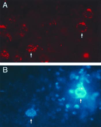
Neurobiology. In the article “Structural recovery in lesioned adult mammalian spinal cord by x-irradiation of the lesion site” by Nurit Kalderon and Zvi Fuks, which appeared in number 20, October 1, 1996, of Proc. Natl. Acad. Sci. USA (93, 11179–11184), the following correction should be noted. Due to a printer’s error, Fig. 5 A and B was unsatisfactorily reproduced and a better version appears below.
Figure 5.

Degree of recovery of axotomized CS neurons—retrograde double-labeling. Micrographs of a cortical section (A and B) taken from rat with lesioned and irradiated cord; this section was photographed for diI labeling (red) (A) and for FB labeling (blue) (B). Seen in this section are diI–labeled CS cells (i.e., axotomized) (A) but only 2 of them (arrows) are double-labeled; these 2 cells (in B) are also labeled by FB (arrows) (i.e., regrown axons 10 mm into the distal stump). Note the FB-labeled dendrites surrounding the neuron on the right (B). (C) The individual sums of the DL–CS neurons in each of the rats (bars) with lesioned cords which were untreated (n = 8) and treated (n = 23) with different doses of x-rays are plotted. Our measured number of 600 CS neurons projecting normally to the application site of FB was used as the maximal expected sum of DL–CS neurons. Cord injury in the 18.5-Gy-treated group included also the suture-loop procedure, and two rats of the 15-Gy-treated group were analyzed at 41 days PI. Note the increase in the sums of DL–CS in the groups treated with 17.5–20 Gy.


