

Neurobiology. In the article “Severed corticospinal axons recover electrophysiologic control of muscle activity after x-ray therapy in lesioned adult spinal cord” by Nurit Kalderon and Zvi Fuks, which appeared in number 20, October 1, 1996, of Proc. Natl. Acad. Sci. USA (93, 11185–11190), the following correction should be noted. Due to a printer’s error, Fig. 5 was unsatisfactorily reproduced and a better version appears below.
Figure 5.
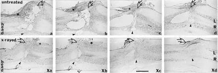
The morphological features of the lesion site in two lesioned cords, unirradiated (a–e) and irradiated (Xa–Xe), obtained from the rats whose EMG recordings are shown in Fig. 3B and Fig. 3C, respectively. Serial reconstruction of the two cords are shown; each panel is a composite of thionin-stained horizontal sections taken from different regions along the dorsal–ventral direction. Indicated are the left (L) and the right (R) hemicords, the extent of incision traversing from left (clear arrow) past the midline and into the right hemicord (arrowhead), and the dorsal nerves (asterisks). Note in the untreated cord the cavitation and tissue degeneration throughout the entire volume surrounding the incision site. In the irradiated cord the incision disappeared and an almost complete structural continuity was established. (Bar = 1 mm.)