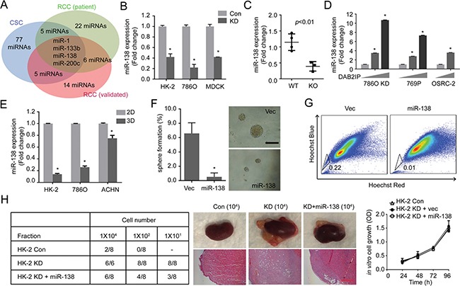
Figure 2. miR-138 is regulated by DAB2IP to suppress stem-like phenotypes in RCC.

(A) Potential miRNAs candidates were analyzed from different data sources: DAB2IP-regulated miRNAs (Blue), Screening of tumor suppressive miRNAs from RCC patients (Green) [39], and Profiling of miRNAs during RCC progression (Red) [40]. (B and C) Expression levels of miR-138 were determined in Con vs. KD renal cells (B), and in wild type (WT) vs. DAB2IP knock-out (KO) mice tissues (C). (D) Effect of DAB2IP on the expression levels of miR-138 expression. (E) miR-138 expressions were compared between monolayer (2D) and sphere (3D) culture condition of HK-2, 786O or ACHN cells. (F and G) HK-2 KD cells were transfected with miR-138 expression vector. The sphere forming ability (E), and SP (G) were determined. (H) Cancer-initiating ability of each subline was determined using orthotopic xenograft model after 8 weeks post-injection. Scale bar, 250 μm.
