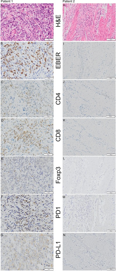
Figure 1. Representative examples of H&E staining, EBER (Epstein-Barr Virus-encoded RNA) in situ hybridization and CD4 (cluster of differentiation 4), CD8 (cluster of differentiation 8), Foxp3 (Forkhead box P3), PD-1 (programmed death 1), and PD-L1 (programmed death ligand-1) immunohistochemistry staining in 2 patients.

Patient 1 (A, B, C, D, E, F and G) was positive for EBER, CD4, CD8, Foxp3, PD-1 and PD-L1 (Shown at ×200 original magnification), whereas patient 2 (H, I, J, K, L, M and N) was negative for EBER, CD4, CD8, Foxp3, PD-1 and PD-L1 (shown at ×100 original magnification).
