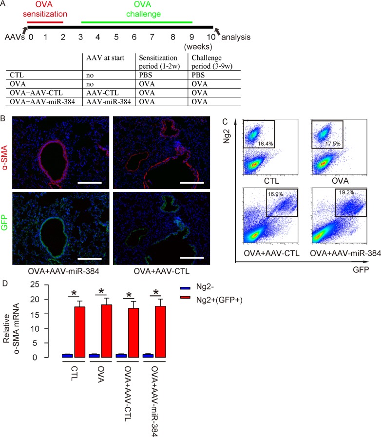
Figure 4. Successful in vivo re-expression of miR-384 in ASM cells.
(A) Schematic of the experiment: AAVs were used to treat mice at the beginning of OVA sensitization. Four group of mice of 10 of each were included in this experiment. Group 1, the mice received saline only as control for OVA (CTL). Group 2, mice received OVA treatment only (OVA). Group 3, mice received OVA and intranasal injection of AAV-CTL (OVA+AAV-CTL). Group 4, mice received OVA and intranasal injection of AAV-miR-384 (OVA+AAV-miR-384). (B) Immunostaining for α-SMA and GFP in AAVs/OVA-treated mice. Nuclei were stained with DAPI. (C) (Transduced) ASM cells were thus isolated from 4 groups, shown by representative flow charts. (D) RT-qPCR for α-smooth muscle actin (α-SMA) in Ng2+(GFP+) and Ng2- cells. *p<0.05. NS: non-significant. N=10. Scale bars are 100 μm.

