

Trichorrhexis nodosa (TN) is a well-known entity which affects hair shafts. Clinically, it presents as minute nodular concretions along the hair shaft. This is caused by the loss of cuticle and cortical fibers. On light microscopy, tiny nodules appear as “thrust paint brushes” as if two brushes are thrust into each other.[1]
Trichoscopy is a noninvasive diagnostic tool. It allows the detailed visualization of hair with respect to structure and size, perifollicular areas, and scalp.[2] It provides clues for inherited and acquired causes of hair loss, and helps in the diagnosis of several hair shaft disorders.[3] Here, the authors describe the importance of trichoscopy in the diagnosis of an isolated TN.
A 38-year-old male presented to the Dermatology outpatient department with a feeling that something was there at the tips of the eyebrow hairs since 1 year [Figure 1]. There was no history of trauma, itching, or topical application. Patient denied any manipulation of eyebrows. Examination revealed tiny white-to-brownish nodes on the tips of hairs of bilateral eyebrows. No similar findings were found in the eyelashes, scalp, or other hairy areas of the body. There was no scaling in the eyebrows or scalp. Systemic examination was unremarkable. Trichoscopy was performed using videodermoscopy, which showed that hair shafts were broken at multiple places with “paint brush” like ends [Figure 2; 20×]. Both proximal and distal hairs were affected resulting in hair shaft breakage giving characteristic “thrust paint brush” appearance [Figure 3; 70×]. Based on trichoscopic examination, a diagnosis of TN isolated to eyebrows was made.
Figure 1.
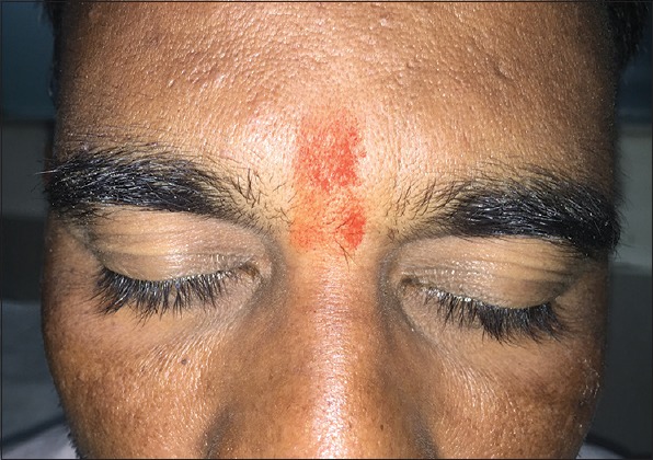
Clinical image showing tiny white-to-brownish nodes on the tips of hairs of eyebrows
Figure 2.

Trichoscopy using videodermoscopy shows broken hair shafts with frayed ends appearing as a paint brush. These are suggestive of tiny nodes clinically. (Nonpolarized mode, 20× magnifications; FotoFinder®, Germany)
Figure 3.
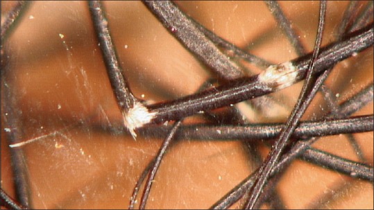
Trichoscopy using videodermoscopy shows “thrust paint brushes” pattern as if two brushes are thrust into each other. (Nonpolarized mode, 70× magnifications; FotoFinder®, Germany)
Trichoscopically, TN, trichorrhexis invaginata, and hair casts were considered as possible differential diagnosis. Trichorrhexis invaginata is caused by brittle hairs and is characteristic of hair shaft change in Netherton syndrome. It appears as “ball-and-socket” under trichoscopy. Hair casts are remnants of inner root sheaths and appear as cylindrical transulant casings around the hair shaft in trichoscopy.[4,5] In our case, trichoscopy showed “thrust paint brush” pattern confirming the diagnosis. TN can be associated with argininosuccinic aciduria, Menkes’ kinky hair syndrome, Netherton's syndrome, hypothyroidism, or trichothiodystrophy.[6]
Hence, trichoscopy is a useful diagnostic tool in TN; in this case, it diagnosed isolated TN which was confined to eyebrows. Authors suggest the usage of trichoscopy in daily practice, especially in hair disorders.
Financial support and sponsorship
Nil.
Conflicts of interest
There are no conflicts of interest.
References
- 1.Kharkar V, Gutte R, Thakkar V, Khopkar U. Trichorrhexis nodosa with nail dystrophy: Diagnosis by dermoscopy. Int J Trichol. 2011;3:105–6. doi: 10.4103/0974-7753.90823. [DOI] [PMC free article] [PubMed] [Google Scholar]
- 2.Tosti A, Ross EK. Patterns of scalp and hair disease revealed by videodermoscopy. In: Tosti A, editor. Dermoscopy of hair and scalp disorders. 1st ed. London: Informa healthcare; 2007. pp. 1–14. [Google Scholar]
- 3.Rudnicka L, Rakowska A, Kerzeja M, Olszewska M. Hair shafts in trichoscopy: Clues for diagnosis of hair and scalp diseases. Dermatol Clin. 2013;31:695–708. doi: 10.1016/j.det.2013.06.007. [DOI] [PubMed] [Google Scholar]
- 4.Pinheiro AM. Acquired Trichorrhexis Nodosa in a Girl: The Use of Trichoscopy for Diagnosis. J Dermatolog Clin Res. 2016;4:1064. [Google Scholar]
- 5.Adya AK, Inamadar AC, Palit A, Shivanna R, Deshmukh NS. Light Microscopy of the Hair: A Simple Tool to “Untangle” Hair Disorders. Int J Trichol. 2011;3:46–56. doi: 10.4103/0974-7753.82124. [DOI] [PMC free article] [PubMed] [Google Scholar]
- 6.Rakowska A, Slowinska M, Kowalska-Oledzka EL. Trichoscopy in genetic hair shaft abnormalities. J Dermatol Case Rep. 2008;2:14–20. doi: 10.3315/jdcr.2008.1009. [DOI] [PMC free article] [PubMed] [Google Scholar]