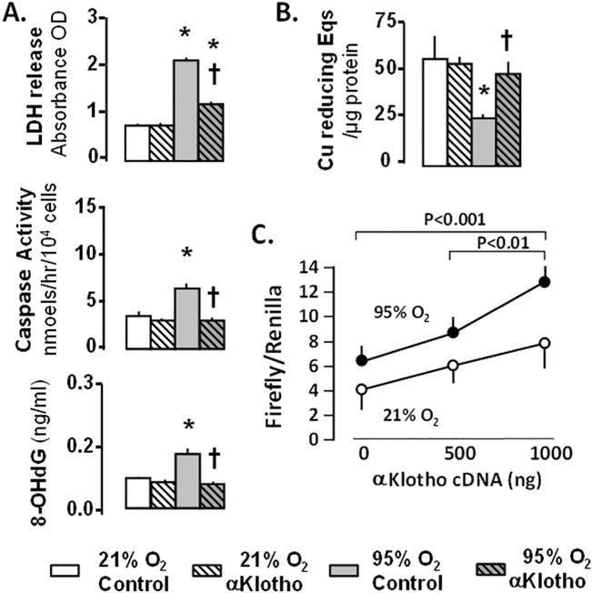
Fig. 4.

A549 lung epithelial cells exposed to 21% or 95% O2 with/without αKlotho. A. Cell death measured by lactate dehydrogenase (LDH); apoptosis measured by caspase-8; oxidative damage measured by 8-hydroxy-deoxyguanosine (8-OHdG). B. Antioxidant capacity measured by copper (Cu) reducing equivalents (Eqs). * 95% vs. 21% O2 at the same αKlotho state; † vs. Control treatment at the same O2. C. A549 cells transfected with αKlotho cDNA: Luciferase antioxidant response element (ARE) reporter assay. Data are from [3].
