

In the August 1998 issue, attribution for the cover artwork and accompanying legend was inadvertently omitted. Both were generously provided by Benjamin Glick of the University of Chicago. The figure and legend are reprinted here. The cover figure (Friend, D.S. and Murray, M.J. [1965]. Am J Anat 117, 135–150. Plate 2) was reprinted with permission of Wiley-Liss Inc., a division of John Wiley & Sons, Inc.
Cover
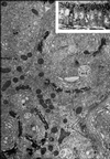
This year marks the 100th anniversary of the discovery by Camillo Golgi of an “internal reticular apparatus” in eukaryotic cells. The cover figure, from a classic paper by Friend and Murray (Friend, D.S., and Murray, M.J. [1965]. Am. J. Anat. 117, 135–150), highlights the pivotal role played by electron microscopy in developing the modern view of the Golgi apparatus. In the earlier phase of Golgi research, this organelle has been visualized by light microscopy, often using osmium as a specific stain. For example, the inset of the figure is a light micrograph showing mouse epithelial cells that were stained with osmium and then sectioned parallel to their long axis. The Golgi apparatus appears as a reticulum above the nucleus. The remainder of the figure shows the same preparation of osmicated cells, except in this case they were sectioned perpendicular to their long axis and viewed by electron microscopy. Such images revealed that the Golgi reticulum comprises multiple interconnected stacks of membrane cisternae. Remarkably, Friend and Murray found that osmium selectively stains the cisternae on one side of each Golgi stack. This result provided some of the first concrete evidence that the Golgi apparatus is biochemically polarized. Today, a third of a century later, cell biologists are still striving to understand the mechanisms that generate polarized Golgi stacks.—Benjamin Glick