Summary
Background
Renal and bladder ultrasound (RBUS) is recommended in evaluation of children after an initial, febrile urinary tract infection. Although it is not uncommon to observe debris within the bladder lumen on sonography, the significance of this finding is uncertain. Debris may be interpreted as an indication of ongoing infection, but there have been no studies to date investigating the association of bladder debris with a positive culture.
Objective
The aim of this study was to evaluate the association of bladder debris noted at the time of RBUS with positive urine culture results obtained from a catheterized specimen, among patients undergoing RBUS and voiding cystourethrogram (VCUG) on the same day.
Study design
We performed a retrospective cross-sectional study of 3,995 patients who presented for same-day RBUS and VCUG. RBUS reports were reviewed for the presence of bladder debris, and analysis was limited to patients under 60 months of age with a catheterized urine specimen sent for culture at the time of the studies. Those with prior postnatal imaging or a diagnosis of prenatal hydronephrosis or other GU abnormalities were excluded. Thirty-four subjects with bladder debris on RBUS were identified and matched to 155 controls based on age, gender, circumcision status, and presence of vesicoureteral reflux. A positive urine culture was defined as ≥50,000 colony forming units per mL of at least one organism. A conditional logistic regression model was used to evaluate the association between debris on RBUS and positive urine culture results.
Results
In conditional logistic regression stratifying by matching age, gender, circumcision status, and presence of vesicoureteral reflux, there was a statistically significant association between bladder debris on RBUS and positive urine culture result collected on the same day during VCUG (OR 7.88, 95% CI 1.88–33.04, p=0.0048). This corresponds to a 688% increase in odds of positive urine culture for patients with debris (Table).
Discussion
This is the first study to evaluate the association between bladder debris on RBUS and positive urine culture, and it should serve as a starting point for future investigations. The study is limited in its generalizability to the sampled population; further work should evaluate the predictive value of RBUS debris among children without UTI history, with prior imaging or known genitourinary anomalies, or older children.
Conclusion
Among children younger than 60 months old undergoing initial imaging for history of UTI, there is a significant association between bladder debris and a positive urine culture.
Introduction
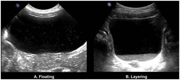
Renal and bladder ultrasound (RBUS) is recommended in the evaluation of children after an initial, febrile urinary tract infection (UTI) [1]. As a part of RBUS, the American College of Radiology recommends that the bladder walls and lumen be evaluated [2,3]. Findings may include stones, masses, or mobile debris in the lumen (Fig. 1). Bladder debris in particular is observed commonly, and is often attributed to infection. Small studies in symptomatic adult women have found a relatively high prevalence of debris (61.6–72.0%) [4,5], but data regarding children and asymptomatic adults are lacking. Thus, while debris is commonly considered to be abnormal, the clinical significance of this finding, and in particular its relationship with urinary tract infection, remains uncertain.
Figure 1.
Representative ultrasound images of patients with floating and layering debris.
In some cases, intraluminal echoes on sonography may be technical artifacts. These can result from scanning technique or settings, or from physical limitations of the imaging modality itself [6]. Of greater concern is the scenario in which echogenic debris reflects a true pathologic process. Patients with bladder mucus, gross hematuria, inflammation of the genitourinary system, or sloughing of urothelium can demonstrate debris and linear stranding on ultrasound [7]. Such echogenic debris also has been reported in the setting of acute UTI with associated pyuria [8].
The management of patients in whom bladder debris is seen on RBUS is also uncertain and may be based on many clinical factors (e.g. ongoing fever, urinary symptoms, etc.). One recent study in adults concluded that bladder debris is not significantly associated with an abnormal urinalysis [3]. In that study, the imaging and urine sample were obtained asynchronously – within 1 week - and in a non-sterile fashion. Furthermore, active symptomatology at the time of study was not considered. Two other adult studies of symptomatic individuals suggested no association between sonographic debris and positive urine culture [4,5]. To date, the pediatric literature lacks any investigation of a potential association between bladder debris on RBUS and UTI (as indicated by positive urine culture). This is of particular importance for children undergoing initial work-up of a urinary tract infection (UTI), for whom the risk of UTI may be elevated, and in whom bladder debris may be an indicator of persistent or recurrent infection.
At our institution, it is standard practice to send a catheterized urine specimen for culture on all patients undergoing voiding cystourethrogram (VCUG), and many children undergo VCUG and RBUS on the same day as part of their work-up for a prior UTI. The aim of this study was to evaluate the association of bladder debris noted at the time of RBUS with positive urine culture results from catheterized VCUG specimens, among patients undergoing both studies on the same day. We hypothesized that the presence of sonographic debris would be significantly associated with a positive synchronous urine culture in otherwise asymptomatic children undergoing evaluation for a prior UTI.
Methods
We performed a retrospective, cross-sectional study of all patients presenting for same-day RBUS and VCUG, after obtaining institutional review board approval with a waiver of consent.
Data source and patient selection
We reviewed institutional billing records between Jan 1, 2006 and Dec 31, 2010 to identify encounters during which patients underwent both a RBUS (Current Procedural Technology [CPT] code 76700, 76705, 76770, 76775, 76856, or 76857) and VCUG (CPT code 74455) on the same day. For all patients, RBUS was completed according to a standardized, institutional protocol requiring transverse and sagittal views of the bladder. The results of these studies were obtained from the text of radiology reports, and clinical information, including the results of urine cultures obtained at the time of VCUG, was abstracted from the electronic medical record. The RBUS data were collected as a part of two, previously published studies [9,10] and were secondarily analyzed with VCUG culture results, which were collected for this study.
The cohort analyzed in the present study included all patients <60 months who underwent both initial RBUS and VCUG on the same day for a history of UTI. We excluded those patients with prior, postnatal GU imaging, as well as those who lacked a urine culture obtained at time of VCUG. Furthermore, we excluded children with a history of prenatal hydronephrosis or other prenatal GU abnormalities.
Imaging data abstraction and urine culture classification
Recorded RBUS and VCUG findings were based on radiology reports. When a finding was not commented on, it was assumed not to have been present. The presence of debris was based on inclusion of this finding in the results or impression section of the report. Vesicoureteral reflux status was based on its presence in the results or impression section of the VCUG report from the same day. For urine cultures, colony counts and speciation were recorded. Based on the American Academy of Pediatrics Guidelines on UTI [11], we defined the threshold for a “positive” urine culture as the presence of ≥ 50,000 colony forming units/mL [1].
Data analysis
Patients identified with bladder debris were matched to controls based on age, gender, circumcision status, and presence of vesicoureteral reflux. Age-matching of controls to subjects was conducted within a range of ±1 month, except for one subject (aged 47 months) who was matched to two controls aged 50 and 53 months, as no closer matches were available. When possible (24 cases), a 5:1 ratio of controls to cases was used for matching. Alternatively, 4:1 (seven cases), 3:1 (one case), and 2:1 (two cases) ratios of controls to cases were used when necessary. Descriptive statistics were calculated for the debris and non-debris groups. Conditional logistic regression stratified by matching on age, gender, circumcision status, and presence of reflux was used to determine whether positive urine culture was associated with bladder debris. Analyses were performed using SAS 9.4 (SAS Institute Inc; Cary, NC) and R v.2.15.2 (http://www.R-project.org).
Ethical approval
Approval was obtained from the institutional review board.
Results
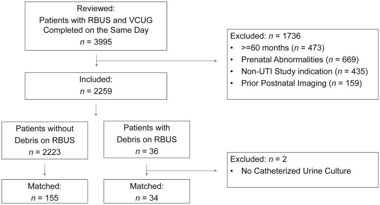
A total of 3,995 patients were identified who had a RBUS and VCUG completed on the same day during the study period. Of these, 1,736 were initially excluded who met any of the following criteria: ≥60 months, prior postnatal imaging, prior diagnosis of prenatal GU abnormalities, or a non-UTI study indication. The 2259 remaining patients were further reviewed, and of these, 36 (1.6%) were identified with debris on RBUS. Of these, two patients with bladder debris were excluded because of lack of a catheterized urine specimen. A total of 34 patients with bladder debris were therefore successfully matched to 155 controls for the final analysis (Fig. 2).
Figure 2. Patient Selection for Study Inclusion and Matching.
The median age of the cohort was 18 months (IQR 5–35), and 128 (67.7%) of the included patients were female (Table 1). Of the 61 males, 41 (67.2%) were uncircumcised, five (8.2%) were circumcised, and 15 (24.6%) were of unknown circumcision status. Among all included patients, eight (4.2%) of the collected urine specimens met the AAP threshold of 50,000 CFU/mL to qualify as a positive culture (summary Table). Of patients with debris, 5/34 (14.7%) demonstrated a positive urine culture, with 3/155 (1.9%) patients without debris demonstrating a positive urine culture. Escherichia coli (2/8 (25%)), Enterococcus (2/8 (25%)), and Proteus mirabilis (2/8 (25%)) were the most frequent organisms, while Enterobacter Cloacae (1/8 (12.5%)) and Proteus vulgaris (1/8 (12.5%)) populated the remaining cultures (summary Table).
Table 1. Characteristics of children <60 months undergoing renal bladder ultrasound (RBUS) and voiding cystourethrogram (VCUG) on the same day for a history of UTI.
| Children with bladder debris on RBUS n=34 | Children without bladder debris on RBUS n=155 | All included children n=189 | |
|---|---|---|---|
| Gender (%) | |||
| Female | 22 (64.7) | 106 (68.4) | 128 (67.7) |
| Male: | |||
| circumcised | 1 (2.9) | 4 (2.6) | 5 (2.7) |
| Male: | |||
| uncircumcised | 8 (23.5) | 33 (21.3) | 41 (21.7) |
| Male: status | |||
| unknown | 3 (8.8) | 12 (7.7) | 15 (7.9) |
| Age, months | |||
| Median (IQR) | 18 (4.8–36) | 18 (5–35) | 18 (5–35) |
| 0–1 (%) | 4 (11.8) | 13 (8.4) | 17 (9.0) |
| 2–6 | 8 (23.5) | 40 (25.8) | 48 (25.4) |
| 7–12 | 3 (8.8) | 16 (10.3) | 19 (10.0) |
| 13–18 | 2 (5.9) | 10 (6.5) | 12 (6.4) |
| 19–24 | 2 (5.9) | 11 (7.1) | 13 (6.9) |
| 25–59 | 15 (44.1) | 65 (41.9) | 80 (42.3) |
| Vesicoureteral reflux (%) | |||
| Yes | 19 (55.9) | 88 (56.8) | 107 (56.6) |
| No | 15 (44.1) | 67 (43.2) | 82 (43.4) |
Summary Table.
| Children with Bladder Debris on RBUS n=34 | Children without Bladder Debris on RBUS n=155 | All Included Children n=189 | |
|---|---|---|---|
| Gender (%) | |||
| Female | 22 (64.7) | 106 (68.4) | 128 (67.7) |
| Male: Circumcised | 1 (2.9) | 4 (2.6) | 5 (2.7) |
| Male: Uncircumcised | 8 (23.5) | 33 (21.3) | 41 (21.7) |
| Male: Status Unknown | 3 (8.8) | 12 (7.7) | 15 (7.9) |
| Age | |||
| Median (IQR) | 18 (4.8-36) | 18 (5-35) | 18 (5-35) |
| 0-1 mo (%) | 4 (11.8) | 13 (8.4) | 17 (9.0) |
| 2-6 mo | 8 (23.5) | 40 (25.8) | 48 (25.4) |
| 7-12 mo | 3 (8.8) | 16 (10.3) | 19 (10.0) |
| 13-18 mo | 2 (5.9) | 10 (6.5) | 12 (6.4) |
| 19-24 mo | 2 (5.9) | 11 (7.1) | 13 (6.9) |
| 25-59 mo | 15 (44.1) | 65 (41.9) | 80 (42.3) |
| Vesicoureteral Reflux (%) | |||
| Yes | 19 (55.9) | 88 (56.8) | 107 (56.6) |
| No | 15 (44.1) | 67 (43.2) | 82 (43.4) |
In conditional logistic regression stratifying by matching age, gender, circumcision status, and presence of vesicoureteral reflux, there was a statistically significant association between bladder debris on RBUS and positive urine culture result collected on the same day during VCUG (OR 7.88, 95% CI 1.88–33.04, p=0.0048). This corresponds to a 688% increase in odds of positive urine culture for patients with debris.
Discussion
Despite the prevalence of bladder debris seen in clinical practice, surprisingly little has been written on its significance, including its potential association with positive urine cultures. A recent study in the radiologic literature failed to correlate intraluminal debris with “abnormal” urinalysis results; however, urine specimens were collected asynchronously with the bladder ultrasound [3]. Our ability to review a large number of catheterized urine specimens, collected nearly simultaneously with the RBUS, is a unique strength. We conclude that among a select pediatric population (<60 months, presenting for initial work-up of a prior UTI, and without prior postnatal imaging or prenatal GU abnormalities) there exists a significant association between bladder debris and a positive urine culture. In conditional logistic regression stratifying by matching age, gender, circumcision status, and presence of vesicoureteral reflux, children with debris demonstrate a 688% increased odds of a positive urine culture compared with those children without debris.
These results support our hypothesis that there is an association between bladder debris and positive urine culture. Previous authors have commented on the lack of sensitivity and specificity of echogenic debris in the renal collecting system for the diagnosis of pyonephrosis [12]. Just as sonographic echoes can be seen in uninfected hydronephrosis, it seems logical to assume that not all cases of pyuria can be differentiated by such echogenic features on ultrasound. Scanning technique or machine settings can lead to artifact in some cases [6]. Even in scenarios in which there is true particulate matter in the setting of a recent infection, like many of the patients in our cohort, it is attractive to dismiss debris as a product of inflammation: sloughed urothelium that is otherwise sterile. However, the results of our study suggest that these patients are at increased risk of having a positive culture, if a specimen is sent.
At our institution, all children undergoing VCUG have a culture routinely sent from the initial catheterization, if urine is obtained. We do not, however, routinely culture children based on RBUS findings alone. The overall positive culture rate for children undergoing VCUG and RBUS on the same day is approximately 4% (unpublished data), which is much lower than that seen in children with bladder debris on RBUS. Thus, one might argue that the routine ordering of cultures for all subjects undergoing this imaging should be abandoned, while culture should be actively sought in those patients with findings predictive of positive culture, such as debris. It would clearly be helpful to identify specific populations of children undergoing cystography in whom a culture is most likely to be positive, as well as children in whom a culture may be safely omitted. Sonographic debris may be one helpful predictor in appropriately identifying such “at risk” patients.
The results of this study should be interpreted in light of its limitations. Methodologically, this study represents a secondary analysis of data collected for another study [9,10]. Given the specific inclusion and exclusion criteria, this sample is not representative of all pediatric patients evaluated at our institution, or even of all pediatric patients undergoing RBUS. Strictly speaking, these findings are applicable only to patients undergoing RBUS and VCUG for evaluation of prior UTI, and may not be generalizable to other populations. Extrapolation of these findings to older children, children with no UTI history, those who are undergoing initial imaging for prenatal hydronephrosis, or children with known GU anomalies or conditions undergoing follow-up imaging, should be approached with caution. We further recognize that such a secondary analysis limits the covariates available for consideration in our model. Nonetheless, we were able to control for several factors which have been associated with UTIs. Circumcision status in particular has a well-established relationship to febrile UTIs, with uncircumcised boys <1 years of age having an increased risk [1].
It is uncertain whether the association of debris with positive culture would be different among children undergoing imaging when they have active signs and symptoms of infection. It is also important to note that these data are based on the radiology report as dictated; we did not independently review the images to determine the presence of bladder debris. These findings were somewhat subjective, and we did not establish a priori criteria for identifying debris. It is possible that radiologists at other institutions might interpret these same images differently, with resulting impact on the analysis.
Despite these limitations, this remains the only study to consider the association of bladder debris on RBUS with synchronous positive culture among children, and we hope that this serves as a useful starting point for future investigations. Specifically, there is further work to be done to determine how the finding of bladder debris on ultrasound should inform clinical decision-making with respect to urine cultures and other testing.
Conclusion
Among children younger than 60 months old undergoing initial imaging for history of UTI, there is a significant association between bladder debris and a positive urine culture, with children with debris over seven times more likely to have a positive culture than those without debris.
Table. Urine culture results for patients with and without bladder debris on renal bladder ultrasound (p=0.0048).
| Bladder debris (n=34) | No bladder debris (n=155) | ||||
|---|---|---|---|---|---|
| Urine culture | Positive | 5 (14.7%) |
E. coli (1) Enterococcus (1) P. mirabilis (2) P. vulgaris (1) |
3 (1.9%) |
E. coli (1) Enterococcus (1) E. cloacae (1) |
| Negative | 29 (85.3%) | 152 (98.1%) | |||
Acknowledgments
Funding: None.
Footnotes
Conflict of interest: None.
Publisher's Disclaimer: This is a PDF file of an unedited manuscript that has been accepted for publication. As a service to our customers we are providing this early version of the manuscript. The manuscript will undergo copyediting, typesetting, and review of the resulting proof before it is published in its final citable form. Please note that during the production process errors may be discovered which could affect the content, and all legal disclaimers that apply to the journal pertain.
References
- 1.Wan J, Skoog SJ, Hulbert WC, Casale AJ, Greenfield SP, Cheng EY, et al. Section on Urology response to new Guidelines for the diagnosis and management of UTI. Pediatrics. 2012;129:e1051–3. doi: 10.1542/peds.2011-3615. [DOI] [PubMed] [Google Scholar]
- 2.American College of Radiology. ACR-AIUM-SPR-SRU Practice Parameter for the Performance of an Ultrasound Examination of the Abodmen and/or Retroperitoneum. Diagnostic Radiology: Genitourinary Imaging Practice Parameters. 2012 [Google Scholar]
- 3.Cheng SN, Phelps A. Correlating the Sonographic Finding of Echogenic Debris in the Bladder Lumen With Urinalysis. J Ultrasound Med. 2016;35:1533–40. doi: 10.7863/ultra.15.09024. [DOI] [PubMed] [Google Scholar]
- 4.Wachsberg RH, Festa S, Samaan P, Estrada HJ, Baker SR. Particulate Echoes within the Bladder Detected with Transvaginal Sonography: A Sign of Urinary Tract Infection? Emergency Radiology. 1998;5:137–9. [Google Scholar]
- 5.Wilches C, Gallo A, Moreno A, Rivero O, Romero J. Particulate Echoes within the Bladder: Does this Finding Correlate with Urinary Tract Infection? Rev Colomb Radiol. 2011;22:3334–40. [Google Scholar]
- 6.Feldman MK, Katyal S, Blackwood MS. US artifacts. Radiographics. 2009;29:1179–89. doi: 10.1148/rg.294085199. [DOI] [PubMed] [Google Scholar]
- 7.Hertzberg BS, Bowie JD, King LR, Webster GD. Augmentation and replacement cystoplasty: sonographic findings. Radiology. 1987;165:853–6. doi: 10.1148/radiology.165.3.3317511. [DOI] [PubMed] [Google Scholar]
- 8.Subramanyam BR, Raghavendra BN, Bosniak MA, Lefleur RS, Rosen RJ, Horii SC. Sonography of pyonephrosis: a prospective study. AJR Am J Roentgenol. 1983;140:991–3. doi: 10.2214/ajr.140.5.991. [DOI] [PubMed] [Google Scholar]
- 9.Nelson CP, Johnson EK, Logvinenko T, Chow JS. Ultrasound as a screening test for genitourinary anomalies in children with UTI. Pediatrics. 2014;133:e394–403. doi: 10.1542/peds.2013-2109. [DOI] [PMC free article] [PubMed] [Google Scholar]
- 10.Logvinenko T, Chow JS, Nelson CP. Predictive value of specific ultrasound findings when used as a screening test for abnormalities on VCUG. J Pediatr Urol. 2015;11:176 e1–7. doi: 10.1016/j.jpurol.2015.03.006. [DOI] [PMC free article] [PubMed] [Google Scholar]
- 11.Subcommittee on Urinary Tract Infection SCoQI, Management. Roberts KB. Urinary tract infection: clinical practice guideline for the diagnosis and management of the initial UTI in febrile infants and children 2 to 24 months. Pediatrics. 2011;128:595–610. doi: 10.1542/peds.2011-1330. [DOI] [PubMed] [Google Scholar]
- 12.Jeffrey RB, Laing FC, Wing VW, Hoddick W. Sensitivity of sonography in pyonephrosis: a reevaluation. AJR Am J Roentgenol. 1985;144:71–3. doi: 10.2214/ajr.144.1.71. [DOI] [PubMed] [Google Scholar]


