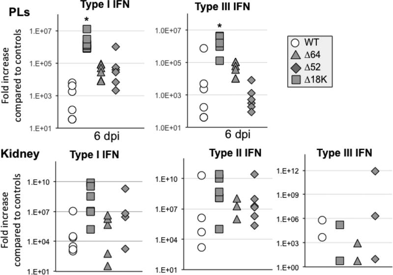
Fig. 8. Changes in expression by RT-qPCR of type I and III IFN genes in adult frog PLs and kidneys during infection with Δ52L- or Δ64R- and Δ18K-FV3 compared to WT-FV3.
Outbred adult frogs (6 individuals per group) were infected by i.p. injection of 1 ×10⁶ PFU of each virus type for 6 days (dpi). Results are average ± SEM fold increase relative to uninfected controls. **; P <0.001 and *; P <0.05 significant differences between WT- and KO-FV3 using one-way ANOVA test and Tukey’s post hoc test.

