Abstract
Background
Blepharospasm can be present as an isolated dystonia or in conjunction with other forms of cranial dystonia, causing significant disability.
Case Report
We report a case of a 69-year-old male with craniocervical dystonia, manifesting primarily as incapacitating blepharospasm refractory to medical treatments. He underwent bilateral globus pallidus (GP) deep brain stimulation (DBS) with complete resolution of his blepharospasm and sustained benefit at 12 months postoperatively.
Discussion
This case illustrates successful treatment of blepharospasm with pallidal stimulation. GP-DBS should be considered a reasonable therapeutic option for intractable blepharospasm.
Keywords: Deep brain stimulation, blepharospasm, cranial dystonia, globus pallidus internus, pallidal stimulation
Introduction
Deep brain stimulation (DBS) has an established role for treatment of primary generalized and cervical dystonia, but historically it has been considered to be relatively less effective for cranial and axial dystonia.1–4 Cranial dystonia has variable clinical manifestations, either blepharospasm alone, oromandibular dystonia alone, or both in combination, with the last one being termed Meige syndrome. Pallidal DBS for Meige syndrome has previously been described in case reports or small case series, with improvement in dystonia rating scales ranging from 45% to 89%, and with follow up ranging from 1 to 6 years.5–10 The results of these studies encourage the use of DBS in this population of patients.
Whether blepharospasm is focal or part of Meige syndrome, it can be the most incapacitating symptom. Blepharospasm is characterized by prolonged tonic and clonic spasms of eye closure, caused by recurrent contractions of orbicularis oculi.11 Blepharospasm can also less frequently resemble apraxia of eyelid opening with inability to open the eyes. There have been rare case reports of using globus pallidus interna (GPi) DBS for isolated blepharospasm.12,13 Table 1 summarizes the results of pallidal DBS in cases of isolated blepharospasm and the results of DBS on blepharospasm specifically in cases of Meige syndrome. Although traditionally it is rare to perform DBS for patients whose only major dystonic feature is blepharospasm, we demonstrate another successful outcome of pallidal stimulation for primarily blepharospasm in a patient with Meige syndrome.
Table 1. Published Cases of Pallidal DBS in Meige Syndrome and Isolated Blepharospasm.
| Number of Patients | Indication for DBS | Follow-up Duration (months) | Improvement in Blepharospasm (%)1 | Reference no. |
|---|---|---|---|---|
| 1 | Meige | 6 | Improved but not quantified | 7 |
| 11 | Meige | 12 | 63 | 8 |
| 5 | Meige | 10–124 | 88 | 9 |
| 6 | Meige | 6 | 79 | 5 |
| 6 | Meige | 12 | Improved in all patients but not quantified | 10 |
| 12 | Meige | 12–78 | 55 | 6 |
| 1 | Isolated blepharospasm | 1 | 63 | 12 |
| 1 | Isolated blepharospasm | 15 | 88 | 13 |
Abbreviations: DBS, Deep Brain Stimulation.
Each reference describes improvement in the Burke–Fahn–Marsden Dystonia Rating Scale.
Case report
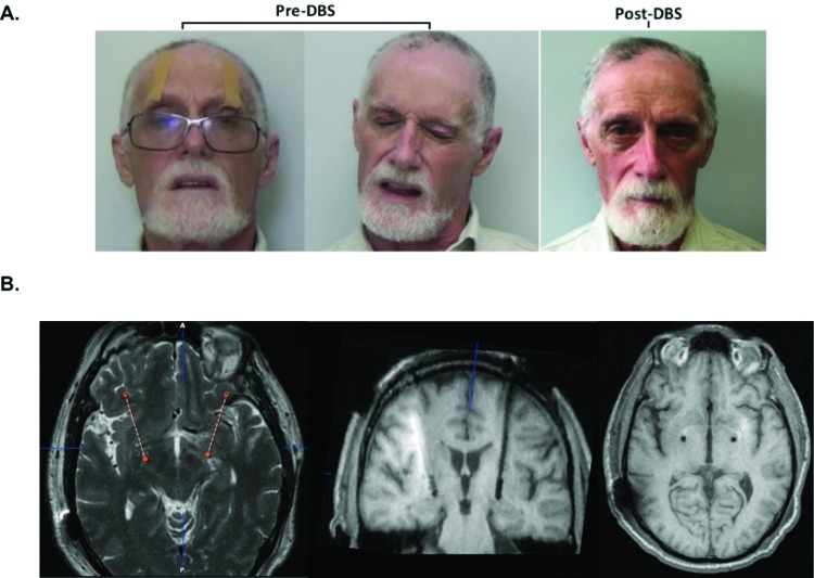
A 69-year-old male presented with an 8–9-year history of blepharospasm that began gradually and became debilitating. He had subtle intermittent activation of lower facial muscles and slight torticollis, prompting the diagnosis of Meige syndrome. His primarily tonic/clonic blepharospasm progressed until he was functionally blind unless he manually held his eyelids or taped them open (Figure 1A). He wore ptosis crutches mounted on eyeglasses with minimal relief. Over the course of 6 years, he was injected numerous times with botulinum toxin but with minimal benefit. A variety of toxins, including onabotulinumtoxinA (up to 75 units), incobotulinumtoxinA (up to 100 units), and abobotulinumtoxinA (up to 280 units), had been injected in the upper face muscles (orbicularis oculi, pretarsal orbicularis, corrugator, procerus, and frontalis). Clonazepam, up to 2 mg total daily, provided no appreciable benefit, and the dose could not be increased dose further because of sedation. He was reluctant to try other oral medications because of concern about similar side effects. His severe blepharospasm progressed, significantly impairing his quality of life, as he could no longer do activities he enjoyed such as driving, sailing, scuba diving, and biking.
Figure 1. Subject Photographs and MRI Imaging. (A) Subject’s blepharospasm pre- and post implantation of DBS. Pre-DBS, the subject used tape to hold his eyelids open. (B) T2-weighted MRI showing trajectory planning and T1-weighted images showing electrode placement in bilateral GP. DBS, Deep Brain Stimulation; GP, Globus Pallidus; MRI, Magnetic Resonance Imaging.
The patient was evaluated for DBS and had neuropsychological testing showing only mild deficits in attention and processing speed, possibly because of distraction from blepharospasm, without other significant concerns. He had a history of depression but was stable on an antidepressant. Magnetic resonance imaging (MRI) of the brain was normal and did not reveal an alternate etiology.
He underwent bilateral pallidal DBS placement using 1.5 T MRI-based direct targeting and microelectrode recordings with intraoperative test stimulation to determine thresholds for stimulation-induced adverse effects, as has been previously described.14 Medtronic DBS leads model 3387 (Medtronic Inc., Minneapolis, MN) were implanted, with the lowest contacts corresponding to the last recorded cell of the globus pallidus interna (GPi) and the highest contacts in the dorsal globus pallidus externa (GPe). Lead tip location was measured relative to the midcommissural point. A dual channel pulse generator (Medtronic Activa PC) was implanted in the chest wall. A postoperative MRI scan was performed to verify electrode position within the pallidum. No complications were observed during or after the operation.
Initial DBS programming occurred on postoperative day 10. After monopolar review, optimal DBS settings were found to be with monopolar stimulation using contact 1 and 9, pulse width 90 μs, frequency 150 Hz, and voltage 3.5 volts bilaterally. The coordinates of the active contacts with respect to the midcommissural point for left and right GPi respectively were lateral 22.2 mm and 20.9 mm, anterior–posterior 4.8 mm and 6.5 mm, and ventral –4.2 mm and –2.0 mm. Postoperative images of electrode placement are shown in Figure 1B.
The patient’s pre- and postoperative scores for the Burke–Fahn–Marsden Dystonia Rating Scale (BFMDRS) and Jankovic Rating Scale (JRS) are described in Table 2. Over the first month, he had 50–60% improvement in dystonia (BFMDRS total motor score improved from 15 at baseline to 6; BFMDRS blepharospasm score improved from 8 at baseline to 4; JRS improved from 8 to 4). At his 1-year follow-up visit, he reported 100% improvement in blepharospasm (BFMDRS blepharospasm and JRS scores improved to 0). BFMDRS total motor score improved to 1.5 or by 90% compared with baseline, and BFMDRS disability score improved from 8 at baseline to 2 or by 80% compared with baseline. His postoperative photograph and video show complete resolution of blepharospasm (Figure 1A, Video 1). He also had 88% improvement in his mild baseline cervical dystonia, with BFMDRS neck subscore improving from 4 at baseline to 0.5 at 1- month and 1-year follow ups. He no longer requires oral medications or botulinum toxin injections for blepharospasm. He did not report any stimulation-induced side effects over one year and has not required programming adjustments since his 1-month postoperative visit.
Table 2. Dystonia Severity at Baseline and Follow-up.
| Baseline | 1 Month Post Operation (improvement from baseline) | 1 Year Post Operation (improvement from baseline) | |
|---|---|---|---|
| BFMDRS total movement score | 15 | 6 (60%) | 1.5 (90%) |
| BFMDRS blepharospasm score | 8 | 4 (50%) | 0 (100%) |
| BFMDRS neck score | 4 | 0.5 (88%) | 0.5 (88%) |
| BFMDRS disability score | 10 | 2 (80%) | 2 (80%) |
| JRS score | 8 | 4 (50%) | 0 (100%) |
Abbreviations: BFMDRS, Burke–Fahn–Marsden Dystonia Rating Scale; JRS, Jankovic Rating Scale.
Video 1. Segment 1A. Examination of the Subject’s Blepharospasm Prior to Deep Brain Stimulation. There are frequent spasms and difficulty keeping eyes open. Patient requires tape or use of hands to hold his eyelids open. Segment 1B. Examination of Patient’s Blepharospasm 1 Year after Deep Brain Stimulation Implantation. The patient is able to hold his eyes open easily and there are no spasms or contractions seen.
Discussion
This case demonstrates successful use of pallidal DBS for intractable and debilitating blepharospasm. While botulinum toxin injections remain the first-line treatment for blepharospasm and many patients respond well to it, there are limited alternative options available if injections fail. This case illustrates that pallidal DBS should be considered in patients refractory to medical treatments and experiencing significant disability.
Müller et al.15 have reported poor health-related quality of life and depression in patients with blepharospasm. Patients report not only a decline in physical functioning but also significant social stigma from blepharospasm. Improvement from commonly used drugs such as anticholinergics, benzodiazepines, baclofen, and tetrabenazine is modest at best and can cause problematic side effects such as sedation. Patient satisfaction with botulinum toxin injections is usually very high, but most patients feel a considerable decline in treatment effects at 8–10 weeks after injection.16 With repeated use of botulinum toxin injections, there is also the risk of developing immunity to the drug.
DBS has emerged as a treatment option for Meige syndrome, although the number of patients studied is small. Most prior studies describe patients with craniocervical dystonia treated with DBS, often with the most significant improvement in cervical dystonia and an overall improvement of 50–75% on dystonia rating scales.5–10,17,18 The specific improvement in blepharospasm in these cases ranges from 55% to 88% (Table 1). Our results show similar and substantial improvement of 100% at 12 months. While GPi is the most commonly used target for Meige syndrome, subthalamic nucleus DBS has also been reported.17 Reports of patients receiving DBS for isolated blepharospasm or primarily blepharospasm are rare. Santos et al.13 described pallidal stimulation for isolated blepharospasm, with loss of benefit unilaterally at 7 months postoperatively, which was attributed to a more laterally placed electrode in the GPe. Troubleshooting the electrode with interleaved programming resulted in 63% improvement in blepharospasm. Yamada et al.12 reported the second case of pallidal stimulation for isolated blepharospasm with 88% improvement at 15 months. Our results are similar to the above two case reports for isolated blepharospasm.
Given the previously reported benefit of DBS for Meige syndrome and the recent case reports of pallidal stimulation for isolated blepharospasm, DBS should be considered a potential therapy for medically refractory blepharospasm. A larger case series with extended follow-up will be helpful in determining the long-term efficacy of DBS for blepharospasm.
Acknowledgments
The authors are grateful to Dr. Lin Zhang and colleagues at University of California, Davis for the initial referral of this patient to our center.
Footnotes
Funding: None.
Financial Disclosures: Dr. Ostrem received research and fellowship grant support from Medtronic, Boston Scientific, St Jude Medical, AbbVie, Cala Health, Veril, Samgamo. She received consulting fees from Medtronic. Dr. Starr received consulting fees from Boston Scientific.
Conflict of Interest: The authors report no conflict of interest.
Ethics Statement: All patients that appear on video have provided written informed consent; authorization for the videotaping and for publication of the videotape was provided.
References
- 1.Kupsch A, Benecke R, Müller J, Trottenberg T, Schneider G-H, Poewe W, et al. Deep-Brain Stimulation for Dystonia Study Group. Pallidal deep-brain stimulation in primary generalized or segmental dystonia. N Engl J Med. 2006;355:1978–1990. doi: 10.1056/NEJMoa063618. doi: 10.1056/NEJMoa063618. [DOI] [PubMed] [Google Scholar]
- 2.Coubes P, Cif L, El Fertit H, Hemm S, Vayssiere N, Serrat S, et al. Electrical stimulation of the globus pallidus internus in patients with primary generalized dystonia: long-term results. J Neurosurg. 2004;101:189–194. doi: 10.3171/jns.2004.101.2.0189. doi: 10.3171/jns.2004.101.2.0189. [DOI] [PubMed] [Google Scholar]
- 3.Volkmann J, Mueller J, Deuschl G, Kühn AA, Krauss JK, Poewe W, et al. DBS Study Group for Dystonia. Pallidal neurostimulation in patients with medication-refractory cervical dystonia: a randomised, sham-controlled trial. Lancet Neurol. 2014;13:875–884. doi: 10.1016/S1474-4422(14)70143-7. doi: 10.1016/S1474-4422(14)70143-7. [DOI] [PubMed] [Google Scholar]
- 4.Ostrem JL, San Luciano M, Dodenhoff KA, Ziman N, Markun LC, Racine CA, et al. Subthalamic nucleus deep brain stimulation in isolated dystonia: a 3-year follow-up study. Neurology. 2017;88:25–35. doi: 10.1212/WNL.0000000000003451. doi: 10.1212/WNL.0000000000003451. [DOI] [PubMed] [Google Scholar]
- 5.Ostrem J, Marks W, Volz M, Heath S, Starr P. Pallidal deep brain stimulation in patients with cranial-cervical dystonia (Meige syndrome) Mov Disord. 2007;22:1885–1891. doi: 10.1002/mds.21580. doi: 10.1002/mds.21580. [DOI] [PubMed] [Google Scholar]
- 6.Reese R, Gruber D, Schoenecker T, Bäzner H, Blahak C, Capelle HH, et al. Long-term clinical outcome in Meige syndrome treated with internal pallidum deep brain stimulation. Mov Disord. 2011;26:691–698. doi: 10.1002/mds.23549. doi: 10.1002/mds.23549. [DOI] [PubMed] [Google Scholar]
- 7.Markaki E, Kefalopoulou Z, Georgiopoulos M, Paschali A, Constantoyannis C. Meige’s syndrome: a cranial dystonia treated with bilateral pallidal deep brain stimulation. Clin Neurol Neurosurg. 2010;112:344–346. doi: 10.1016/j.clineuro.2009.12.005. doi: 10.1016/j.clineuro.2009.12.005. [DOI] [PubMed] [Google Scholar]
- 8.Ghang JY, Lee MK, Jun SM, Ghang CG. Outcome of pallidal deep brain stimulation in meige syndrome. J Korean Neurosurg Soc. 2010;48:134–138. doi: 10.3340/jkns.2010.48.2.134. doi: 10.3340/jkns.2010.48.2.134. [DOI] [PMC free article] [PubMed] [Google Scholar]
- 9.Sako W, Morigaki R, Mizobuchi Y, Tsuzuki T, Ima H, Ushio Y, et al. Bilateral pallidal deep brain stimulation in primary Meige syndrome. Parkinsonism Relat Disord. 2011;17:123–125. doi: 10.1016/j.parkreldis.2010.11.013. doi: 10.1016/j.parkreldis.2010.11.013. [DOI] [PubMed] [Google Scholar]
- 10.Limotai N, Go C, Oyama G, Hwynn N, Zesiewicz T, Foote K, et al. Mixed results for GPi-DBS in the treatment of cranio-facial and cranio-cervical dystonia symptoms. J Neurol. 2011;258:2069–2074. doi: 10.1007/s00415-011-6075-0. doi: 10.1007/s00415-011-6075-0. [DOI] [PubMed] [Google Scholar]
- 11.Tolosa E, Marti M. Blepharospasm-oromandibular dystonia syndrome (Meige’s syndrome): clinical aspects. Adv Neurol. 1987;49:73–84. [PubMed] [Google Scholar]
- 12.Yamada K, Shinojima N, Hamasaki T, Kuratsu J. Pallidal stimulation for medically intractable blepharospasm. BMJ Case Rep. 2016 doi: 10.1136/bcr-2015-214241. pii: bcr2015214241. doi: 10.1136/bcr-2015-214241. [DOI] [PMC free article] [PubMed] [Google Scholar]
- 13.Santos A, Veiga A, Augusto L, Vaz R, Rosas M, Volkmann J. Successful treatment of blepharospasm by pallidal neurostimulation. Mov Disord Clin Pract. 2016;3:409–441. doi: 10.1002/mdc3.12297. doi: 10.1002/mdc3.12297. [DOI] [PMC free article] [PubMed] [Google Scholar]
- 14.Starr PA, Turner RS, Rau G, Lindsey N, Heath S, Volz M, et al. Microelectrode-guided implantation of deep brain stimulators into the globus pallidus internus for dystonia: techniques, electrode locations, and outcomes. J Neurosurg. 2006;104:488–501. doi: 10.3171/jns.2006.104.4.488. doi: 10.3171/jns.2006.104.4.488. [DOI] [PubMed] [Google Scholar]
- 15.Müller J, Kemmler G, Wissel J, Schneider A, Voller B, Grossmann J, et al. The impact of blepharospasm and cervical dystonia on health-related quality of life and depression. J Neurol. 2002;249:842–846. doi: 10.1007/s00415-002-0733-1. doi: 10.1007/s00415-002-0733-1. [DOI] [PubMed] [Google Scholar]
- 16.Fezza J, Burns J, Woodward J, Truong D, Hedges T, Verma A. A cross-sectional structured survey of patients receiving botulinum toxin type A treatment for blepharospasm. J Neurol Sci. 2016;367:56–62. doi: 10.1016/j.jns.2016.05.033. doi: 10.1016/j.jns.2016.05.033. [DOI] [PubMed] [Google Scholar]
- 17.Lyons MK, Birch BD, Hillman RA, Boucher OK, Evidente VG. Long-term follow-up of deep brain stimulation for Meige syndrome. Neurosurg Focus. 2010;29:E5. doi: 10.3171/2010.4.FOCUS1067. doi: 10.3171/2010.4.FOCUS1067. [DOI] [PubMed] [Google Scholar]
- 18.Sobstyl M, Ząbek M, Mossakowski Z, Zaczyński A. Pallidal deep brain stimulation in the treatment of Meige syndrome. Neurol Neurochir Pol. 2014;48:196–199. doi: 10.1016/j.pjnns.2014.05.008. [DOI] [PubMed] [Google Scholar]


