Abstract
Background
Acquired hepatocerebral degeneration (AHD) refers to a chronic neurological syndrome in patients with advanced hepatobiliary diseases. This comprehensive review focuses on the pathomechanism and neuroimaging findings in AHD.
Methods
A PubMed search was performed using the terms “acquired hepatocerebral degeneration,” “chronic hepatocerebral degeneration,” “Non-Wilsonian hepatocerebral degeneration,” “cirrhosis-related parkinsonism,” and “manganese and liver disease.”
Results
Multiple mechanisms involving the accumulation of toxic substances such as ammonia or manganese and neuroinflammation may lead to widespread neurodegeneration in AHD. Clinical characteristics include movement disorders, mainly parkinsonism and ataxia-plus syndrome, as well as cognitive impairment with psychiatric features. Neuroimaging studies of AHD with parkinsonism show hyperintensity in the bilateral globus pallidus on T1-weighted magnetic resonance images, whereas molecular imaging of the presynaptic dopaminergic system shows variable findings. Ataxia-plus syndrome in AHD may demonstrate high-signal lesions in the middle cerebellar peduncles on T2-weighted images.
Discussion
Future studies are needed to elucidate the exact pathomechanism and neuroimaging findings of this heterogeneous syndrome.
Keywords: Acquired hepatocerebral degeneration, liver cirrhosis, manganese, parkinsonism, neuroinflammation, dopamine transporter imaging
Introduction
Acquired hepatocerebral degeneration (AHD) refers to a neurological syndrome consisting of various movement disorders and cognitive impairment in advanced liver cirrhosis (LC) or portosystemic shunt. Since the first detailed report by Victor et al. in 1965,1 AHD is now more widely recognized by physicians. Over the past several decades, many nomenclatures have been used for this heterogeneous syndrome, adding to the difficulty in understanding AHD. Furthermore, in the era when computed tomography was the only imaging modality available, the diagnosis of AHD could only be made with reference to the patient’s neurologic manifestations, laboratory findings, and the presence of LC. The diagnosis was challenging because it was sometimes difficult to differentiate from Wilson disease (WD). It was not until about 20 years ago that the discovery of symmetric hyperintensities in the bilateral globus pallidus and putamina on T1-weighted magnetic resonance imaging (MRI) sparked research into AHD. Neurological symptoms and signs may be attributed to the accumulation of toxic substances resulting from dysfunctional clearance as found in diverse chronic hepatobiliary diseases. More specifically, manganese accumulation has been recognized as a key feature in the pathomechanism of AHD, mainly presenting with parkinsonism. The development of neuroimaging modalities to evaluate the integrity of presynaptic dopamine neurons provided new clues to the mechanism in which accumulating manganese acts on dopaminergic motor pathways, leading to parkinsonism. In contrast, AHD with ataxia-plus syndrome may have different pathomechanisms. These patients exhibit hyperintensities in the cerebellum and middle cerebellar peduncles (MCPs) on T2-weighted MRI. There are many unresolved issues related to this disease including its clinical characteristics, neuroimaging findings, and treatment strategies.
This review article addresses the basic concepts and recent updates on AHD, mainly focusing on the pathomechanism and neuroimaging findings associated with each distinct clinical syndrome.
Methods
We conducted a search for articles using the PubMed database from November 2016 to June 2017. The specific search terms included “acquired hepatocerebral degeneration” (81 articles), “chronic hepatocerebral degeneration” (49 articles), “Non-Wilsonian hepatocerebral degeneration” (25 articles), “manganese and liver disease” (69 articles), “portosystemic encephalopathy” (21 articles), “cirrhosis-related parkinsonism (8 articles),” and “hepatic myelopathy” (43 articles). We reviewed the published articles by search terms and selected 188 articles published in English using the following criteria: 1) chronic neurologic manifestations, 2) the presence of hepatobiliary diseases or portosystemic shunt, and 3) co-existing acute hepatic encephalopathy. Duplicated articles, articles about WD, and articles about pediatric patients with AHD were excluded. The articles were published between 1965 and 2017. Randomized controlled studies are rare; most publications on AHD are reviews, case series, and retrospective clinical studies. Regarding the frequencies of clinical symptoms, we cited the data presented in the literature when available. For symptoms for which the frequency was not provided in previous reports, we analyzed the frequencies of symptoms by reviewing 76 reports (including 374 patients) that provided detailed descriptions of the clinical features.
AHD
Epidemiology
The exact prevalence and incidence of AHD are unknown because its epidemiology has rarely been reported. The prevalence of AHD in chronic liver disease is estimated to be 1–2%.2–4 AHD prevalence has been reported as higher in males than in females,1,2,4–7 but others reported conflicting results.3,8 This discrepancy in the reported prevalence rates between sexes may be attributable to the prevalence of LC, which is more common in males (72.7%) than in females (27.3%).9 Furthermore, male sex may itself be a risk factor for AHD.2
A variety of movement disorders, mainly parkinsonism and cerebellar ataxia, have been found to occur in about 60% of patients with AHD.3,7 Parkinsonism is present in 10.5–25% of AHD patients.3,5,7 In addition, parkinsonism is present in 3.5–4.2% of patients with LC.10,11 The frequencies of symptoms in AHD or LC are variable, which may explain the variation in the characteristics of the reported patient groups and methodologies used in different studies.
Etiology and pathology
AHD occurs in a huge variety of advanced hepatobiliary diseases. Portosystemic shunting is an important predisposing factor for AHD development because its presence may allow toxic substances to enter the brain via the systemic circulation, ultimately resulting in toxic substance accumulation in the brain.12 No relationship has been found between the type of hepatobiliary disease and AHD.13 Patients with AHD have moderate to severe LC (Child-Pugh class B and C);2,3,5 however, disease severity may not be associated with AHD development given that liver function may be normal in the presence of portosystemic shunting.13
The duration between the diagnoses of liver disease and a neurologic syndrome varies widely from 1 to 33 years,3,8 suggesting that duration may not be associated with AHD development. Acute hepatic encephalopathy (HE) can occur before and after AHD onset. History of acute HE, which has been reported to be present in 24 out of 27 patients,1 has been suggested as a risk factor for AHD development. AHD seems to follow prolonged time in a coma or multiple episodes of severe HE,14 but a relationship between HE severity or frequency and AHD has not been established.13
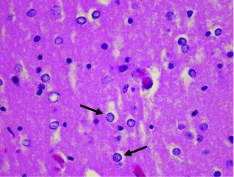
The extent of pathologic involvement in AHD is wide and diffuse and includes any areas from the cerebral cortices to the brainstem and cerebellum. Although only 32 autopsy cases have been reported, the pathologic findings were similar. Pathologic features do not necessarily correlate with clinical findings.1 Neuronal cell loss and spongy degeneration frequently affect deep cortical areas, especially layers III–V of the frontal, parietal, and occipital areas, while sparing the temporal cortex.15 In the cortices, laminar and pseudolaminar necroses are typical pathologic features. Neuronal loss and atrophy are observed throughout the deep nuclei including the lenticular, caudate, thalamic, and subthalamic nuclei and cerebellum.16 Microscopically, polymicrocavitation is found in cortical areas and the basal ganglia. Alzheimer type II astrocytosis invariably occurs in areas affected by polymicrocavitation, as well as those that appear macroscopically normal (Figure 1).12,13,15 Postmortem examination of patients with HE revealed central pontine myelinolysis and extrapontine myelinolysis combined with classic pathological findings of AHD.17,18 Furthermore, subacute combined degeneration may be related to white matter lesions in patients with AHD.19
Figure 1. Pathology of AHD. Alzheimer type II astrocyte showing large pale nuclei with basophilic nuclei. Reproduced with permission from Ferrara et al.12 AHD, Acquired Hepatocerebral Degeneration.
Pathomechanism
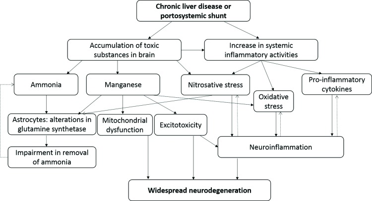
The exact pathophysiology of AHD has yet to be fully elucidated. Among multiple hypotheses that have been proposed, none embraces all the plausible mechanisms that are thought to be involved in its development (Figure 2). Accumulation of toxic substances like manganese or ammonia secondary to defective liver clearance mechanisms may facilitate AHD development. Although hyperammonemia was the focus of attention in early research on AHD, the manganese theory has gained more support over the past two decades. Hyperammonemia may play a role in AHD development due to its influence on astrocyte function. Astrocytes have a homeostatic function in glutamate uptake/release through glutamine synthetase.20 Increased concentrations of ammonia in chronic liver disease inhibit glutamate removal by glutamine synthetase in astrocytes, which leads to glutamine accumulation and low-grade brain edema.21 A higher ammonia concentration can activate nitric oxide synthase signal transduction, which implicates nitrosative stress in the central nervous system (CNS). Effects of an increased ammonia level may synergize with other actions stimulated by pro-inflammatory cytokines and toxic metals like manganese.22
Figure 2. The Putative Pathomechanism in AHD. The exact pathomechanism in AHD remains unclear, but the putative pathomechanism includes complex actions between toxic substance accumulation, neuroinflammation, oxidative stress, and inducible nitric oxide (nitrosative stress). AHD, Acquired Hepatocerebral Degeneration.
Accumulation of manganese may play a major role in AHD pathophysiology. The manganese theory was supported by MRI findings including high signal intensities in the bilateral globus pallidus and adjacent areas on T1-weighted images (T1WI). Such features are identical to those found in patients with high exposure to manganese in association with occupational manganism and total parenteral nutrition.23 There are also similar clinical features between manganism and AHD, such as increased manganese levels in whole blood and cerebrospinal fluid. High concentrations of manganese in brain tissue support the theory that neurotoxicity by manganese has a key role in the pathomechanism of AHD.24 In contrast, increased manganese levels in blood do not predict neurological impairment in AHD, indicating a lack of a cause-effect relationship. Despite this finding, manganese may be responsible for parkinsonism in AHD by significantly altering the dopaminergic neurotransmission.25 In the human diet, manganese intake comes from sources such as nuts, rice, whole grains, and multivitamins. Only 3–5 % of ingested manganese is absorbed through the gastrointestinal tract, which is mainly excreted through the biliary system. In advanced hepatobiliary disease, dysfunctional manganese removal leads to its accumulation in the brain. Iron deficiency anemia, a common nutritional problem in the general population and patients with liver disease, may worsen manganese accumulation because manganese and iron share common uptake transporters.26 Recently, progress has been made in understanding the mechanisms of manganese uptake into the CNS. Neurotoxicity occurs with high intracellular concentrations of manganese, so it is essential to understand the actions of manganese transporters in manganese-induced neurotoxicity. Membrane importers of manganese into the cell include the divalent metal transporter 1 (DMT1), zinc transporters ZIP8 and ZIP14, citrate transporter, choline transporter, dopamine transporter (DAT), transferrin receptor (TfR), and calcium channels. Both the DMT1 and TfR are the primary transporters of manganese into the cell. Furthermore, manganese has high affinity for DMT1, which is highly expressed in the basal ganglia including the globus pallidus and striatum.27 As a result, the basal ganglia is more susceptible to intracellular accumulation of manganese and subsequent neurotoxicity. Proteins involved in manganese efflux include SLC30A10, ATP13A2 (also known as PARK9), ferroportin, and SPCA1.28 Of these, SLC30A10 and ATP13A2/PARK9 have been associated with the involvement of manganese in AHD. SLC30A10 is mainly expressed in the basal ganglia and liver, and mutations in the SLC30A10 gene are associated with familial parkinsonism, dystonia, hypermanganesemia polycythemia, and LC.29 ATP13A2 mutations cause Kufor-Rakeb syndrome, characterized by early onset parkinsonism, spasticity, supranuclear gaze palsy, and dementia.30 Based on these findings, membrane transporters of manganese may play key roles in the regional specificity and neurologic manifestations of manganese accumulation in the brain.
Neuroinflammation may contribute to AHD development.22,31 In the acute phase of neuronal injuries cause by metabolic insults, low-to-intermediate inflammation has positive effects on neuronal injuries because it facilitates debris removal. Conversely, low-to-high intensity of inflammation in the chronic phase such as in chronic liver disease leads to greater release of pro-inflammatory cytokines and reactive oxygen species. This maladaptive inflammatory response hinders recovery of neuronal damage and synaptic plasticity. Chronic neuroinflammation may contribute to widespread neurodegeneration, which reinforces a chronic inflammatory state. Multiple mechanisms including toxic substance accumulation, neuroinflammation, and oxidative and nitrosative stress may act synergistically to cause neurodegeneration in AHD.
Clinical characteristics
The age of onset, which may be related to the duration of underlying hepatobiliary disease, is variable. Symptom onset is generally insidious despite previous reports describing exceptional cases with acute symptom development.32,33 In contrast to the reversible course of acute HE, AHD is generally characterized by a chronic, progressive, and irreversible course without spontaneous recovery,1,5,13 with the exception of cases that recover after liver transplantation (LT) (see “Treatment”).34,35
Neurologic symptoms and signs include the three following features: 1) movement disorders, mainly parkinsonism and ataxia-plus syndrome, 2) cognitive impairment and psychiatric symptoms, and 3) myelopathy.
Parkinsonism and other movement disorders
Movement disorders are the most common neurologic manifestations of AHD. About 20% of AHD patients have parkinsonism.5 AHD-related parkinsonism is mostly characterized by symmetric parkinsonism, early gait disturbance or falls, prominent postural or action tremor, mild cognitive impairment, and variable response to levodopa therapy (Video 1 & 2).5,8 The clinical course can vary; some patients may rapidly worsen, while others may be stable. Both isolated parkinsonism and parkinsonism with other accompanying movement disorders can be seen in AHD. These features of parkinsonism may distinguish AHD from typical Parkinson’s disease (PD), which is characterized by the asymmetric, pill-rolling type of rest tremor, late postural instability or gait disturbance, and excellent response to levodopa. Only rare reports on AHD have described parkinsonism resembling typical PD.13,36 Rest tremor is rarely observed in parkinsonism related to AHD and is mostly accompanied by postural or action tremor in one or both hands.5,13,37 Rest tremor without prominent postural tremor is much rarer than rest tremor with postural or action tremor. Postural and intentional tremors are commonly seen and characterized by a large amplitude and frequency of 4–7 Hz.13 Dysarthria related to parkinsonism in AHD is characterized by slow, monotonous, and slurred speech. Chorea,1,12,13 orolingual dyskinesia,15 dystonia,5,6 and pyramidal tract signs1 can be seen in AHD patients, whereas ballism has rarely been reported.38 Our analysis of 76 studies including 374 patients showed that 36 (9.6%) had chorea and 28 (7.5%) had dystonia. Cranial dyskinesia may occur alone or together with chorea in the limbs, being observed in 7.5% (28) of all 374 patients and in 77.8% (28) of the 36 patients with chorea. Cranial dyskinesia, characterized by protrusion and retraction of the tongue and lips accompanied by grimacing movements of the face, may be the characteristic motor phenotype in AHD.12,15 Generalized choreoathetotic movements in AHD may resemble those seen in Huntington’s chorea. The severity, extent, and accompanying features of hyperkinetic movements vary.1 Hyperkinetic movement disorders usually occur together but can occur separately. Abnormal movements are frequently accompanied by ataxia, but in our analysis, it was absent in 27.8% of patients with chorea and 32.1% of those with dystonia. No report to date has described ballism accompanied by ataxia, but this may be due to selection bias as these movement disorders are rarely observed in patients with AHD. Myoclonus/asterixis is less common in AHD than in acute HE and can be seen in AHD associated with HE.3,38
Video 1. Case 1: A 63-Year-Old Male with a 3-Year History of Tremor. He presented with masked face, hypophonia, micrographia, mild action tremor of hands, and mild rigidity and bradykinesia. With levodopa 300 mg/day, there was marked improvement of tremor, micrographia, and bradykinesia.
Video 2. Case 2: A 38-Year-Old Male with Severe Tremors of the Tongue, Jaw, and Both Hands. He presented with severe resting and postural tremors in the arms, masked face, mild bradykinesia, and rigidity, which were not responsive to levodopa.
Ataxia-plus syndrome
Ataxia-plus syndrome, referring to predominant ataxia combined with other signs, is commonly observed in AHD and is characterized by ataxic dysarthria, absence of nystagmus, and limb and gait ataxia.1,3,39 The frequency of ataxia-plus syndrome has not been reported. Our analysis of 374 patients in 76 studies revealed that 114 (30.5%) had ataxia-plus syndrome, whereas Victor et al. reported that all patients except one had cerebellar ataxia.1 Nystagmus may occasionally be present but rarely so.1 Dysarthria associated with ataxia-plus syndrome is common in AHD and possesses characteristic features of scanning, slurred, staccato, and explosive speech.39 Postural tremor, dystonia, or chorea co-occur with ataxia. Pure cerebellar syndrome with cerebellar atrophy has been reported in rare instances.40 Ataxia may be attributable to lesions in the cerebellum1 and MCPs.39
Cognitive impairment and psychiatric features
Global cognitive functions are largely preserved,5,6 and cognitive impairment is generally mild if present.6 Disorientation,41 inattentiveness,42,43 and psychomotor retardation43 are common,13 but they are not specific for AHD. Impairment in memory,41 frontal function,5,35 visuospatial function,6,35 and verbal fluency41 may be present, but focal cortical dysfunctions such as agnosia, apraxia, or aphasia are usually absent.6,41 One case report used the term “hepatic dementia” to emphasize that AHD is associated with a treatable dementia syndrome.41 However, this term is rarely used because the neuropsychological deficits in AHD do not correspond with the commonly accepted definition of dementia.6 Neuropsychiatric features such as disinhibition, aggression, apathy, or paranoia may be accompanying44 or presenting features.33,41,41,6,41AHD may be superimposed on an acute confusional state or acute HE. Under general circumstances, minimal HE would not be diagnosed as AHD, but minimal HE accompanied by various movement disorders may be given such a diagnosis based on its chronic, progressive course.
Hepatic myelopathy
Hepatic myelopathy, which is characterized by progressive spastic paraparesis, rare sensory impairment, rare sphincter dysfunction, and normal spinal imaging, has rarely been reported in previous AHD studies.2,45 Excluding more common causes of spastic paraparesis is the first step in diagnosing hepatic myelopathy,46 which seems to occur more frequently in males than in females.1,47 In a report describing 49 cases of hepatic myelopathy, episodes of HE were reported in about 80% of patients,47 suggesting an association between a clinical history of HE and hepatic myelopathy.
Differential diagnosis for Wilson disease and chronic manganism
Similarities and differences exist among neurologic manifestations of AHD, chronic manganism, and WD (Table 1).48 All three conditions share clinical characteristics of various movement disorders and neuropsychiatric symptoms. Limb or truncal ataxia is common in both WD and AHD, whereas dystonia is commonly present in chronic manganism.49 The Kayser-Fleischer (K-F) ring is a pathognomonic sign in WD, while it is absent in AHD and chronic manganism. Hepatic myelopathy may be present in AHD.46 A scoring system for WD diagnosis includes K-F rings, neurological symptoms, hemolytic anemia, 24-h urinary copper excretion, hepatic copper values, rhodamine-positive hepatocytes, serum ceruloplasmin concentration, and ATP7B gene mutation analysis.50 Scores ≥4 suggest a high likelihood of WD, scores of 2–3 points suggest probable WD, and scores of 0–1 points indicate that WD is unlikely. The diagnosis of AHD and chronic manganism is based upon a relevant history (history of liver disease or high exposure to manganese) and supportive laboratory and imaging features.
Table 1. Comparison between Wilson disease, AHD, and Chronic Manganism.
| Wilson Disease | AHD | Chronic Manganism | |
|---|---|---|---|
| Etiology or risk factors | Causative gene: ATP7B (Family history) | Chronic liver failure, portosystemic shunt | Occupational exposures: welders, miners |
| Possible pathomechanism | Accumulation of copper | Synergistic actions of multiple mechanisms
|
Accumulation of manganese |
| Clinical characteristics |
|
|
|
| Laboratory findings |
|
|
|
| Neuroimaging findings |
|
|
|
| Pathology | Opalski cells, Alzheimer type II astrocyte, cavitations | Alzheimer type II astrocyte, polymicrocavitation, CPM/EPM | Alzheimer type II astrocyte |
| Management |
|
No established treatments
|
|
Abbreviations: BCAA, Branched-Chain Amino Acid; BRTO, Balloon-Occluded Retrograde Transvenous Obliteration; CaNa2EDTA, Calcium Disodium Salt Ethylene Diamine Tetraacetic Acid; CPM, Central Pontine Myelinolysis; CSF, Cerebrospinal Fluid; DAT, Dopamine Transporter; EPM, Extrapontine Myelinolysis; F-DOPA, Fluorodopa; K-F rings, Kayser-Fleischer Rings; MCP, Middle Cerebellar Peduncles; m/c, Most Common; PAS, Para-Aminosalicylic Acid; T1-MRI, T1-Weighted Magnetic Resonance Imaging; T2-MRI, T2-Weighted Magnetic Resonance Imaging.
Brain Imaging
Brain imaging related to parkinsonism
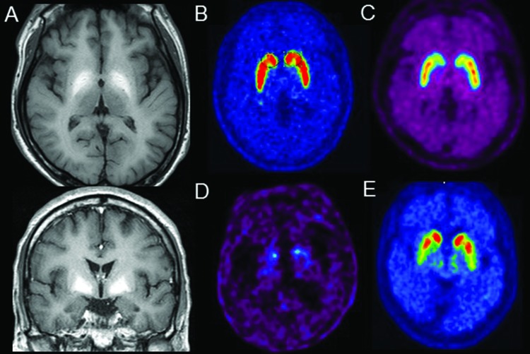
Symmetric high signal intensity on T1WI in the globus pallidus has been consistently reported as a typical finding linked to parkinsonism in AHD patients (Figure 3A).3,12,42,43,51–56 High signal intensity on T1WI may extend to adjacent areas including the putamen, caudate nucleus, and midbrain, but it is rarely seen in the cerebellum.37,57 The high signal lesions on T1WI are thought to be the result of manganese accumulation.23,58 Similarity in the high signal intensity on T1WI, as well as overlapping dopamine-deficient clinical syndrome in patients with AHD and those with manganese intoxication, suggest that manganese may play an identical role in these two conditions.59 Manganese is a paramagnetic material that shortens the T1 relaxation time, resulting in hyperintensity on T1WI without altering signal intensity on T2-weighted images (T2WI).60 The concentration of manganese in the basal ganglia, which corresponds to the high signal intensity on T1WI, was higher in AHD patients than in controls.8,52,61,62 Despite contradictory results,42 high serum manganese levels in patients with hyperintense lesions in the basal ganglia on T1WI8 support the hypothesis that manganese plays a key role in the development of high signal intensity on T1WI in AHD patients.23,58 The causal relationship between MRI lesions and clinical symptoms is not clear. Approximately 60–70% of patients with increased signal intensities in the globus pallidus on T1WI did not develop parkinsonism. This finding was similar to that in asymptomatic welders in whom T1WI MRI showed a high pallidal signal.42,63 Patients who have lesions with high signal intensity on T1WI do not necessarily develop symptoms of AHD. The exact role of manganese in the development of signal alterations on MRI and its effects on neurological symptoms remain to be determined.
Figure 3. Neuroimaging Studies in AHD with Parkinsonism. (A) Brain MRI in AHD. High signal intensities in the bilateral globus pallidus on T1-weighted images, (B-E) (18F FP-CIT PET findings. (B) Normal controls; (C) AHD patients with parkinsonism; (D) AHD patients with parkinsonism; (E) Idiopathic Parkinson diseases. AHD, Acquired Hepatocerebral Degeneration; 18F FP-CIT PET, 18F-N-3-Fluoropropyl-2β-Carboxymethoxy-3β-(4-iodophenyl)-Nortropane Positron Emission Tomography; MRI, Magnetic Resonance Imaging.
18F-Dopa (F-DOPA) positron emission tomography (PET) and DAT scans have been studied in to evaluate presynaptic dopaminergic neuronal degeneration in AHD patients with parkinsonism. F-DOPA has been used as a PET radioligand to evaluate presynaptic dopaminergic neuron integrity by measuring dopamine precursor uptake. DAT scans shows presynaptic dopaminergic neuronal integrity by measuring the degree of uptake of radioligands that bind to DAT. DAT single-photon emission computed tomography (SPECT) has been widely used to diagnose presynaptic dopaminergic neuronal degeneration. DAT PET was recently introduced to evaluate presynaptic dopaminergic neuron integrity. Because the resolution of PET imaging is higher than that of SPECT, DAT PET may be superior to SPECT to support diagnosing parkinsonian syndromes, although these imaging tools have not been directly compared.64,65 On the other hand, PET imaging using radiotracers including 123I-iodobenzamide,11C-raclopride, and 18F-fallypride have been applied to evaluate postsynaptic dopaminergic receptor density.66 Previous AHD pathology studies have demonstrated the loss of D2 dopaminergic receptor binding, and PET studies have shown decreased uptake of radioligands that bind to D2 receptors. Based on such findings, dysfunction of postsynaptic dopamine receptors related to manganese accumulation has been implicated in parkinsonism in AHD patients.10,67,68 There is growing evidence from F-DOPA PET, DAT SPECT, and DAT PET that supports a role for presynaptic dopaminergic degeneration in AHD patients. Although few studies have described F-DOPA results in patients with AHD, they have reported diffuse reduced uptake throughout the striatum in F-DOPA PET, in addition to showing typical patterns of PD. Three different patterns have been described in DAT scans of patients with AHD. The first is normal DAT uptake, which supports the hypothesis that manganese does not cause a loss of presynaptic dopaminergic nerve terminals (Figure 3B–C).12,37,69 Secondly, AHD patients have shown scattered or diffusely decreased putaminal radioligand uptake on DAT scans (Figure 3D). Patients with intact dopaminergic nerve terminals or diffuse dopaminergic deficits that differ from the typical PD pattern exhibit symmetric parkinsonism and early gait disturbance with limited levodopa responsiveness. Abnormal DAT uptake in the striatum compatible with PD (Figure 3E) has been reported in AHD patients.13,36,69 Patients with PD patterns on DAT scans were originally considered to have PD superimposed on AHD, owing to clinical signs identical to those in levodopa-responsive PD patients.36,69 However, recent evidence has led to the hypothesis that manganese in AHD may be associated with presynaptic dopaminergic degeneration.70 Studies of manganese-induced parkinsonism may provide clues to parkinsonism pathophysiology in patients with AHD. Manganese may aggravate alpha-synuclein dynamics and promote its aggregation.71 Moreover, progression of manganese-induced parkinsonism was found to depend on the dose of manganese exposure.72 Taken together, existing findings indicate that manganese accumulation may lead to progressive degeneration of presynaptic dopaminergic nerve terminals. Moreover, manganese exposure may be a risk factor for the development of PD, although the relationship between this heavy metal and PD is still unclear.73,74 Further imaging studies that evaluate the pathomechanism in patients with manganese intoxication and AHD may provide clues to the diverging findings of F-DOPA PET and DAT scans in AHD patients.
Brain imaging related to ataxia-plus syndrome
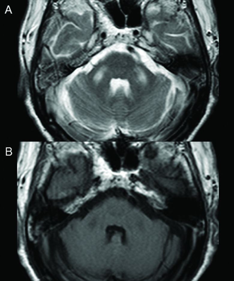
Predominant gait ataxia with other movement disorders has been frequently reported in AHD patients. Lesions in the cerebellum and MCP are thought to be responsible for ataxia-plus syndrome in AHD patients.18,39,75,76 These lesions appear as high signal intensity on T2WI (Figure 4A) with low or isodense signal intensity on T1WI (Figure 4B). This suggests that lesions in the cerebellum and MCP may be caused by mechanisms other than manganese accumulation. Patients with alcoholic LC exhibit distinct signal intensity changes in the corticocerebellar tract on T2WI; these are different from the usual patterns seen in the normal elderly population.19 Dysfunctional methionine metabolism and dysregulation of cytokine production have been proposed as possible pathomechanisms for white matter changes in patients with AHD. High signal intensity on T2WI is not limited to the cerebellum and MCP; signal changes on T2WI have also been found in the basal ganglia and inferior corona radiata.19,43,58 Besides high signal lesions on T2WI, isolated cerebellar degeneration has been reported in relation to ataxia-plus syndrome in an AHD patient.40
Figure 4. Brain MRI findings in AHD with Ataxia-Plus Syndrome. (A) High signal lesion on T2-weighted image and (B) low signal lesion on T1-weighted image in the middle cerebellar peduncles Reproduced with permission from Ishii K et al.75 AHD, Acquired Hepatocerebral Degeneration; MRI, Magnetic Resonance Imaging.
Other imaging studies
Diffuse cerebral atrophy has been observed in patients with chronic liver disease.55,77–79 Results of voxel-based analysis of MRI showed differences in regional gray and white matter volumes between patients with liver cirrhosis and those with AHD.77,80 The clinical significance of cerebral atrophy or volume changes in certain brain regions in AHD patients remains to be clarified.
Signal changes in the striatum can be detected earlier on proton density-weighted (PDW) MRI than on T1WI.81 The clinical usefulness of PDW imaging in patients with AHD remains unclear due to the lack of studies.
MR spectroscopy (MRS), a clinically useful tool for assessing alterations in brain metabolites, has been used to assess patients with HE.82,83 Typical findings include reductions in myoinositol/creatine and choline/creatine ratios and an increased glutamine/glutamate ratio.82,83 Similar MRS findings have been observed in patients with AHD.84
Treatment
Liver transplantation
LT is the ultimate treatment for liver cirrhosis. Favorable outcomes of AHD following LT have been continuously reported.2,4,8,34,35,85,86 A study comparing the outcomes of LT and conservative management in AHD patients found that LT improved neurological deficits and reversed MRI abnormalities, whereas conservative medical treatment alone did not improve outcome.34 In one case series, three patients with AHD showed improvements in cognitive dysfunction and parkinsonism.35 In contrast, many studies have reported less favorable outcomes after LT, including transient improvement followed by AHD recurrence,87,88 unchanged neurological state,3 and newly developed AHD.3,87–89 These poor outcomes following LT were mostly caused by failure of the transplanted liver. The mechanism by which LT reverses neurological deficits in AHD has not been established. Larger prospective studies are needed to assess the effects of LT in patients with AHD.
Symptomatic management
Levodopa has been empirically administered to manage parkinsonism symptoms in AHD.5,8,10,90 Modest to dramatic improvement has been observed, especially in AHD patients who had parkinsonian symptoms identical to PD. The effects of levodopa in these patients, along with asymmetrically decreased DAT uptake with a rostrocaudal gradient, support the presence of presynaptic dopaminergic degeneration in AHD patients. Dopamine receptor antagonists including haloperidol and quetiapine, as well as tetrabenazine and anticholinergics, have been administered to AHD patients to treat hyperkinetic movement disorders.38,88
Other treatments
Many anecdotal treatments have been applied to AHD patients. In one report, trientine was administered to chelate manganese. It was found to improve parkinsonian symptoms and eliminate abnormal signal intensity on brain MRI.37 Branched-chain amino acids (BCAAs) are thought to improve neurological deficits in HE patients by competing with aromatic amino acids (AAAs) for blood brain transport and reducing AAA entry into the brain.91,92 In a case report, BCAAs were administered to two patients with AHD, one with parkinsonism and another with cerebellar ataxia,93 both of whom showed improvements in imaging findings and neurological deficits.93 Balloon-occluded retrograde transvenous obliteration (BRTO) is an elective treatment modality for gastric varices in patients with LC.92 A case report described that BRTO in a patient with AHD improved ataxia and parkinsonism and reversed abnormal brain MRI signals. In addition, portosystemic shunt obliteration via percutaneous transvenous embolization in a patient with AHD was found to improve both cerebellar symptoms and abnormal brain MRI findings.94
Conclusions
AHD has various clinical spectrums, each with its own distinct pathophysiological mechanisms and neuroimaging findings. AHD can be classified into subtypes rather than being considered a single disease entity. LT is the most promising therapeutic strategy to ultimately eliminate the source of the disease. Additional clinical studies and pathophysiological and neuroimaging research including large numbers of patients with chronic liver disease are needed to clarify the exact nature of AHD.
Acknowledgments
We would like to thank Sun J. Chung, MD, PhD for providing two video cases to help demonstrate the clinical characteristics of AHD.
Footnotes
Funding: This study was supported by a grant from Research year of Inje University in 20150606.
Financial Disclosures: None.
Conflicts of interest: The authors report no conflict of interest.
Ethics Statement: All patients that appear on video have provided written informed consent; authorization for the videotaping and publication of the videotape was provided.
References
- 1.Victor M, Adams RD, Cole M. The acquired (non-Wilsonian) type of chronic hepatocerebral degeneration. Medicine (Baltimore) 1965;44:345–396. doi: 10.1097/00005792-196509000-00001. doi: http://dx.doi.org/10.1097/00005792-196509000-00001. [DOI] [PubMed] [Google Scholar]
- 2.Pinarbasi B, Kalymakoglu S, Matur Z, Akyuz F, Demir K, Besisik F, et al. Are acquired hepatocerebral degeneration and hepatic myelopathy reversible? J Clin Gastroenterol. 2009;43:176–181. doi: 10.1097/MCG.0b013e318150d399. doi: http://dx.doi.org/10.1097/MCG.0b013e318150d399. [DOI] [PubMed] [Google Scholar]
- 3.Fernandez-Rodriguez R, Contreras A, de Villoria JG, Grandas F. Acquired hepatocerebral degeneration: clinical characteristics and MRI findings. Eur J Neurol. 2010;17:1463–1470. doi: 10.1111/j.1468-1331.2010.03076.x. doi: http://dx.doi.org/10.1111/j.1468-1331.2010.03076.x. [DOI] [PubMed] [Google Scholar]
- 4.Qavi AH, Hammad S, Rana AI, Salih M, Shah NH, Dar FS, et al. Reversal of acquired hepatocerebral degeneration with living donor liver transplantation. Liver Transpl. 2016;22:125–129. doi: 10.1002/lt.24306. doi: http://dx.doi.org/10.1002/lt.24306. [DOI] [PubMed] [Google Scholar]
- 5.Burkhard PR, Delavelle J, Du Pasquier R, Spahr L. Chronic parkinsonism associated with cirrhosis: a distinct subset of acquired hepatocerebral degeneration. Arch Neurol. 2003;60:521–528. doi: 10.1001/archneur.60.4.521. doi: http://dx.doi.org/10.1001/archneur.60.4.521. [DOI] [PubMed] [Google Scholar]
- 6.Stracciari A, Mattarozzi K, D'Alessandro R, Baldin E, Guarino M. Cognitive functioning in chronic acquired hepatocerebral degeneration. Metab Brain Dis. 2008;23:155–160. doi: 10.1007/s11011-008-9088-3. doi: http://dx.doi.org/10.1007/s11011-008-9088-3. [DOI] [PubMed] [Google Scholar]
- 7.Methawasin K, Chonmaitree P, Wongjitrat C, Rattanamongkolgul S, Asawavichienjinda T. Movement Disorders in Non-Wilsonian Cirrhotic Patients: A Report of the Prevalence and Risk Factors from a Study Done in a Medical School in an Agricultural-Based Community. J Mov Disord. 2016;9:28–34. doi: 10.14802/jmd.15034. doi: http://dx.doi.org/10.14802/jmd.15034. [DOI] [PMC free article] [PubMed] [Google Scholar]
- 8.Klos KJ, Ahlskog JE, Josephs KA, Fealey RD, Cowl CT, Kumar N. Neurologic spectrum of chronic liver failure and basal ganglia T1 hyperintensity on magnetic resonance imaging: probable manganese neurotoxicity. Arch Neurol. 2005;62:1385–1390. doi: 10.1001/archneur.62.9.1385. doi: http://dx.doi.org/10.1001/archneur.62.9.1385. [DOI] [PubMed] [Google Scholar]
- 9.Scaglione S, Kliethermes S, Cao G, Shoham D, Durazo R, Luke A, et al. The Epidemiology of Cirrhosis in the United States: A Population-based Study. J Clin Gastroenterol. 2015;49:690–696. doi: 10.1097/MCG.0000000000000208. doi: http://dx.doi.org/10.1097/MCG.0000000000000208. [DOI] [PubMed] [Google Scholar]
- 10.Tryc AB, Goldbecker A, Berding G, Rümke S, Afshar K, Shahrezaei GH, et al. Cirrhosis-related Parkinsonism: prevalence, mechanisms and response to treatments. J Hepatol. 2013;58:698–705. doi: 10.1016/j.jhep.2012.11.043. doi: http://dx.doi.org/10.1016/j.jhep.2012.11.043. [DOI] [PubMed] [Google Scholar]
- 11.Kang JH, Tsai MC, Lin CC, Lin HL, Lin HC. Increased risk of Parkinsonism among patients with cirrhosis: a 7-year follow-up study. Liver Int. 2011;31:685–691. doi: 10.1111/j.1478-3231.2010.02432.x. doi: http://dx.doi.org/10.1111/j.1478-3231.2010.02432.x. [DOI] [PubMed] [Google Scholar]
- 12.Ferrara J, Jankovic J. Acquired hepatocerebral degeneration. J Neurol. 2009;256:320–332. doi: 10.1007/s00415-009-0144-7. doi: http://dx.doi.org/10.1007/s00415-009-0144-7. [DOI] [PubMed] [Google Scholar]
- 13.Jog MS, Lang AE. Chronic acquired hepatocerebral degeneration: case reports and new insights. Mov Disord. 1995;10:714–722. doi: 10.1002/mds.870100604. doi: http://dx.doi.org/10.1002/mds.870100604. [DOI] [PubMed] [Google Scholar]
- 14.Butterworth RF. Metal toxicity, liver disease and neurodegeneration. Neurotox Res. 2010;18:100–105. doi: 10.1007/s12640-010-9185-z. doi: http://dx.doi.org/10.1007/s12640-010-9185-z. [DOI] [PubMed] [Google Scholar]
- 15.Finlayson MH, Superville B. Distribution of cerebral lesions in acquired hepatocerebral degeneration. Brain. 1981;104:79–95. doi: 10.1093/brain/104.1.79. doi: http://dx.doi.org/10.1093/brain/104.1.79. [DOI] [PubMed] [Google Scholar]
- 16.Butterworth R. Neuronal cell death in hepatic encephalopathy. Metab Brain Dis. 2007;22:309–320. doi: 10.1007/s11011-007-9072-3. doi: http://dx.doi.org/10.1007/s11011-007-9072-3. [DOI] [PubMed] [Google Scholar]
- 17.Kleinschmidt-DeMasters BK, Filley CM, Rojiani AM. Overlapping features of extrapontine myelinolysis and acquired chronic (non-Wilsonian) hepatocerebral degeneration. Acta Neuropathol. 2006;112:605–616. doi: 10.1007/s00401-006-0112-6. doi: http://dx.doi.org/10.1007/s00401-006-0112-6. [DOI] [PubMed] [Google Scholar]
- 18.Lee J, Lacomis D, Comu S, Jacobsohn J, Kanal E. Acquired hepatocerebral degeneration: MR and pathologic findings. AJNR Am J Neuroradiol. 1998;19:485–487. [PMC free article] [PubMed] [Google Scholar]
- 19.Hathout L, Huang J, Zamani A, Morioka C, El-Saden S. White matter changes in chronic alcoholic liver disease: Hypothesized association and putative biochemical mechanisms. Med Hypotheses. 2015;85:825–834. doi: 10.1016/j.mehy.2015.10.002. doi: http://dx.doi.org/10.1016/j.mehy.2015.10.002. [DOI] [PubMed] [Google Scholar]
- 20.Rose CF, Verkhratsky A, Parpura V. Astrocyte glutamine synthetase: pivotal in health and disease. Biochem Soc Trans. 2013;41:1518–1524. doi: 10.1042/BST20130237. doi: http://dx.doi.org/10.1042/BST20130237. [DOI] [PubMed] [Google Scholar]
- 21.Zwingmann C, Butterworth R. An update on the role of brain glutamine synthesis and its relation to cell-specific energy metabolism in the hyperammonemic brain: further studies using NMR spectroscopy. Neurochem Int. 2005;47:19–30. doi: 10.1016/j.neuint.2005.04.003. doi: http://dx.doi.org/10.1016/j.neuint.2005.04.003. [DOI] [PubMed] [Google Scholar]
- 22.Butterworth RF. Pathogenesis of hepatic encephalopathy in cirrhosis: the concept of synergism revisited. Metab Brain Dis. 2016;31:1211–1215. doi: 10.1007/s11011-015-9746-1. doi: http://dx.doi.org/10.1007/s11011-015-9746-1. [DOI] [PubMed] [Google Scholar]
- 23.Pomier-Layrargues G, Spahr L, Butterworth RF. Increased manganese concentrations in pallidum of cirrhotic patients. Lancet. 1995;345:735. doi: 10.1016/s0140-6736(95)90909-5. doi: http://dx.doi.org/10.1016/S0140-6736(95)90909-5. [DOI] [PubMed] [Google Scholar]
- 24.Hauser RA, Zesiewicz TA, Rosemurgy AS, Martinez C, Olanow CW. Manganese intoxication and chronic liver failure. Ann Neurol. 1994;36:871–875. doi: 10.1002/ana.410360611. doi: http://dx.doi.org/10.1002/ana.410360611. [DOI] [PubMed] [Google Scholar]
- 25.Butterworth RF, Spahr L, Fontaine S, Layrargues GP. Manganese toxicity, dopaminergic dysfunction and hepatic encephalopathy. Metab Brain Dis. 1995;10:259–267. doi: 10.1007/BF02109357. doi: http://dx.doi.org/10.1007/BF02109357. [DOI] [PubMed] [Google Scholar]
- 26.Chen P, Chakraborty S, Mukhopadhyay S, Lee E, Paoliello MM, Bowman AB, et al. Manganese homeostasis in the nervous system. J Neurochem. 2015;134:601–610. doi: 10.1111/jnc.13170. doi: http://dx.doi.org/10.1111/jnc.13170. [DOI] [PMC free article] [PubMed] [Google Scholar]
- 27.Huang E, Ong WY, Connor JR. Distribution of divalent metal transporter-1 in the monkey basal ganglia. Neuroscience. 2004;128:487–496. doi: 10.1016/j.neuroscience.2004.06.055. doi: http://dx.doi.org/10.1016/j.neuroscience.2004.06.055. [DOI] [PubMed] [Google Scholar]
- 28.Peres TV, Schettinger MR, Chen P, Carvalho F, Avila DS, Bowman AB, et al. “Manganese-induced neurotoxicity: a review of its behavioral consequences and neuroprotective strategies”. BMC Pharmacol Toxicol. 2016;17:57. doi: 10.1186/s40360-016-0099-0. doi: http://dx.doi.org/10.1186/s40360-016-0099-0. [DOI] [PMC free article] [PubMed] [Google Scholar]
- 29.Lechpammer M, Clegg MS, Muzar Z, Huebner PA, Jin LW, Gospe SM., Jr Pathology of inherited manganese transporter deficiency. Ann Neurol. 2014;75:608–612. doi: 10.1002/ana.24131. doi: http://dx.doi.org/10.1002/ana.24131. [DOI] [PubMed] [Google Scholar]
- 30.Park JS, Blair NF, Sue CM. The role of ATP13A2 in Parkinson's disease: Clinical phenotypes and molecular mechanisms. Mov Disord. 2015;30:770–779. doi: 10.1002/mds.26243. doi: http://dx.doi.org/10.1002/mds.26243. [DOI] [PubMed] [Google Scholar]
- 31.Wright G, Swain M, Annane D, Saliba F, Samuel D, Arroyo V, et al. Neuroinflammation in liver disease: sessional talks from ISHEN. Metab Brain Dis. 2016;31:1339–1354. doi: 10.1007/s11011-016-9918-7. doi: http://dx.doi.org/10.1007/s11011-016-9918-7. [DOI] [PubMed] [Google Scholar]
- 32.Smita B, Gafoor VA, Saifudheen K, Jose J. Acute stroke-like presentation of acquired hepatocerebral degeneration. Ann Indian Acad Neurol. 2014;17:204–206. doi: 10.4103/0972-2327.132631. doi: http://dx.doi.org/10.4103/0972-2327.132631. [DOI] [PMC free article] [PubMed] [Google Scholar]
- 33.Gleason A, Hayhow B, Walterfang M, Evans A, Mocellin R, Gates P, et al. Neuropsychiatric symptoms as the presenting feature of acquired hepatocerebral degeneration. Aust N Z J Psychiatry. 2014;48:959–960. doi: 10.1177/0004867414531079. doi: http://dx.doi.org/10.1177/0004867414531079. [DOI] [PubMed] [Google Scholar]
- 34.Salehi SD, Shawcross DL. Reversal of acquired hepatocerebral degeneration with living donor liver transplantation. Liver Transpl. 2016;22:693. doi: 10.1002/lt.24423. doi: http://dx.doi.org/10.1002/lt.24423. [DOI] [PubMed] [Google Scholar]
- 35.Stracciari A, Baldin E, Cretella L, Delaj L, D'Alessandro R, Guarino M. Chronic acquired hepatocerebral degeneration: effects of liver transplantation on neurological manifestations. Neurol Sci. 2011;32:411–415. doi: 10.1007/s10072-011-0481-z. doi: http://dx.doi.org/10.1007/s10072-011-0481-z. [DOI] [PubMed] [Google Scholar]
- 36.Kim JM, Kim JS, Jeong SH, Kim YK, Kim SE, Kim SH, et al. Dopaminergic neuronal integrity in parkinsonism associated with liver cirrhosis. Neurotoxicology. 2010;31:351–355. doi: 10.1016/j.neuro.2010.05.002. doi: http://dx.doi.org/10.1016/j.neuro.2010.05.002. [DOI] [PubMed] [Google Scholar]
- 37.Park HK, Kim SM, Choi CG, Lee MC, Chung SJ. Effect of trientine on manganese intoxication in a patient with acquired hepatocerebral degeneration. Mov Disord. 2008;23:768–770. doi: 10.1002/mds.21957. doi: http://dx.doi.org/10.1002/mds.21957. [DOI] [PubMed] [Google Scholar]
- 38.Zaman Q, Ahmad A, Khokar N, Khan MF. Bilateral ballismus as a presenting feature of acquired hepatocerebral degeneration. Parkinsonism Relat Disord. 2016;25:104–105. doi: 10.1016/j.parkreldis.2016.02.014. doi: http://dx.doi.org/10.1016/j.parkreldis.2016.02.014. [DOI] [PubMed] [Google Scholar]
- 39.Park SA, Heo K. Prominent cerebellar symptoms with unusual magnetic resonance imaging findings in acquired hepatocerebral degeneration. Arch Neurol. 2004;61:1458–1460. doi: 10.1001/archneur.61.9.1458. doi: http://dx.doi.org/10.1001/archneur.61.9.1458. [DOI] [PubMed] [Google Scholar]
- 40.Melzer N, Grimm A, Meuth SG, Solymosi L, Stoll G. A pure cerebellar syndrome with corresponding ponto-cerebellar atrophy in acquired hepatocerebral degeneration. J Neurol Sci. 2010;292:96–98. doi: 10.1016/j.jns.2010.02.022. doi: http://dx.doi.org/10.1016/j.jns.2010.02.022. [DOI] [PubMed] [Google Scholar]
- 41.Mendez MF. Hepatic dementia or acquired hepatocerebral degeneration. J Am Geriatr Soc. 1989;37:259–260. doi: 10.1111/j.1532-5415.1989.tb06817.x. doi: http://dx.doi.org/10.1111/j.1532-5415.1989.tb06817.x. [DOI] [PubMed] [Google Scholar]
- 42.Maffeo E, Montuschi A, Stura G, Giordana MT. Chronic acquired hepatocerebral degeneration, pallidal T1 MRI hyperintensity and manganese in a series of cirrhotic patients. Neurol Sci. 2014;35:523–530. doi: 10.1007/s10072-013-1458-x. doi: http://dx.doi.org/10.1007/s10072-013-1458-x. [DOI] [PubMed] [Google Scholar]
- 43.Renjen PN, Khanna L, Rastogi R, Khan NI. Acquired hepatocerebral degeneration. BMJ Case Rep. 2013:2013. doi: 10.1136/bcr-2013-009387. doi: http://dx.doi.org/10.1136/bcr-2013-009387. [DOI] [PMC free article] [PubMed] [Google Scholar]
- 44.Ghys C, Kong MF, Glibert G, Lasser L, Karmali R. A lady with NASH and choreoathetosis. Acta Clin Belg. 2015;70:375–376. doi: 10.1179/2295333715Y.0000000027. doi: http://dx.doi.org/10.1179/2295333715Y.0000000027. [DOI] [PubMed] [Google Scholar]
- 45.Campellone JV, Lacomis D, Giuliani MJ, Kroboth FJ. Hepatic myelopathy. Case report with review of the literature. Clin Neurol Neurosurg. 1996;98:242–246. doi: 10.1016/0303-8467(96)00018-2. doi: http://dx.doi.org/10.1016/0303-8467(96)00018-2. [DOI] [PubMed] [Google Scholar]
- 46.Utku U, Asil T, Balci K, Uzunca I, Celik Y. Hepatic myelopathy with spastic paraparesis. Clin Neurol Neurosurg. 2005;107:514–516. doi: 10.1016/j.clineuro.2004.10.002. doi: http://dx.doi.org/10.1016/j.clineuro.2004.10.002. [DOI] [PubMed] [Google Scholar]
- 47.Mendoza G, Marti-Fabregas J, Kulisevsky J, Escartin A. Hepatic myelopathy: a rare complication of portacaval shunt. Eur Neurol. 1994;34:209–212. doi: 10.1159/000117040. doi: http://dx.doi.org/10.1159/000117040. [DOI] [PubMed] [Google Scholar]
- 48.Bandmann O, Weiss KH, Kaler SG. Wilson's disease and other neurological copper disorders. Lancet Neurol. 2015;14:103–113. doi: 10.1016/S1474-4422(14)70190-5. doi: http://dx.doi.org/10.1016/S1474-4422(14)70190-5. [DOI] [PMC free article] [PubMed] [Google Scholar]
- 49.Pal PK, Samii A, Calne DB. Manganese neurotoxicity: a review of clinical features, imaging and pathology. Neurotoxicology. 1999;20:227–238. [PubMed] [Google Scholar]
- 50.Ferenci P, Caca K, Loudianos G, Mieli-Vergani G, Tanner S, Sternlieb I, et al. Diagnosis and phenotypic classification of Wilson disease. Liver Int. 2003;23:139–142. doi: 10.1034/j.1600-0676.2003.00824.x. doi: http://dx.doi.org/10.1034/j.1600-0676.2003.00824.x. [DOI] [PubMed] [Google Scholar]
- 51.Damasio J, Ramos C, Miranda H, Magalhaes M. Acquired hepatocerebral degeneration and Wilson's disease: Differential diagnosis. Movement Disord. 2009;24:S400–S400. [Google Scholar]
- 52.Klos KJ, Ahlskog JE, Kumar N, Cambern S, Butz J, Burritt M, et al. Brain metal concentrations in chronic liver failure patients with pallidal T1 MRI hyperintensity. Neurology. 2006;67:1984–1989. doi: 10.1212/01.wnl.0000247037.37807.76. doi: http://dx.doi.org/10.1212/01.wnl.0000247037.37807.76. [DOI] [PubMed] [Google Scholar]
- 53.Maeda H, Sato M, Yoshikawa A, Kimura M, Sonomura T, Terada M, et al. Brain MR imaging in patients with hepatic cirrhosis: relationship between high intensity signal in basal ganglia on T1-weighted images and elemental concentrations in brain. Neuroradiology. 1997;39:546–550. doi: 10.1007/s002340050464. doi: http://dx.doi.org/10.1007/s002340050464. [DOI] [PubMed] [Google Scholar]
- 54.Pujol A, Pujol J, Graus F, Rimola A, Peri J, Mercader JM, et al. Hyperintense globus pallidus on T1-weighted MRI in cirrhotic patients is associated with severity of liver failure. Neurology. 1993;43:65–69. doi: 10.1212/wnl.43.1_part_1.65. doi: http://dx.doi.org/10.1212/WNL.43.1_Part_1.65. [DOI] [PubMed] [Google Scholar]
- 55.Kulisevsky J, Pujol J, Junque C, Deus J, Balanzo J, Capdevila A. MRI pallidal hyperintensity and brain atrophy in cirrhotic patients: two different MRI patterns of clinical deterioration? Neurology. 1993;43:2570–2573. doi: 10.1212/wnl.43.12.2570. doi: http://dx.doi.org/10.1212/WNL.43.12.2570. [DOI] [PubMed] [Google Scholar]
- 56.Pujol A, Graus F, Peri J, Mercader JM, Rimola A. Hyperintensity in the globus pallidus on T1-weighted and inversion-recovery MRI: a possible marker of advanced liver disease. Neurology. 1991;41:1526–1527. doi: 10.1212/wnl.41.9.1526. doi: http://dx.doi.org/10.1212/WNL.41.9.1526. [DOI] [PubMed] [Google Scholar]
- 57.Saito H, Ejima A. Liver dysfunction and probable manganese accumulation in the brainstem and basal ganglia. J Neurol Neurosurg Psychiatry. 1995;58:760–761. doi: 10.1136/jnnp.58.6.760-a. doi: http://dx.doi.org/10.1136/jnnp.58.6.760-a. [DOI] [PMC free article] [PubMed] [Google Scholar]
- 58.Nagappa M, Sinha S, Saini JS, Kallolimath P, Singh N, Kumar A, et al. Non-Wilsonian hepatolenticular degeneration: Clinical and MRI observations in four families from south India. J Clin Neurosci. 2016;27:91–94. doi: 10.1016/j.jocn.2015.06.035. doi: http://dx.doi.org/10.1016/j.jocn.2015.06.035. [DOI] [PubMed] [Google Scholar]
- 59.Cersosimo MG, Koller WC. The diagnosis of manganese-induced parkinsonism. Neurotoxicology. 2006;27:340–346. doi: 10.1016/j.neuro.2005.10.006. doi: http://dx.doi.org/10.1016/j.neuro.2005.10.006. [DOI] [PubMed] [Google Scholar]
- 60.Sung JH, Kim CY, Yang SO, Khang HS, Cheong HK, Lee JS, et al. Changes in blood manganese concentration and MRI t1 relaxation time during 180 days of stainless steel welding-fume exposure in cynomolgus monkeys. Inhal Toxicol. 2007;19:47–55. doi: 10.1080/08958370600985834. doi: http://dx.doi.org/10.1080/08958370600985834. [DOI] [PubMed] [Google Scholar]
- 61.Spahr L, Butterworth RF, Fontaine S, Bui L, Therrien G, Milette PC, et al. Increased blood manganese in cirrhotic patients: relationship to pallidal magnetic resonance signal hyperintensity and neurological symptoms. Hepatology. 1996;24:1116–1120. doi: 10.1002/hep.510240523. doi: http://dx.doi.org/10.1002/hep.510240523. [DOI] [PubMed] [Google Scholar]
- 62.Layrargues GP, Shapcott D, Spahr L, Butterworth RF. Accumulation of manganese and copper in pallidum of cirrhotic patients: role in the pathogenesis of hepatic encephalopathy? Metab Brain Dis. 1995;10:353–356. doi: 10.1007/BF02109365. doi: http://dx.doi.org/10.1007/BF02109365. [DOI] [PubMed] [Google Scholar]
- 63.Kim Y. Neuroimaging in manganism. Neurotoxicology. 2006;27:369–372. doi: 10.1016/j.neuro.2005.12.002. doi: http://dx.doi.org/10.1016/j.neuro.2005.12.002. [DOI] [PubMed] [Google Scholar]
- 64.Ravina B, Eidelberg D, Ahlskog JE, Albin RL, Brooks DJ, Carbon M, et al. The role of radiotracer imaging in Parkinson disease. Neurology. 2005;64:208–215. doi: 10.1212/01.WNL.0000149403.14458.7F. doi: http://dx.doi.org/10.1212/01.WNL.0000149403.14458.7F. [DOI] [PubMed] [Google Scholar]
- 65.Cummings JL, Henchcliffe C, Schaier S, Simuni T, Waxman A, Kemp P. The role of dopaminergic imaging in patients with symptoms of dopaminergic system neurodegeneration. Brain. 2011;134:3146–3166. doi: 10.1093/brain/awr177. doi: http://dx.doi.org/10.1093/brain/awr177. [DOI] [PubMed] [Google Scholar]
- 66.Nikolaus S, Antke C, Kley K, Poeppel TD, Hautzel H, Schmidt D, et al. Investigating the dopaminergic synapse in vivo. I. Molecular imaging studies in humans. Rev Neurosci. 2007;18:439–472. doi: 10.1515/revneuro.2007.18.6.439. [DOI] [PubMed] [Google Scholar]
- 67.Weissenborn K, Berding G, Kostler H. Altered striatal dopamine D2 receptor density and dopamine transport in a patient with hepatic encephalopathy. Metab Brain Dis. 2000;15:173–178. doi: 10.1007/BF02674526. [DOI] [PubMed] [Google Scholar]
- 68.Mousseau DD, Perney P, Layrargues GP, Butterworth RF. Selective loss of pallidal dopamine D2 receptor density in hepatic encephalopathy. Neurosci Lett. 1993;162:192–196. doi: 10.1016/0304-3940(93)90593-a. doi: http://dx.doi.org/10.1016/0304-3940(93)90593-A. [DOI] [PubMed] [Google Scholar]
- 69.Kim J, Kim JM, Kim YK, Shin JW, Choi SH, Kim SE, et al. Dopamine transporter SPECT of a liver cirrhotic with atypical parkinsonism. Ind Health. 2007;45:497–500. doi: 10.2486/indhealth.45.497. doi: http://dx.doi.org/10.2486/indhealth.45.497. [DOI] [PubMed] [Google Scholar]
- 70.Kim Y, Kim JM, Kim JW, Yoo CI, Lee CR, Lee JH, et al. Dopamine transporter density is decreased in parkinsonian patients with a history of manganese exposure: what does it mean? Mov Disord. 2002;17:568–575. doi: 10.1002/mds.10089. doi: http://dx.doi.org/10.1002/mds.10089. [DOI] [PubMed] [Google Scholar]
- 71.Peres TV, Parmalee NL, Martinez-Finley EJ, Aschner M. Untangling the Manganese-alpha-Synuclein Web. Front Neurosci. 2016;10:364. doi: 10.3389/fnins.2016.00364. doi: http://dx.doi.org/10.3389/fnins.2016.00364. [DOI] [PMC free article] [PubMed] [Google Scholar]
- 72.Racette BA, Nielsen SS, Criswell SR, Sheppard L, Seixas N, Warden MN, et al. Dose-dependent progression of parkinsonism in manganese-exposed welders. Neurology. 2017;88:344–351. doi: 10.1212/WNL.0000000000003533. doi: http://dx.doi.org/10.1212/WNL.0000000000003533. [DOI] [PMC free article] [PubMed] [Google Scholar]
- 73.Jankovic J. Searching for a relationship between manganese and welding and Parkinson's disease. Neurology. 2005;64:2021–2028. doi: 10.1212/01.WNL.0000166916.40902.63. doi: http://dx.doi.org/10.1212/01.WNL.0000166916.40902.63. [DOI] [PubMed] [Google Scholar]
- 74.Powers KM, Smith-Weller T, Franklin GM, Longstreth WT, Jr, Swanson PD, Checkoway H. Parkinson's disease risks associated with dietary iron, manganese, and other nutrient intakes. Neurology. 2003;60:1761–1766. doi: 10.1212/01.wnl.0000068021.13945.7f. doi: http://dx.doi.org/10.1212/01.WNL.0000068021.13945.7F. [DOI] [PubMed] [Google Scholar]
- 75.Ishii K, Shioya A, Fukuda K, Mori K, Tamaoka A. Acquired hepatocerebral degeneration with middle cerebellar peduncles lesions: case report and review of the literature. Clin Neurol Neurosurg. 2012;114:1361–1364. doi: 10.1016/j.clineuro.2012.03.032. doi: http://dx.doi.org/10.1016/j.clineuro.2012.03.032. [DOI] [PubMed] [Google Scholar]
- 76.Furukawa R, Sakakibara R, Hosoe N, Kishi M, Ogawa E, Suzuki Y. Ataxia and middle cerebellar peduncle lesions in hepatic encephalopathy. Neuroradiology. 2009;51:273–274. doi: 10.1007/s00234-009-0515-x. doi: http://dx.doi.org/10.1007/s00234-009-0515-x. [DOI] [PubMed] [Google Scholar]
- 77.Iwasa M, Mifuji-Moroka R, Kuroda M, Moroka H, Fujita N, Kobayashi Y, et al. Regional reduction in gray and white matter volume in brains of cirrhotic patients: voxel-based analysis of MRI. Metab Brain Dis. 2012;27:551–557. doi: 10.1007/s11011-012-9314-x. doi: http://dx.doi.org/10.1007/s11011-012-9314-x. [DOI] [PubMed] [Google Scholar]
- 78.Chen HJ, Zhu XQ, Shu H, Yang M, Zhang Y, Ding J, et al. Structural and functional cerebral impairments in cirrhotic patients with a history of overt hepatic encephalopathy. European J Radiol. 2012;81:2463–2469. doi: 10.1016/j.ejrad.2011.10.008. doi: http://dx.doi.org/10.1016/j.ejrad.2011.10.008. [DOI] [PubMed] [Google Scholar]
- 79.Melzer N, Grimm A, Meuth SG, Solymosi L, Stoll G. A pure cerebellar syndrome with corresponding ponto-cerebellar atrophy in acquired hepatocerebral degeneration. J Neurol Sci. 2010;292:96–98. doi: 10.1016/j.jns.2010.02.022. doi: http://dx.doi.org/10.1016/j.jns.2010.02.022. [DOI] [PubMed] [Google Scholar]
- 80.Zhang LJ, Qi R, Zhong J, Xu Q, Zheng G, Lu GM. The effect of hepatic encephalopathy, hepatic failure, and portosystemic shunt on brain volume of cirrhotic patients: a voxel-based morphometry study. PloS One. 2012;7:e42824. doi: 10.1371/journal.pone.0042824. doi: http://dx.doi.org/10.1371/journal.pone.0042824. [DOI] [PMC free article] [PubMed] [Google Scholar]
- 81.Erro R, Vitale C, Picillo M, Barone P, Pellecchia MT. Early MRI findings in acquired hepatocerebral degeneration. Neurol Sci. 2013;34:589–591. doi: 10.1007/s10072-012-1087-9. doi: http://dx.doi.org/10.1007/s10072-012-1087-9. [DOI] [PubMed] [Google Scholar]
- 82.Kreis R, Ross BD, Farrow NA, Ackerman Z. Metabolic disorders of the brain in chronic hepatic encephalopathy detected with H-1 MR spectroscopy. Radiology. 1992;182:19–27. doi: 10.1148/radiology.182.1.1345760. doi: http://dx.doi.org/10.1148/radiology.182.1.1345760. [DOI] [PubMed] [Google Scholar]
- 83.Tarasów E, Panasiuk A, Siergiejczyk L, Orzechowska-Bobkiewicz A, Lewszuk A, Walecki J, et al. MR and 1H MR spectroscopy of the brain in patients with liver cirrhosis and early stages of hepatic encephalopathy. Hepatogastroenterology. 2003;50:2149–2153. [PubMed] [Google Scholar]
- 84.Long LL, Li XR, Huang ZK, Jiang YM, Fu SX, Zheng W. Relationship between changes in brain MRI and (1)H-MRS, severity of chronic liver damage, and recovery after liver transplantation. Exp Biol Med (Maywood) 2009;234:1075–1085. doi: 10.3181/0903-RM-118. doi: http://dx.doi.org/10.3181/0903-RM-118. [DOI] [PMC free article] [PubMed] [Google Scholar]
- 85.Powell EE, Pender MP, Chalk JB, Parkin PJ, Strong R, Lynch S, et al. Improvement in chronic hepatocerebral degeneration following liver transplantation. Gastroenterology. 1990;98:1079–1082. doi: 10.1016/0016-5085(90)90036-z. doi: http://dx.doi.org/10.1016/0016-5085(90)90036-Z. [DOI] [PubMed] [Google Scholar]
- 86.Shulman LM, Minagar A, Weiner WJ. Reversal of parkinsonism following liver transplantation. Neurology. 2003;60:519. doi: 10.1212/wnl.60.3.519. doi: http://dx.doi.org/10.1212/WNL.60.3.519. [DOI] [PubMed] [Google Scholar]
- 87.Chen Y, Haque M, Yoshida EM. Transient improvement of acquired hepatocerebral degeneration with parkinsonian symptoms after failed liver transplant: case report and literature review. Exp Clin Transplant. 2011;9:363–369. [PubMed] [Google Scholar]
- 88.Papapetropoulos S, Singer C. Management of the extrapyramidal syndrome in chronic acquired hepatocerebral degeneration (CAHD) Mov Disord. 2005;20:1088–1089. doi: 10.1002/mds.20585. doi: http://dx.doi.org/10.1002/mds.20585. [DOI] [PubMed] [Google Scholar]
- 89.Servin-Abad L, Tzakis A, Schiff ER, Regev A. Acquired hepatocerebral degeneration in a patient with HCV cirrhosis: complete resolution with subsequent recurrence after liver transplantation. Liver Transpl. 2006;12:1161–1165. doi: 10.1002/lt.20815. doi: http://dx.doi.org/10.1002/lt.20815. [DOI] [PubMed] [Google Scholar]
- 90.Butterworth RF. Parkinsonism in cirrhosis: pathogenesis and current therapeutic options. Metab Brain Dis. 2013;28:261–267. doi: 10.1007/s11011-012-9341-7. doi: http://dx.doi.org/10.1007/s11011-012-9341-7. [DOI] [PubMed] [Google Scholar]
- 91.Dejong CH, van de Poll MC, Soeters PB, Jalan R, Olde Damink SW. Aromatic amino acid metabolism during liver failure. J Nutr. 2007;137:1579S–1585S. doi: 10.1093/jn/137.6.1579S. discussion 1597S–1598S. [DOI] [PubMed] [Google Scholar]
- 92.Fukui H, Saito H, Ueno Y, Uto H, Obara K, Sakaida I, et al. Evidence-based clinical practice guidelines for liver cirrhosis 2015. Journal Gastroenterol. 2016;51:629–650. doi: 10.1007/s00535-016-1216-y. doi: http://dx.doi.org/10.1007/s00535-016-1216-y. [DOI] [PubMed] [Google Scholar]
- 93.Ueki Y, Isozaki E, Miyazaki Y, Koide R, Shimizu T, Yagi K, et al. Clinical and neuroradiological improvement in chronic acquired hepatocerebral degeneration after branched-chain amino acid therapy. Acta Neurol Scand. 2002;106:113–116. doi: 10.1034/j.1600-0404.2002.01230.x. doi: http://dx.doi.org/10.1034/j.1600-0404.2002.01230.x. [DOI] [PubMed] [Google Scholar]
- 94.Hisahara S, Matsushita T, Kitamura M, Mezawa S, Nonaka M, Imai T, et al. Long-term clinical and radiological improvement of chronic acquired hepatocerebral degeneration after obliteration of portosystemic shunt: Report of a case. J Neurol Sci. 2014;346:303–306. doi: 10.1016/j.jns.2014.07.068. doi: http://dx.doi.org/10.1016/j.jns.2014.07.068. [DOI] [PubMed] [Google Scholar]


