Abstract
The vermiform appendix (whether inflamed or not) within a hernia is very rare occurrence. We present the unprecedented case of a normal appendix found within a Pfannenstiel incisional hernia. A diagnostic laparoscopy was performed as appendicitis was suspected. However, the tip of a normal appendix was visualised within a previous Pfannenstiel incision. Laparoscopic appendicectomy was carried successfully and the patient was discharged. The patient later returned for a successful elective laparoscopic incisional hernia repair.
Keywords: General Surgery, Radiology
Background
Appendicitis is a common surgical diagnosis, but appendicitis within a hernia is a much rarer diagnosis with the the most common being appendicitis within an inguinal/inguinoscrotal hernia,1 commonly due to the close relation of the anatomy. We describe the unprecedented case of a misdiagnosed appendicitis as an incarcerated normal appendix within a Pfannenstiel incision.
Case presentation
We report the case of a 48-year-old woman who presented with a 6-day history of gradual onset abdominal pain localised to the right iliac fossa. She described a dull aching pain without radiation that was transient in nature (in increments of minutes) with no exacerbating or relieving factors. She also complained of vomiting twice, anorexia and a severity score of 7/10. There were no urinary or gynaecological symptoms.
Her medical history revealed a total abdominal hysterectomy (open) 18 months prior, fibroids, hypercholesterolaemia, sensorineural hearing loss, axillary cyst removal and laparoscopic adjustable gastric banding.
On examination, there was a clear Pfannenstiel incision, the abdomen was soft but tender in the right iliac fossa with guarding.
Investigations
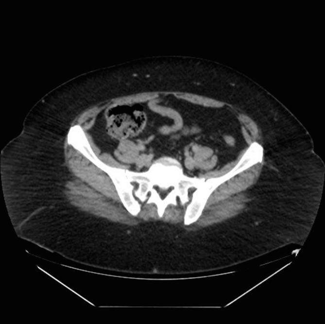
Blood results and urinalysis were normal. CT abdomen pelvis (CTAP) (figure 1) showed an abnormal area adjacent to the caecum likely representing an inflamed appendix, therefore the diagnostic conclusion was likely appendicitis.
Figure 1.

CT of abdomen and pelvis showing dilated appendix.
Differential diagnosis
Having acquired the CTAP results, our main differential diagnosis was appendicitis.
Treatment
Based on these findings, a diagnostic laparoscopy was performed with a view to appendicectomy.
Outcome and follow-up
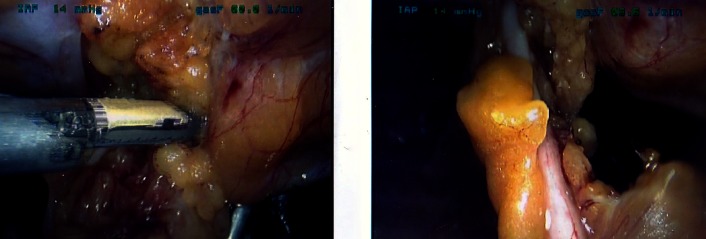
On entry into the abdomen, it was clear the tip of the appendix was adherent and within an incisional hernia of the previous Pfannenstiel incision (figure 2). The appendix did not appear to be inflamed and was dissected from the incisional hernia using a Thunderbeat energy device. It was then ligated at the base, which was found to be healthy, using endoloops. The patient did well postoperatively and was discharged with 2 days of oral antibiotics and analgesia.
Figure 2.
Intraoperative picture of non-inflamed appendix incarcerated in Pfannenstiel incisional hernia.
Histology revealed an atrophic and uninflamed appendix measuring 70×5 mm, and microscopically appeared uninflamed with extensive fibrofatty obliteration of the lumen. We submit that this is likely due to incarceration. The final diagnosis was incarcerated normal vermiform appendix within Pfannenstiel incisional hernia.
Following discharge, the patient continued to have a dull pain which she described as severe, and on examination was maximally tender within her Pfannenstiel incision, although clinically no hernia was palpable due to her body habitus. The patient returned 5 months later for an uncomplicated elective laparoscopic mesh repair of the incisional hernia with and was discharged the following day.
Discussion
The presence of appendices within hernias is historically documented in the literature originally by Amyand. Overall, it is uncommon to find an appendiceal hernia with figures reported as low 0.51% (out of 1950 patients) with a male to female ratio of 2:1. However, it is even more uncommon to find appendicitis within a hernia.2
Galiananes and Ramaswamy3 described an appendicitis found within a Pfannenstiel incisional hernia in 2012, which extended into the preperitoneal area held by adhesions. However, this appendix’s body initially appeared non-inflamed but after dissection, the tip was visualised as inflamed and later confirmed by pathology as appendicitis (unlike our case). Although there have been reports of appendices within hernias in the literature, only Galinanes and Ramaswamy3 detail an appendix found within a Pfannenstiel incisional hernia.3
The mechanism of the patient’s pain does not exactly fit with the presentation of acute appendicitis nor a symptomatic incarcerated hernia but can be attributed to two mechanisms. As the tip of the appendix was stuck in the incisional hernia, the appendix itself was behaving as an adhesional band responsible for an element of traction on the caecum. Additionally, the appendix itself could have been the cause of an internal hernia leading to intermittent episodes of abdominal pain and explaining the episodes of vomiting at presentation.
A Pfannenstiel incisional hernia is a rare complication reported as low as 3.5%.4 Hermann Johannes Pfannenstiel initially described closure of the abdomen in four layers (peritoneum, rectus muscle, transversely incised fascia and skin), but it is not common practice to plicate the rectus muscle as originally described, with gynaecologists reporting only 5.6% closing the rectus muscle in a case series by Patil et al.5 Low rates have been cited due to lack of evidence in literature to support this step, but this case series found all incisional hernias occurred where the rectus had not been closed. Similarly in this case, the rectus was not plicated in closure of Pfannenstiel incision of the hysterectomy 18 months prior.6
Incidental appendicectomy was performed in this case as the dissection of the appendix from the incisional hernia required multiple manipulations and traction on the base of the appendix. The tip of the appendix appeared macroscopically unhealthy due to the incarceration and it was difficult to exclude inflammation. Appendectomy was performed to avoid progression to complicated appendicitis7 8 as a macroscopic view cannot confirm appendicitis.3
Despite the literature advocating primary hernia repair for non-inflamed appendix,1 repair was deferred to avoid the risk of infection of the mesh as microscopic infection could not be ruled out (having visualised the unhealthy looking tip). Although associated with higher risk of hernia recurrence, a direct hernia repair could have been performed; however, the appendix was considered the main source of patient symptoms in this circumstance and the hernia was addressed at later stage.
Appendicitis within groin hernias have a variable presentation and do not present as a classical acute appendicitis but rather with symptoms of an incarcerated hernia. This can lead to a delayed perioperative diagnosis of appendicitis. However, our case did present with a typical right tender iliac fossa, but instead, there was simply an incarcerated appendix rather than appendicitis.
Learning points.
CT scanning of the abdomen is not a definitive diagnosis for appendicitis. Direct visualisation and histology will the reveal the final diagnosis.
The rectus muscle should be plicated in Pfannenstiel incision closure to avoid incisional hernia.
Right iliac fossa pain should not always be dismissed as appendicitis, as there are many atypical presentations of appendicitis and similarly other differential diagnoses of right iliac fossa pain.
Acknowledgments
none
Footnotes
Contributors: AK: acquired all of the information, drafted the article and revised it after corrections and review from other authors. NH: assisted in drafting the article and assisted in revision. SS: assisted in the drafting of the article and acquiring of the patient information. RS: approved the final version for submission.
Competing interests: None declared.
Patient consent: Obtained.
Provenance and peer review: Not commissioned; externally peer reviewed.
References
- 1.Sharma H, Gupta A, Shekhawat NS, et al. Amyand's hernia: a report of 18 consecutive patients over a 15-year period. Hernia 2007;11:31–5. 10.1007/s10029-006-0153-8 [DOI] [PubMed] [Google Scholar]
- 2.Gurer A, Ozdogan M, Ozlem N, et al. Uncommon content in groin hernia sac. Hernia 2006;10:152–5. 10.1007/s10029-005-0036-4 [DOI] [PubMed] [Google Scholar]
- 3.Galiñanes EL, Ramaswamy A. Appendicitis found in an incisional hernia. J Surg Case Reports. 2012. [DOI] [PMC free article] [PubMed] [Google Scholar]
- 4.Luijendijk RW, Jeekel J, Storm RK, et al. The low transverse Pfannenstiel incision and the prevalence of incisional hernia and nerve entrapment. Ann Surg 1997;225:365–9. 10.1097/00000658-199704000-00004 [DOI] [PMC free article] [PubMed] [Google Scholar]
- 5.Patil A, Davies HO, Coulston J, et al. Atypical incisional hernia following Pfannenstiel incision. Hernia 2013;17:479–82. 10.1007/s10029-012-0994-2 [DOI] [PubMed] [Google Scholar]
- 6.Meinke AK. Review article: appendicitis in groin hernias. J Gastrointest Surg 2007;11:1368–72. 10.1007/s11605-007-0160-9 [DOI] [PubMed] [Google Scholar]
- 7.Phillips AW, Jones AE, Sargen K. Should the macroscopically normal appendix be removed during laparoscopy for acute right iliac fossa pain when no other explanatory pathology is found? Surg Laparosc Endosc Percutan Tech 2009;19:392–4. 10.1097/SLE.0b013e3181b71957 [DOI] [PubMed] [Google Scholar]
- 8.Grunewald B, Keating J. Should the 'normal' appendix be removed at operation for appendicitis? J R Coll Surg Edinb 1993;38:158–60. [PubMed] [Google Scholar]


