Abstract
The DNA repair enzyme polynucleotide kinase/phosphatase (PNKP) protects genome integrity by restoring ligatable 5’-phosphate and 3’-hydroxyl termini at single-strand breaks (SSBs). In humans, PNKP mutations underlie the neurological disease known as MCSZ, but these individuals are not predisposed for cancer, implying effective alternative repair pathways in dividing cells. Homology-directed repair (HDR) of collapsed replication forks was proposed to repair SSBs in PNKP-deficient cells, but the critical HDR protein Rad51 is not required in PNKP-null (pnk1Δ) cells of Schizosaccharomyces pombe. Here, we report that pnk1Δ cells have enhanced requirements for Rad3 (ATR/Mec1) and Chk1 checkpoint kinases, and the multi-BRCT domain protein Brc1 that binds phospho-histone H2A (γH2A) at damaged replication forks. The viability of pnk1Δ cells depends on Mre11 and Ctp1 (CtIP/Sae2) double-strand break (DSB) resection proteins, Rad52 DNA strand annealing protein, Mus81-Eme1 Holliday junction resolvase, and Rqh1 (BLM/WRN/Sgs1) DNA helicase. Coupled with increased sister chromatid recombination and Rad52 repair foci in pnk1Δ cells, these findings indicate that lingering SSBs in pnk1Δ cells trigger Rad51-independent homology-directed repair of collapsed replication forks. From these data, we propose models for HDR-mediated tolerance of persistent SSBs with 3’ phosphate in pnk1Δ cells.
Author summary
DNA is constantly damaged by normal cellular metabolism, for example production of reactive oxygen species, or from exposure to external DNA damaging sources, such as radiation from the sun or chemicals in the environment. These genotoxic agents create thousands of single-strand breaks/cell/day in the human body. An essential DNA repair protein known as polynucleotide kinase/phosphatase (PNKP) makes sure the single-strand breaks have 5’ phosphate and 3’ hydroxyl ends suitable for healing by DNA ligase. Mutations that reduce PNKP activity cause a devastating neurological disease but surprisingly not cancer, suggesting that other DNA repair mechanisms step into the breach in dividing PNKP-deficient cells. One popular candidate was homology-directed repair (HDR) of replication forks that collapse at single-strand breaks, but the crucial HDR protein Rad51 was found to be non-essential in PNKP-deficient cells of fission yeast. In this study, Sanchez and Russell revive the HDR model by showing that SSBs in PNKP-deficient cells are repaired by a variant HDR mechanism that bypasses the requirement for Rad51. Notably, Mus81 endonuclease that resolves sister chromatid recombination structures formed during HDR of collapsed replication forks was found to be essential in PNKP-deficient cells.
Introduction
Maintenance of genome integrity depends on the accurate repair of DNA lesions that sever one or both strands of the double-helix. Single-strand breaks (SSBs) are by far the most abundant DNA scission, occurring at frequencies of thousands/cell/day in proliferating human cells [1]. SSBs are formed by many mechanisms, including oxidative attack of the sugar-phosphate backbone by endogenous reactive oxygen species (ROS), by base and nucleotide excision repair, through the activity of anti-cancer drugs such as camptothecin or bleomycins, or by exposure to other DNA damaging agents. These SSBs often have 5’-hydroxyl or 3’-phosphate termini that prevent ligation. Polynucleotide kinase phosphatase (PNKP) is a bifunctional enzyme that restores 5’-phosphate and 3’-hydroxyl to these DNA ends [2, 3]. PNKP’s importance is indicated by its conservation throughout eukaryotic evolution, although some species such as Saccharomyces cerevisiae have only retained the phosphatase domain [4].
The consequences of eliminating PNKP activity varies dramatically in eukaryotes. At one extreme, deleting the PNKP gene in mice causes early embryonic lethality [5]. PNKP probably plays an equally important role in humans, as a rare autosomal recessive disease characterized by microcephaly, early-onset intractable seizures and developmental delay (denoted MCSZ) was traced to partial loss-of-function mutations in the PNKP gene [6–8]. MCSZ is not associated with cancer; indeed, neurodegeneration in the absence of cancer predisposition appears to be a typical consequence of SSB repair defects in humans [9]. In contrast to mammals, S. cerevisiae cells lacking the DNA 3’ phosphatase encoded by TPP1 display no obvious phenotypes or sensitivity to DNA damaging agents [10]. However, requirements for Tpp1 are revealed when other DNA repair pathways are inactivated. Most notably, in cells lacking the apurinic/apyrimidinic (AP) endonucleases Apn1 and Apn2, deletion of TPP1 increases cellular sensitivity to several DNA damaging agents, including the DNA alkylating agent methyl methanesulfonate (MMS) and the topoisomerase I inhibitor camptothecin (CPT) [10, 11]. These AP endonucleases process DNA ends with various 3’-terminal blocking lesions, including 3’ phosphoglycolate (3’-PG), 3′‐unsaturated aldehydic, α,β‐4‐hydroxy‐2‐pentenal (3′‐dRP), and 3’-phosphates. PNKP is not essential in the fission yeast Schizosaccharomyces pombe, but pnk1Δ cells are sensitive to a variety of DNA damaging agents, most notably CPT [12–14]. These phenotypes were attributed to loss of Pnk1 phosphatase activity, as they are rescued by expression of TPP1 or kinase-null mutations of pnk1, but not pnk1 alleles that eliminate phosphatase activity [14]. In contrast to S. cerevisiae, in which tpp1Δ apn1Δ apn2Δ cells display no obvious growth defect [10], in S. pombe pnk1Δ apn2Δ cells are inviable [14].
If SSBs with 5’-hydroxyl or 3’-phosphate are left unrepaired in PNKP-deficient cells, progression through S-phase should lead to replication fork collapse by replication runoff, resulting in one-ended double-strand breaks (DSBs) [1]. These DNA lesions are subject to homology-directed repair (HDR), which initiates when an endonuclease consisting of Mre11-Rad50-Nbs1 (MRN) protein complex and Ctp1 (CtIP/Sae2) binds the DSB and progressively clips the 5’ strand, generating a 3’ single-strand DNA (ssDNA) overhang [15–17]. This ssDNA is coated with Replication Protein A (RPA), which is then replaced by Rad51 recombinase by a mechanism requiring Rad52 strand-annealing protein [18]. Rad51 catalyzes the homology search and invasion of the intact sister chromatid, culminating in restoration of the replication fork. This fork repair mechanism produces a DNA joint molecule (JM), either a D-loop or Holliday junction (HJ), that must be resolved to allow chromosome segregation during mitosis. Replication-coupled single-strand break repair (RC-SSBR) as outlined above has been widely proposed as an alternative mechanism for repairing SSBs in PNKP-deficient cells [1, 19, 20]. However, data supporting this model are weak. Notably, Rad51 is not required in pnk1Δ mutants of fission yeast [14]. Nor does elimination of TPP1 cause any reported phenotype in rad52Δ cells of budding yeast, although significant growth defects appear when AP endonucleases are also eliminated in this genetic background [10]. Most critically, it is unknown whether HJ resolvases are required in PNKP-deficient cells, which is a decisive prediction of the RC-SSBR model.
Brc1 is a fission yeast protein with 6 BRCT (BRCA1 C-terminal) domains that is structurally related to budding yeast Rtt107 and human PTIP [21, 22]. The C-terminal pair of BRCT domains in Brc1 bind phospho-histone H2A (γH2A), equivalent to mammalian γH2AX, which is formed by Tel1 (ATM) and Rad3 (ATR/Mec1) checkpoint kinases at DSBs and damaged or stalled replication forks [22–24]. Brc1 is not required for DSB repair but it plays an important role in recovery from replication fork collapse [13, 23, 25–29]. We recently discovered a synergistic negative genetic interaction involving brc1Δ and pnk1Δ [13], suggesting that pnk1Δ cells suffer increased rates of fork collapse. This result was curious, because as mentioned above, the critical HDR protein Rad51 is not required for the viability of pnk1Δ cells [14]. Here, we investigate the genetic requirements for surviving PNKP deficiency in fission yeast, uncovering crucial roles for key HDR proteins such as Mre11, Rad52 and Mus81 in a variant mechanism of RC-SSBR that does not require Rad51.
Results
Brc1 binding to γH2A is important in pnk1Δ cells
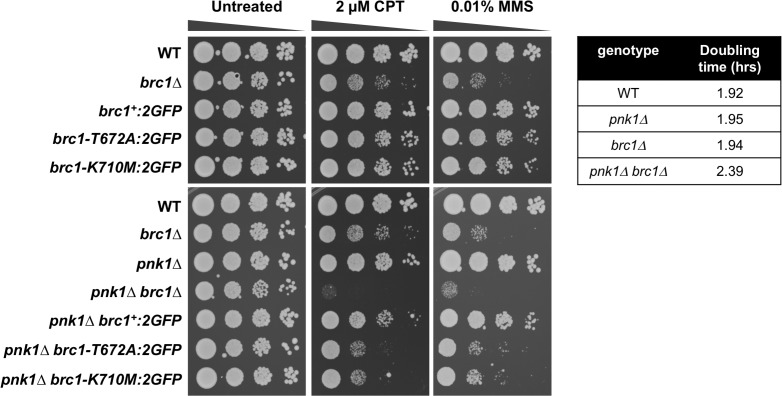
Epistatic mini-array profiling (E-MAP) screens identified synergistic negative genetic interactions involving brc1Δ and pnk1Δ, indicating that Brc1 helps to maintain cell viability when Pnk1 activity is lost [13, 30, 31]. We confirmed this synthetic sick interaction in spot dilution assays in which the colony size of pnk1Δ brc1Δ mutants were reduced compared to either single mutant (Fig 1, untreated panels). The growth defect of pnk1Δ brc1Δ cells was also verified in liquid growth assays that measured doubling times (Fig 1). The growth defect of pnk1Δ brc1Δ cells was enhanced in the presence of MMS or CPT, which produce DNA lesions that can be processed to yield SSBs with 3’ phosphate (Fig 1). Failure to repair these SSBs before entry into S-phase would be expected to increase the frequency of replication fork collapse.
Fig 1. Brc1 binding to γH2A is important in pnk1Δ cells.
Tenfold serial dilutions of cells were exposed to the indicated DNA damaging agents. Plates were incubated at 30°C for 3 to 4 days. Doubling times were determined with cells grown in liquid YES media at 32°C. Note that the brc1-T672A and brc1-K710M alleles contain a C-terminal 2GFP tag, which under some conditions can be observed to partially impair Brc1, thus strains with these alleles should be compared to wild type Brc1 tagged with 2GFP (brc1:2GFP).
Brc1 is thought to act as a scaffold protein to promote replication fork stability and repair [22, 29]. These activities partially depend on the ability of Brc1 to bind γH2A through its C-terminal pair of BRCT domains. The crystal structure of these domains bound to γH2A peptide allowed us to design T672A and K710M mutations that specifically disrupt the γH2A-binding pocket in Brc1 and abolish Brc1 foci formation [22]. These mutations did not cause an obvious growth defect in the pnk1Δ background but they strongly enhanced sensitivity to MMS or CPT (Fig 1). From these data, we conclude that Brc1 binding to γH2A is critical when pnk1Δ cells are treated with genotoxins that cause formation of SSBs with 3’ phosphate.
ATR/Rad3 and Chk1 checkpoint kinases are crucial in pnk1Δ cells
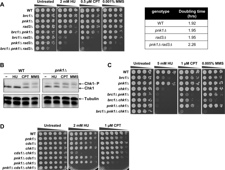
The requirement for Brc1 binding to γH2A in pnk1Δ cells suggested that unrepaired SSBs in these cells triggers a DNA damage response involving the master checkpoint kinase ATR, known as Rad3 in fission yeast [32]. Indeed, pnk1Δ rad3Δ colony size was reduced and doubling time increased compared to either single mutant (Fig 2A). This negative genetic interaction became more obvious when pnk1Δ rad3Δ cells were grown in the presence of CPT, MMS or the replication inhibitor hydroxyurea (HU) (Fig 2A). Elimination of Brc1 further impaired growth in pnk1Δ rad3Δ cells (Fig 2A), which is consistent with previous studies indicating that Brc1 has both Rad3-dependent and independent activities [13].
Fig 2. DNA damage checkpoint activation and requirement for checkpoint proteins in pnk1Δ cells.
A) Effects of combining pnk1Δ with rad3Δ or brc1Δ mutations. Tenfold serial dilutions of cells were exposed to the indicated DNA damaging agents. Plates were incubated at 30°C for 3 to 4 days. B) Chk1 also undergoes activating phosphorylation in untreated and HU-treated pnk1Δ cells, unlike in wild type. Cells were incubated with 10 mM HU, 5 μM CPT, or 0.01% MMS for 3 hours. C) Effects of combining pnk1Δ with chk1Δ or brc1Δ mutations. D) Effects of combining pnk1Δ with chk1Δ or cds1Δ.
Rad3 phosphorylates the checkpoint kinase Chk1 in response to replication fork collapse [33–35]. Immunoblot assays that detect phospho-Chk1 confirmed that Chk1 is activated even in the absence of genotoxin treatment in pnk1Δ cells, providing molecular evidence of increased DNA lesions in these cells (Fig 2B). Chk1 phosphorylation in response to HU treatment was also increased in pnk1Δ cells compared to wild type (Fig 2B). No negative genetic interaction between pnk1Δ and chk1Δ was evident in the absence of genotoxins, indicating that the spontaneous DNA lesions causing Chk1 activation in pnk1Δ cells are efficiently repaired in the time frame of a normal G2 phase (Fig 2C, untreated panel). These data suggest that the Chk1-activiating lesions are occurring early in the cell cycle, such as when replication runoff at lingering SSBs forms Chk1-activating DSBs. However, genotoxin treatment revealed a synergistic negative genetic interaction between pnk1Δ and chk1Δ that was most evident in cells treated with CPT. Chk1 was also critical in pnk1Δ cells treated with HU, which was consistent with the enhanced Chk1 phosphorylation in HU-treated pnk1Δ cells (Fig 2B). Elimination of Chk1 also enhanced the CPT and MMS sensitivity of pnk1Δ brc1Δ cells (Fig 2C).
Rad3 phosphorylates Cds1 checkpoint kinase, homologous to mammalian Chk2, in response to replication fork arrest caused by HU [36]. We did not observe negative genetic interactions between pnk1Δ and cds1Δ, either in the absence or presence of genotoxins (HU or CPT) (Fig 2D). These data indicate that pnk1Δ cells do not suffer from high frequencies of replication fork arrest. However, the triple mutant pnk1Δ cds1Δ chk1Δ displayed modestly increased HU sensitivity relative to cds1Δ chk1Δ, and modestly increased CPT sensitivity relative to pnk1Δ chk1Δ (Fig 2D).
From these results, we conclude that pnk1Δ cells accumulate DNA lesions that activate a Rad3-dependent checkpoint response leading to activation of Chk1. This response becomes especially critical when Brc1 is absent or when cells are treated with genotoxins that create SSBs.
Increased frequency of Rad52 and RPA foci in pnk1Δ cells
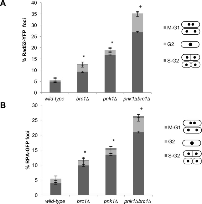
These data indicated that pnk1Δ cells accumulate DNA lesions that activate DNA damage responses. To further test this proposition, we monitored foci formation of Rad52, which is normally essential for all forms of homology-directed repair in fission yeast. Mutants that suffer increased rates of replication fork collapse, or are unable to efficiently repair collapsed forks, typically display increased numbers of Rad52 nuclear foci [37–40]. For these studies, we monitored Rad52 tagged with yellow fluorescent protein (Rad52-YFP) expressed from the endogenous locus. As observed previously [22], the frequency of cells with Rad52-YFP foci was significantly increased in brc1Δ cells (12.5%) compared to wild type (5.6%) (Fig 3A). The incidence of cells with Rad52-YFP foci was higher in the pnk1Δ strain (19%), and there was a further significant increase in the brc1Δ pnk1Δ strain (35.1%) (Fig 3A). Cell cycle phase analysis indicated that in all strains most of the cells with Rad52-YFP foci were in S-phase or early G2 phase, which suggests fork collapse as a primary source of these lesions. It was noteworthy that there was a large increase in mid- to late-G2 phase cells with Rad52 foci in the brc1Δ pnk1Δ strain (8.1%) compared to either single mutant (3.2% or 2.5%), respectively (Fig 3A). These data suggest Brc1 is required to efficiently repair lesions that accumulate in pnk1Δ cells, which could explain why Rad3 and Chk1 are crucial in pnk1Δ brc1Δ cells (Fig 2).
Fig 3. Rad52 and RPA foci increase in pnk1Δ cells and further increase in brc1Δ pnk1Δ cells.
Cells expressing Rad52-YFP (A) or RPA-GFP (B) were cultured in minimal medium at 25°C until mid-log phase. The larger increase of Rad52 foci relative to RPA foci n pnk1Δ brc1Δ cells might reflect foci duration. Cell cycle phase markers (cell length, number and position of nuclei as visualized by nucleoplasmic Rad52-YFP or RPA-GFP, absence or presence of septum) was used to estimate cell cycle position. The columns indicate, from top to bottom, cell cycle phases of cells with foci: M or G1 (M-G1), G2, S or early G2 (S-G2). Error bars correspond to standard deviation of the mean. Asterisk (*) and plus (+) symbols indicate statistically significant differences with wild type or pnk1Δ strains, respectively, as determine by two-tailed Student T-test, p-value ≤ 0.05.
In a separate experiment, we assessed foci formation by RPA, which is the major single-stranded DNA binding activity in eukaryotes. For these studies, we used strains that expressed the largest subunit of RPA, known as Ssb1 or Rad11/Rpa1, with a green fluorescent protein (GFP) tag (Fig 3B). The frequency of RPA-GFP foci was moderately increased in brc1Δ (11.6% versus 5.5% in wild type), further increased in pnk1Δ cells (15.7%), and even further increased in brc1Δ pnk1Δ (26.4%). As seen for Rad52, in all strains the RPA foci were predominantly observed in cells that were in S or early G2 phase, although combining the brc1Δ and pnk1Δ mutations did result in a substantial increase in mid- or late-G2 phase cells with RPA foci (4.8%), versus 1.6% in brc1Δ cells or 1.5% in pnk1Δ cells (Fig 3B).
These results are consistent with the synergistic growth defect and genotoxin sensitivity observed in brc1Δ pnk1Δ cells, and suggest that efficient repair of unligatable SSBs that accumulate in the absence of Pnk1 depends on Brc1.
Mre11 and Ctp1 are crucial in the absence of Pnk1
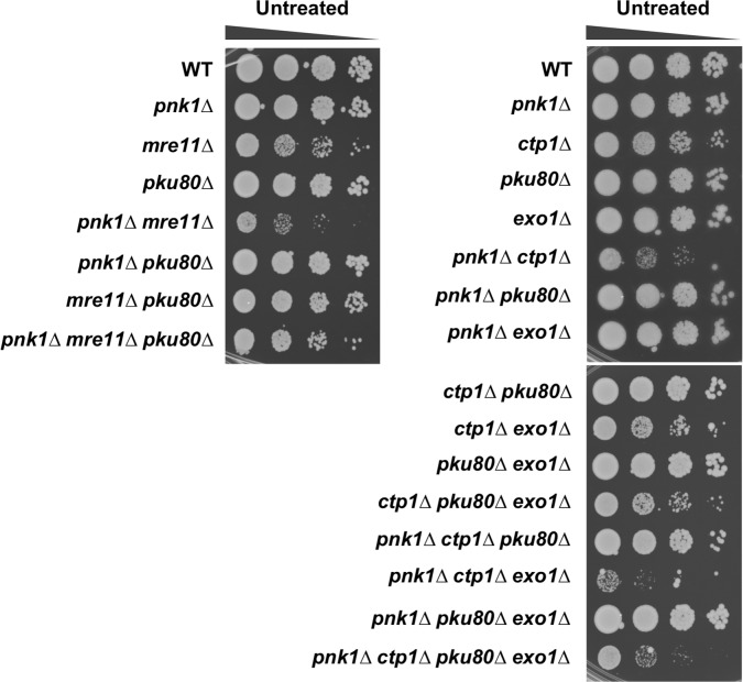
Our studies suggested that lingering SSBs in pnk1Δ cells are converted to DSBs by replication runoff. To further investigate this possibility, we assessed the requirements for the two major DNA end-binding protein complexes in fission yeast. The Ku70/Ku80 heterodimer has a high affinity for DSBs. It promotes nonhomologous end-joining (NHEJ), which is critical for DSB repair in G1 phase when cells lack sister chromatids required for HDR [41]. The pku80Δ mutation did not impair the growth of pnk1Δ cells (Fig 4). These findings show that NHEJ does not play a significant role in an alternative pathway for repairing SSBs in the absence of PNKP. The Mre11-Rad50-Nbs1 (MRN) endonuclease complex also binds DSBs, whereupon it endonucleolytically liberates Ku and initiates 5’-3’ resection to generate ssDNA tails required for HDR [42]. These activities depend on Ctp1 (CtIP/Sae2), which is only expressed in S and G2 phases in S. pombe [43, 44]. E-MAP studies indicated that both Mre11 and Ctp1 are likely to be important in the absence of Pnk1 [31, 45]. Indeed, we found that pnk1Δ mre11Δ and pnk1Δ ctp1Δ double mutants grew very poorly compared to the respective single mutants (Fig 4).
Fig 4. Mre11 and Ctp1 are crucial in pnk1Δ cells.
Effects of eliminating Mre11 or Pku80 in pnk1Δ background. Note that eliminating Ku partially suppresses the poor growth of pnk1Δ mre11Δ and pnk1Δ ctp1Δ cells. In the case of pnk1Δ ctp1Δ pku80Δ cells the data show that suppression requires Exo1. Plates were incubated at 30°C for 3 to 4 days.
The requirement for MRN and Ctp1 to initiate resection of DSBs can be substantially alleviated by genetically eliminating Ku, which allows Exo1 exonuclease to access DSBs and initiate resection [43, 44, 46]. To investigate whether Exo1 effectivity substitutes for MRN-Ctp1 in the absence of Ku, we introduced the pku80Δ mutation into pnk1Δ mre11Δ and pnk1Δ ctp1Δ backgrounds. This analysis revealed that eliminating Ku partially restored growth and genotoxin resistance in these genetic backgrounds (Fig 4). In the case of pnk1Δ ctp1Δ cells, we confirmed that suppression by pku80Δ depended on the presence of Exo1 (Fig 4). These findings indicate that the DSB resection activity of MRN-Ctp1 is critical in pnk1Δ cells.
Spontaneous SSBs in pnk1Δ cells are repaired by Rad52-dependent HDR that does not require Rad51
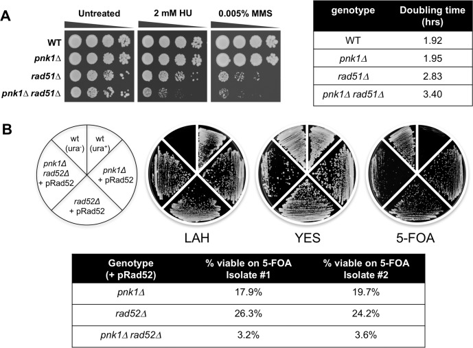
The genetic requirements for Mre11 and Ctp1 strongly suggested that HDR resets replication forks that collapse at lingering SSBs in pnk1Δ cells. However, the critical HDR recombinase Rad51 was reported to be nonessential in these cells [14]. We investigated these seemingly contradictory findings and confirmed that pnk1Δ rad51Δ cells are viable (Fig 5A). Assays that measured doubling times in liquid media indicated a modest growth defect in pnk1Δ rad51Δ cells relative to the single mutants (Fig 5A). Growth in the presence of HU or MMS revealed a more obvious negative genetic interaction between pnk1Δ and rad51Δ (Fig 5A). These findings suggest that many of the spontaneous SSBs with 3’ phosphate that accumulate in pnk1Δ cells are repaired by an MRN-Ctp1-dependent HDR mechanism that does not require Rad51.
Fig 5. Requirement for Rad52 in pnk1Δ cells.
A) A pnk1Δ rad51Δ is viable but it displays increased HU and MMS sensitivity relative to rad51Δ. Tenfold serial dilutions of cells were exposed to the indicated DNA damaging agents. Plates were incubated at 30°C for 3 to 4 days. Doubling times were determined with cells grown in liquid YES media at 32°C. B) Rad52 is crucial for viability in pnk1Δ cells. Strains with pnk1Δ or rad52Δ mutations, or the double mutant, in a ura4-D18 background, were transformed with the pRad52 plasmid containing the rad52+ gene and ura4+ selectable marker. These strains and controls (wild type with ura4+ or ura4-D18) were incubated on rich YES plates (no selection for ura4+), LAH media (selection for ura4+), or 5-FOA plates (counter selection for ura4+). Relative to pnk1Δ or rad52Δ single mutants carrying pRad52, the pnk1Δ rad52Δ, double mutant grew very poorly on 5-FOA plates. These results were confirmed by determining through microscopic observation the percentage of cells that can form colonies on 5-FOA plates.
Previously, Whitby and co-workers reported that ~50% of CPT-induced collapsed replication forks are repaired by a Rad51-independent mechanism of HDR that requires Rad52 [47]. Similarly, we found that elimination of the Swi1-Swi3 replication fork protection complex leads to collapse of replication forks that are repaired by a mechanism requiring Rad52 but not Rad51 [48]. We set out to test whether Rad52 is critical in pnk1Δ cells. Genetic crosses involving rad52Δ are complicated by the frequent appearance of suppressors caused by loss of the F-box helicase Fbh1 [49]. Therefore, we generated pnk1Δ rad52Δ or rad52Δ cells that were complemented by a pRad52 plasmid containing rad52+ and the ura4+ selectable marker (Fig 5B). Both strains grew relatively well in LAH medium that selects for the ura4+ marker, but the pnk1Δ rad52Δ cells grew much more poorly in 5-FOA media that counter-selects against the ura4+ marker (Fig 5B). These data show that Rad52 is critical for cell viability in the pnk1Δ background. These results indicate that many of accumulated spontaneous SSBs in pnk1Δ cells are repaired by a Rad52-dependent mechanism that does not require Rad51.
Mus81-Eme1 Holliday junction resolvase and Rqh1 DNA helicase are critically important in the absence of PNKP
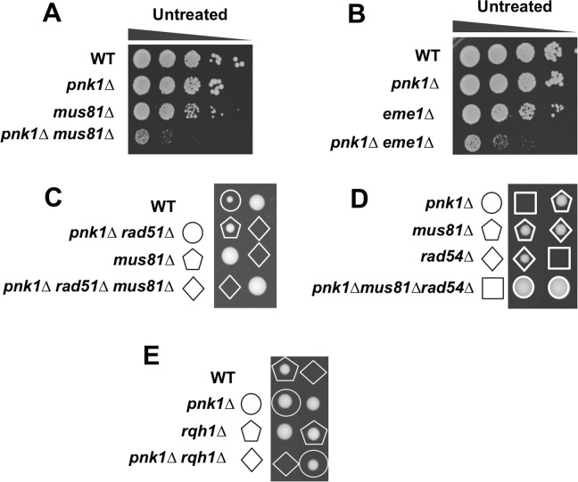
In mitotic fission yeast, homology-directed repair of two-ended DSBs, for example as generated by ionizing radiation (IR), proceeds by synthesis-dependent strand annealing (SDSA). In SDSA, joint molecules do not mature into Holliday junctions, which explains why Mus81-Eme1 resolvase is not required for IR resistance in fission yeast [50–52]. In contrast, HDR-mediated restoration of a broken replication fork produces a joint molecule (JM), either a D-loop or Holliday junction (HJ), that must be resolved to allow chromosome segregation in mitosis, hence the acute requirement for Mus81 in conditions that increase replication fork collapse [47, 51, 53]. We mated pnk1Δ and mus81Δ strains and found that the large majority of double mutant spores failed to yield viable colonies. The few viable double mutants were extremely sick (Fig 6A). We obtained the same results when we attempted to create pnk1Δ eme1Δ strains (Fig 6B).
Fig 6. Mus81-Eme1 resolvase and Rqh1 DNA helicase are essential in pnk1Δ mutant cells.
A) The few viable pnk1Δ mus81Δ viable cells recovered from genetic crosses are very sick compared to single mutants. B) The few viable pnk1Δ eme1Δ viable cells recovered from genetic crosses are very sick compared to single mutants. Tenfold serial dilutions of cells were plated and incubated at 30°C for 3 to 4 days. C) Elimination of Rad51 does not suppress pnk1Δ mus81Δ synthetic lethality. Tetrad analysis of pnk1Δ rad51Δ x mus81Δ cross. D) Elimination of Rad54 does not suppress pnk1Δ mus81Δ synthetic lethality. Tetrad analysis of pnk1Δ rad54Δ x mus81Δ cross. E) Tetrad analysis of mating between pnk1Δ and rqh1Δ strains.
If Rad51 is not required for JM formation during repair of collapsed replication forks in pnk1Δ cells, loss of Rad51 should not rescue the synthetic lethal interaction of pnk1Δ and mus81Δ. Indeed, genetic crosses showed that rad51Δ did not suppress pnk1Δ mus81Δ synthetic lethality (Fig 6C). We also investigated Rad54, which interacts with Rad51 and is required for Rad51-dependent HDR [54], but not the Rad52-dependent HDR of CPT-induced DNA damage that occurs independently of Rad51 [47]. As predicted by our model, elimination of Rad54 failed to rescue the pnk1Δ mus81Δ synthetic lethality (Fig 6D).
Rqh1 is a RecQ family 3’-5’ DNA helicase that is orthologous to human WRN (Werner syndrome) and BLM (Bloom syndrome) DNA helicases, and S. cerevisiae Sgs1 DNA helicase [55, 56]. Rqh1 is involved in multiple genome protection pathways and is particularly notable for its essential function in the absence of Mus81 [51]. Strikingly, we found that Rqh1 is essential in the pnk1Δ background (Fig 6E).
Swi10-Rad16 3’ flap endonuclease is not required in pnk1Δ cells
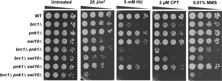
In S. cerevisiae, the viability of tpp1Δ apn1Δ apn2Δ cells depend on Rad10-Rad1 3’ flap endonuclease, which is orthologous to human ERCC1-XPF [11]. These results suggest that Rad10-Rad1 provides an alternative mechanism for eliminating 3’-phosphates from DNA termini through endonucleolytic cleavage of 3’ DNA flaps. In fission yeast, pnk1Δ apn2Δ cells are inviable [14], but it remained possible that the 3’ flap endonuclease Swi10-Rad16 [57, 58], orthologous to budding yeast Rad10-Rad1, played an important role in repairing lingering SSBs in pnk1Δ cells. We found that pnk1Δ swi10Δ cells were viable and displayed no obvious growth defect relative to the respective single mutants (Fig 7). The pnk1Δ swi10Δ strain displayed slightly more sensitivity to HU and CPT but not MMS, but these genetic interactions did not appear to be synergistic. Thus, unlike key HDR proteins, Swi10-Rad16 3’ flap endonuclease is not part of a critical back-up mechanism for repairing SSBs with 3’ phosphate.
Fig 7. Swi10-Rad16 3’ flap endonuclease is not required in pnk1Δ cells.
Tenfold serial dilutions of cells were exposed to the indicated DNA damaging agents. Plates were incubated at 30°C for 3 to 4 days.
Increased spontaneous recombination in pnk1Δ cells
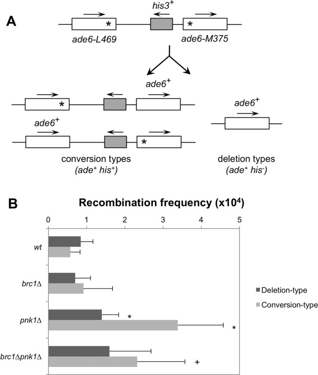
Finally, to explore whether Pnk1 deficiency creates perturbations to replication fork progression that increase recombination, we performed a mitotic intrachromosomal recombination assay. This assay determines the spontaneous frequency of Adenine positive (Ade+) colonies arising by recombination between two ade6 heteroalleles flanking the his3+ gene [59]. Two classes of recombinants can be distinguished: deletion-types (Ade+ His-) and conversion-types (Ade+ His+) (Fig 8A). Total spontaneous recombination frequencies (deletion + conversion types, reported as events per 104 cells) were increased ~3.4-fold in pnk1Δ cells (4.78 ± 1.16) compared with wild-type (1.41 ± 0.57). Although earlier studies indicated that spontaneous recombination frequencies in brc1Δ cells were strongly reduced [29], in our assays the spontaneous recombination frequencies of brc1Δ cells were not significantly different from wild type (Fig 8B). The spontaneous recombination frequency in brc1Δ pnk1Δ cells (3.91 ± 2.3) was moderately decreased compared to pnk1Δ cells (Fig 8B). Interestingly, conversion-type recombinants predominated in pnk1Δ cells even though Rad51 is required for this mechanism of repair [47], suggesting that Rad51 often participates in repair of collapsed forks at this locus even though it is not essential in pnk1Δ cells. The statistically significant decrease of total recombinants in brc1Δ pnk1Δ cells compared to pnk1Δ was caused by a loss of conversion-type recombinants. Collectively, these data indicate that a Pnk1 deficiency increases HDR-mediated genome instability.
Fig 8. Increased spontaneous recombination in pnk1Δ cells.
A) Schematic of the non-tandem direct repeat of ade6- heteroalleles used for measuring spontaneous recombinant frequencies. Conversion types events result in Ade+ His- colonies, whereas deletion types events result in Ade+ His+ colonies. B) Recombination frequencies (per 104 viable cells ± SD) of the following strains: wild type (1.41 ± 0.57), brc1Δ (1.62 ± 1.16), pnk1Δ (4.78 ± 1.62), brc1Δ pnk1Δ (3.91 ± 2.3). Deletion types and conversion types were determined by replica-plating. Error bars correspond to standard deviations of the means. Asterisk depicts statistically significant differences with wild type and + symbol with pnk1Δ, as determined by two-tailed Student T-test, p-value ≤ 0.05.
Discussion
In this study, we have investigated how fission yeast cells tolerate the loss of polynucleotide kinase/phosphatase. In principle, a PNKP deficiency should result in lingering SSBs if there is no other efficient alternative mechanism for repairing SSBs with 3’ phosphate. Note that for this discussion we are presuming that genetic interactions involving pnk1Δ are caused by loss of 3’ phosphatase activity, as this defect is responsible for the DNA damage sensitivities of pnk1Δ cells, although it is formally possible that loss of 5’ kinase activity also contributes to these genetic interactions [14]. SSBs can be converted into broken replication forks during S-phase. Broken forks in PNKP-deficient cells are proposed to be restored by homology-directed repair, also known as RC-SSBR [1], thus key HDR proteins should be critical in the absence of PNKP. Surprisingly, there is scant evidence for this assumption, and that which exists in fission yeast contradicts this model. Notably, the critical HDR protein Rad51 is not required in pnk1Δ mutants of fission yeast [14]. We investigated this conundrum. Our experiments confirm that pnk1Δ rad51Δ cells are viable; indeed, eliminating Pnk1 only moderately impairs growth in the rad51Δ background. However, we have found that other HDR proteins become crucial for cell viability in the absence of Pnk1. Our studies established that Rad52 is critical in pnk1Δ cells. Similarly, pnk1Δ mre11Δ and pnk1Δ ctp1Δ strains grow very poorly. As the principal role of MRN complex and Ctp1 is to initiate resection of DSBs, these data strongly suggest that defective SSB repair in pnk1Δ cells is rescued by a mechanism that involves homology-directed repair of DSBs. Another key finding was the requirement for Mus81-Eme1 resolvase in pnk1Δ cells. As discussed above, Mus81-Eme1 is not required for survival of IR-induced DSBs, but it is crucial for recovery from replication fork breakage [51, 53, 60]. Thus, our data strongly support the idea that lingering SSBs in pnk1Δ cells trigger replication fork collapse. This conclusion is further supported by the large increase in RPA and Rad52 foci in pnk1Δ cells, and cell cycle phase analysis indicating that most of the cells with these foci were in S-phase or early G2 phase.
The nature of the accumulating DNA lesions in pnk1Δ cells are also indicated by the negative genetic interaction with brc1Δ. As previously reported, brc1Δ cells are largely resistant to IR but quite sensitive to CPT, indicating that Brc1 functions in S-phase to assist the repair of collapsed replication forks [22]. Thus, a defect in efficiently repairing collapsed replication forks most likely accounts for the synthetic sickness observed in pnk1Δ brc1Δ cells. Brc1 function partially depends on its ability to bind γH2A [22], hence it is noteworthy that mutations that specifically disrupt this binding show a synergistic negative genetic interaction with pnk1Δ when cells are exposed to CPT. Recent studies with budding yeast revealed that the putative Brc1 ortholog Rtt107 plays a role in promoting the activation of Mus81-Mms4 resolvase (orthologous to fission yeast Mus81-Eme1) by several cell cycle-regulated protein kinases [61, 62]. In fission yeast, Mus81-Eme1 is regulated by the master cell cycle regulator Cdc2 (CDK1) protein kinase and Rad3 (ATR) checkpoint kinase [63]. If Brc1 promotes Cdc2- or Rad3-mediated activation of Mus81-Eme1, this mechanism could partly explain the requirement for Brc1 in pnk1Δ cells.
The absence of an obvious negative genetic interaction involving pnk1Δ and chk1Δ mutations in cells grown without genotoxins, despite the evident activation of Chk1, also provides clues about the DNA lesions that accumulate in the absence of PNKP. Chk1 delays the onset of mitosis by inhibiting Cdc25, which is the activator the cyclin-dependent kinase Cdc2 [64]. Fission yeast has a naturally long G2 phase, thus activating a cell cycle checkpoint that delays mitosis should be less important if all DSBs are formed early in the cell cycle during S-phase. These facts explain why chk1Δ cells are relatively tolerant of moderate doses of genotoxins such as CPT, in which toxicity is mainly caused by breakage of replication forks, unless homology-directed repair is slowed by partial loss-of-function mutations in HDR proteins [65]. These observations are consistent with a model in which replication forks break when they encounter lingering SSBs in pnk1Δ cells.
Neither pnk1Δ or chk1Δ mutants are strongly sensitive to 1 or 2 μM CPT, yet the pnk1Δ chk1Δ double mutant is acutely sensitive (Fig 2C). A similar genetic relationship is observed for pnk1Δ and rad3Δ (Fig 2A). These heightened requirements for checkpoint responses in pnk1Δ cells suggest that alternative repair pathways for repairing SSBs with 3’ phosphate are slow or inefficient. This interpretation is consistent with the increased level of Chk1 phosphorylation observed in the absence of genotoxin exposure in pnk1Δ cells (Fig 2B).
What is the explanation for the Rad51-independent repair of broken replication forks in pnk1Δ cells? As previously proposed, replication fork collapse caused by the replisome encountering a single-strand break or gap can generate a broken DNA end and a sister chromatid with a single-strand gap [47]. This gap will tend to persist when it has a 3’ phosphate, as described below. The ssDNA gap may provide access to a DNA helicase that generates unwound donor duplex that participates in Rad52-mediated strand annealing [47]. This process would not require Rad51. An alternative explanation concerns the location of lingering SSBs in pnk1Δ cells. In fission yeast, the ~150 copies of the ribosomal DNA locus are arranged in tandem repeats at each of chromosome III. We have previously reported that Slx1-Slx4 structure-specific endonuclease helps to maintain rDNA copy number by promoting HDR events during replication of the rDNA [66]. Strikingly, these HDR events require Rad52 but not Rad51. Moreover, Mus81 and Rqh1 have crucial roles in maintaining rDNA in fission yeast [35, 63]. If a large fraction of the lingering SSBs in pnk1Δ cells occur in the rDNA, this property could explain why Rad52, Mus81 and Rqh1 are required in pnk1Δ cells, but Rad51 is dispensable. We note that both gene conversion and deletion types increase between the ade6- heteroalleles in pnk1Δ cells (Fig 8), suggesting that Rad51 often participates in this repair, even if it is not essential in pnk1Δ cells.
The 3’ phosphate responsible for the persistence of a SSB in pnk1Δ cells can itself be a barrier to the completion of homology-directed repair when the SSB is converted to a broken replication fork [67]. Here, we consider models for tolerance of persistent SSBs with 3’ phosphate (Fig 9). When a replication fork collapses upon encountering a SSB with 3’ phosphate in the lagging strand template, the product is a one-ended DSB containing a 3’ phosphate (Fig 9, step 1a). Resection generates a single-strand overhang that invades the sister chromatid, but the 3’ phosphate blocks priming of DNA synthesis and restoration of an active replication fork (step 1b). This barrier to DNA synthesis might favor dissolution of the JM, but its resolution by Mus81-Eme1 would stabilize the sister chromatid junction, allowing completion of replication by the converging fork (step 1c). The final product is a replicated chromosome containing a small ssDNA gap with the 3 ‘phosphate (step 1d). When a replication fork collapses upon encountering a SSB with 3’ phosphate in the leading strand template, the product is a one-ended DSB containing a 3’ hydroxyl opposite a sister chromatid with a ssDNA gap with 3’ phosphate (Fig 9, step 2a). As previously noted [67], the SSB in the sister chromatid will block homology-directed repair, but replication by the converging fork will lead to replication fork collapse, leaving a DSB with a 3’ phosphate (step 2b). At this point repair can proceed by SDSA (step 2c), eventually leading to one intact chromosome and the other containing a single-strand gap with a 3’ phosphate (step 2d). Plans are underway to test these models.
Fig 9. Models for replication-coupled repair of SSBs with 3’ phosphate terminus.
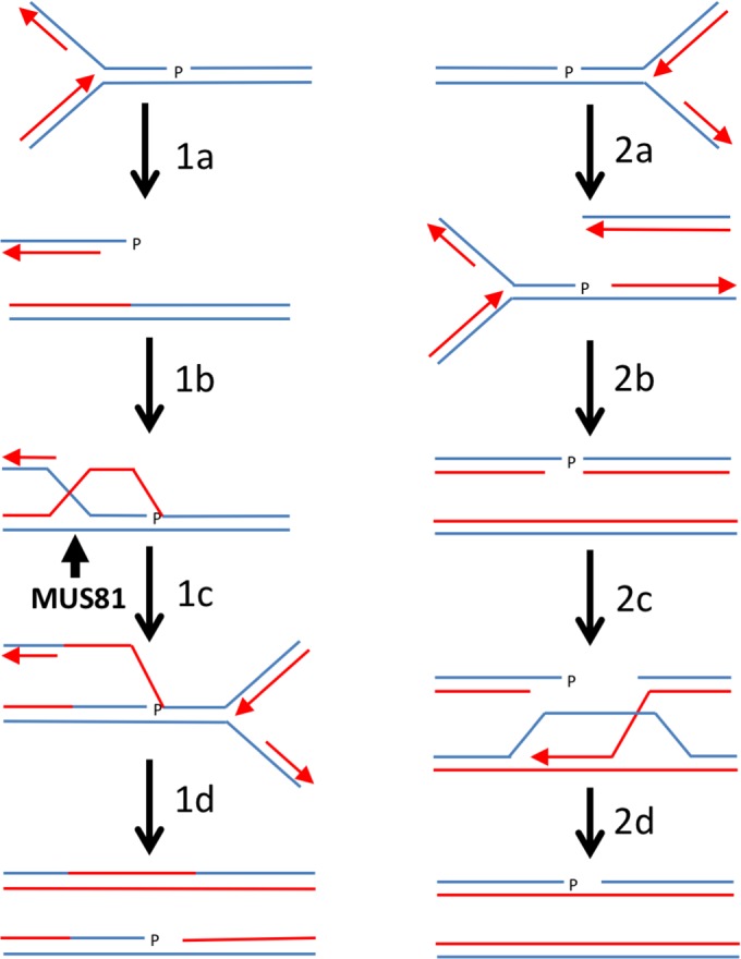
1a, replication fork collapses upon encountering a SSB with 3’ phosphate in the lagging strand template. 1b, resection of DSB followed by strand invasion of the sister chromatid using 3’ single-strand overhang containing 3’ phosphate. 1c, resolution of D-loop or Holliday junction by Mus81-Eme1. 1d, replication by converging fork, leaving a single-strand gap with 3’ phosphate. 2a, replication fork collapses upon encountering a SSB with 3’ phosphate in the leading strand template. 2b, converging fork collapses at SSB, leaving DSB with 3’ phosphate terminus. 2c, resection of DNA end with 3’ hydroxyl, followed by strand invasion of the sister chromatid. 2d, completion of repair by SDSA leaves a single-strand gap with 3’ phosphate.
In summary, these studies establish that polynucleotide/kinase phosphatase plays a crucial role in preventing the accumulation of SSBs that trigger replication fork collapse and genome instability in fission yeast, with the special property that many of these broken replication forks are repaired by an HDR mechanism that requires Mre11, Rad52 and Mus81, but not Rad51. With the recent evidence that Rad52 plays a crucial role in repair of broken replication forks in mammalian cells [68, 69], it will be of special interest to evaluate the importance of Rad52 in PNKP-deficient mammalian cells.
Materials and methods
Strains and genetic methods
The strains used in this study are listed in S1 Table. Standard fission yeast methods were used [70]. Deletion mutations strains were constructed as described [71]. The pnk1::KanMX6 strains were created from the wild-type strains using the PCR-based method and the primers, pnk1.G (5′-GTATGTTATTGAAACCACCCATTTTCATTGCTATGCAATTATAATATAGCTAACTCAATTACCAAGTCCCATTTAGTATTCGGATCCCCGGGTTAATTAA-3′) and pnk1.H (5′-ATAATTTTTATAAACGTTTGGTTTTAGTGGGATCAATAACTATATATTTTTGAAATTAATGCAATTTAATAATTTCTTAG GAATTCGAGCTCGTTTAAAC-3′). The nucleotide sequences in boldface overlap to the KanMX cassette of plasmid pFA6a-kanMX4. Successful deletion of these genes was verified by PCR. Tetrad analysis was performed to construct double mutants and verified by PCR.
Survival and growth assays
DNA damage sensitivity assays were performed by spotting 10-fold serial dilutions of exponentially growing cells onto yeast extract with glucose and supplements (YES) plates, and treated with indicated amounts of hydroxyurea (HU), camptothecin (CPT), and methyl methanesulfonate (MMS). For UV treatment, cells were serially diluted onto YES plates and irradiated using a Stratagene Stratalinker UV source. Cell survival was determined after 3–4 days at 30°C. Doubling times were performed with cell grown in YES liquid media at 32°C. Values are an average 3 cultures.
Immunoblots
For Chk1 shift, whole cells extracts were prepared from exponentially growing cells in standard NP-40 lysis buffer. Protein amounting to ~100 mg was resolved by SDS-PAGE using 10% gels with acrylamide:bis-acrylamide ratio of 99:1. Proteins were transferred to nitrocellulose membranes, blocked with 5% milk in TBST (137 mM Sodium Chloride, 20 mM Tris, pH 7.6, 0.05% Tween-20) and probed with anti-HA (12CA5) antibody (Roche).
Microscopy
Cells were photographed using a Nikon Eclipse E800 microscope equipped with a Photometrics Quantix charge-coupled device (CCD) camera and IPlab Spectrum software. All fusion proteins were expressed at their own genomic locus. Rad52-yellow fluorescence protein (YFP) expressing strains were grown in EMM until mid-log phase for focus quantification assays. Quantification was performed by scoring 500 or more nuclei from three independent experiments.
Recombination assay
Mitotic recombination was assayed by the recovery of Ade+ recombinants from the strains containing the intrachromosomal recombination substrate. Spontaneous recombinant frequencies were measured as described by fluctuation tests [59, 72]. Frequencies of fifteen colonies were averaged to determine the mean recombination frequency. Error bars indicate standard deviation from the mean. Two sample t-test were used to determine the statistical significance of differences in recombination frequencies.
Supporting information
(DOCX)
Acknowledgments
We thank Nick Boddy for helpful discussions.
Data Availability
All relevant data are within the paper and its Supporting Information files.
Funding Statement
This work was supported by grants from the National Institute of General Medical Science (GM059447) and National Cancer Institute (CA077325 and CA117638) awarded to PR. The funders had no role in study design, data collection and analysis, decision to publish, or preparation of the manuscript.
References
- 1.Caldecott KW. Single-strand break repair and genetic disease. Nat Rev Genet. 2008;9(8):619–31. doi: 10.1038/nrg2380 [DOI] [PubMed] [Google Scholar]
- 2.Weinfeld M, Mani RS, Abdou I, Aceytuno RD, Glover JN. Tidying up loose ends: the role of polynucleotide kinase/phosphatase in DNA strand break repair. Trends Biochem Sci. 2011;36(5):262–71. doi: 10.1016/j.tibs.2011.01.006 [DOI] [PMC free article] [PubMed] [Google Scholar]
- 3.Schellenberg MJ, Williams RS. DNA end processing by polynucleotide kinase/phosphatase. Proc Natl Acad Sci U S A. 2011;108(52):20855–6. doi: 10.1073/pnas.1118214109 [DOI] [PMC free article] [PubMed] [Google Scholar]
- 4.Vance JR, Wilson TE. Uncoupling of 3'-phosphatase and 5'-kinase functions in budding yeast. Characterization of Saccharomyces cerevisiae DNA 3'-phosphatase (TPP1). J Biol Chem. 2001;276(18):15073–81. doi: 10.1074/jbc.M011075200 [DOI] [PubMed] [Google Scholar]
- 5.Shimada M, Dumitrache LC, Russell HR, McKinnon PJ. Polynucleotide kinase-phosphatase enables neurogenesis via multiple DNA repair pathways to maintain genome stability. EMBO J. 2015;34(19):2465–80. doi: 10.15252/embj.201591363 [DOI] [PMC free article] [PubMed] [Google Scholar]
- 6.Shen J, Gilmore EC, Marshall CA, Haddadin M, Reynolds JJ, Eyaid W, et al. Mutations in PNKP cause microcephaly, seizures and defects in DNA repair. Nat Genet. 2010;42(3):245–9. doi: 10.1038/ng.526 [DOI] [PMC free article] [PubMed] [Google Scholar]
- 7.Poulton C, Oegema R, Heijsman D, Hoogeboom J, Schot R, Stroink H, et al. Progressive cerebellar atrophy and polyneuropathy: expanding the spectrum of PNKP mutations. Neurogenetics. 2013;14(1):43–51. doi: 10.1007/s10048-012-0351-8 [DOI] [PubMed] [Google Scholar]
- 8.Nakashima M, Takano K, Osaka H, Aida N, Tsurusaki Y, Miyake N, et al. Causative novel PNKP mutations and concomitant PCDH15 mutations in a patient with microcephaly with early-onset seizures and developmental delay syndrome and hearing loss. J Hum Genet. 2014;59(8):471–4. doi: 10.1038/jhg.2014.51 [DOI] [PubMed] [Google Scholar]
- 9.el-Khamisy SF, Caldecott KW. DNA single-strand break repair and spinocerebellar ataxia with axonal neuropathy-1. Neuroscience. 2007;145(4):1260–6. doi: 10.1016/j.neuroscience.2006.08.048 [DOI] [PubMed] [Google Scholar]
- 10.Vance JR, Wilson TE. Repair of DNA strand breaks by the overlapping functions of lesion-specific and non-lesion-specific DNA 3' phosphatases. Mol Cell Biol. 2001;21(21):7191–8. doi: 10.1128/MCB.21.21.7191-7198.2001 [DOI] [PMC free article] [PubMed] [Google Scholar]
- 11.Karumbati AS, Deshpande RA, Jilani A, Vance JR, Ramotar D, Wilson TE. The role of yeast DNA 3'-phosphatase Tpp1 and Rad1/Rad10 endonuclease in processing spontaneous and induced base lesions. J Biol Chem. 2003;278(33):31434–43. doi: 10.1074/jbc.M304586200 [DOI] [PubMed] [Google Scholar]
- 12.Meijer M, Karimi-Busheri F, Huang TY, Weinfeld M, Young D. Pnk1, a DNA kinase/phosphatase required for normal response to DNA damage by gamma-radiation or camptothecin in Schizosaccharomyces pombe. J Biol Chem. 2002;277(6):4050–5. doi: 10.1074/jbc.M109383200 [DOI] [PubMed] [Google Scholar]
- 13.Sanchez A, Roguev A, Krogan NJ, Russell P. Genetic Interaction Landscape Reveals Critical Requirements for Schizosaccharomyces pombe Brc1 in DNA Damage Response Mutants. G3 (Bethesda). 2015;5(5):953–62. [DOI] [PMC free article] [PubMed] [Google Scholar]
- 14.Kashkina E, Qi T, Weinfeld M, Young D. Polynucleotide kinase/phosphatase, Pnk1, is involved in base excision repair in Schizosaccharomyces pombe. DNA Repair (Amst). 2012;11(8):676–83. [DOI] [PubMed] [Google Scholar]
- 15.Cejka P. DNA end resection: nucleases team up with the right partners to initiate homologous recombination. J Biol Chem. 2015;290(38):22931–8. doi: 10.1074/jbc.R115.675942 [DOI] [PMC free article] [PubMed] [Google Scholar]
- 16.Mimitou EP, Symington LS. DNA end resection—unraveling the tail. DNA Repair (Amst). 2011;10(3):344–8. [DOI] [PMC free article] [PubMed] [Google Scholar]
- 17.Heyer WD. Regulation of recombination and genomic maintenance. Cold Spring Harb Perspect Biol. 2015;7(8):a016501 doi: 10.1101/cshperspect.a016501 [DOI] [PMC free article] [PubMed] [Google Scholar]
- 18.Kurokawa Y, Murayama Y, Haruta-Takahashi N, Urabe I, Iwasaki H. Reconstitution of DNA strand exchange mediated by Rhp51 recombinase and two mediators. PLoS Biol. 2008;6(4):e88 doi: 10.1371/journal.pbio.0060088 [DOI] [PMC free article] [PubMed] [Google Scholar]
- 19.Iyama T, Wilson DM 3rd. DNA repair mechanisms in dividing and non-dividing cells. DNA Repair (Amst). 2013;12(8):620–36. [DOI] [PMC free article] [PubMed] [Google Scholar]
- 20.McKinnon PJ. Maintaining genome stability in the nervous system. Nat Neurosci. 2013;16(11):1523–9. doi: 10.1038/nn.3537 [DOI] [PMC free article] [PubMed] [Google Scholar]
- 21.Verkade HM, Bugg SJ, Lindsay HD, Carr AM, O'Connell MJ. Rad18 is required for DNA repair and checkpoint responses in fission yeast. Mol Biol Cell. 1999;10(9):2905–18. [DOI] [PMC free article] [PubMed] [Google Scholar]
- 22.Williams JS, Williams RS, Dovey CL, Guenther G, Tainer JA, Russell P. gammaH2A binds Brc1 to maintain genome integrity during S-phase. EMBO J. 2010;29(6):1136–48. doi: 10.1038/emboj.2009.413 [DOI] [PMC free article] [PubMed] [Google Scholar]
- 23.Rozenzhak S, Mejia-Ramirez E, Williams JS, Schaffer L, Hammond JA, Head SR, et al. Rad3 decorates critical chromosomal domains with gammaH2A to protect genome integrity during S-Phase in fission yeast. PLoS Genet. 2010;6(7):e1001032 doi: 10.1371/journal.pgen.1001032 [DOI] [PMC free article] [PubMed] [Google Scholar]
- 24.Nakamura TM, Du LL, Redon C, Russell P. Histone H2A phosphorylation controls Crb2 recruitment at DNA breaks, maintains checkpoint arrest, and influences DNA repair in fission yeast. Mol Cell Biol. 2004;24(14):6215–30. doi: 10.1128/MCB.24.14.6215-6230.2004 [DOI] [PMC free article] [PubMed] [Google Scholar]
- 25.Sanchez A, Sharma S, Rozenzhak S, Roguev A, Krogan NJ, Chabes A, et al. Replication fork collapse and genome instability in a deoxycytidylate deaminase mutant. Mol Cell Biol. 2012;32(21):4445–54. doi: 10.1128/MCB.01062-12 [DOI] [PMC free article] [PubMed] [Google Scholar]
- 26.Sanchez A, Russell P. Ku stabilizes replication forks in the absence of Brc1. PLoS One. 2015;10(5):e0126598 doi: 10.1371/journal.pone.0126598 [DOI] [PMC free article] [PubMed] [Google Scholar]
- 27.Mejia-Ramirez E, Limbo O, Langerak P, Russell P. Critical Function of gammaH2A in S-Phase. PLoS Genet. 2015;11(9):e1005517 doi: 10.1371/journal.pgen.1005517 [DOI] [PMC free article] [PubMed] [Google Scholar]
- 28.Lee SY, Rozenzhak S, Russell P. gammaH2A-binding protein Brc1 affects centromere function in fission yeast. Mol Cell Biol. 2013;33(7):1410–6. doi: 10.1128/MCB.01654-12 [DOI] [PMC free article] [PubMed] [Google Scholar]
- 29.Bass KL, Murray JM, O'Connell MJ. Brc1-dependent recovery from replication stress. J Cell Sci. 2012;125(Pt 11):2753–64. doi: 10.1242/jcs.103119 [DOI] [PMC free article] [PubMed] [Google Scholar]
- 30.Beltrao P, Trinidad JC, Fiedler D, Roguev A, Lim WA, Shokat KM, et al. Evolution of phosphoregulation: comparison of phosphorylation patterns across yeast species. PLoS Biol. 2009;7(6):e1000134 doi: 10.1371/journal.pbio.1000134 [DOI] [PMC free article] [PubMed] [Google Scholar]
- 31.Roguev A, Bandyopadhyay S, Zofall M, Zhang K, Fischer T, Collins SR, et al. Conservation and rewiring of functional modules revealed by an epistasis map in fission yeast. Science. 2008;322(5900):405–10. doi: 10.1126/science.1162609 [DOI] [PMC free article] [PubMed] [Google Scholar]
- 32.Bentley NJ, Holtzman DA, Flaggs G, Keegan KS, DeMaggio A, Ford JC, et al. The Schizosaccharomyces pombe rad3 checkpoint gene. EMBO J. 1996;15(23):6641–51. [PMC free article] [PubMed] [Google Scholar]
- 33.Lopez-Girona A, Tanaka K, Chen XB, Baber BA, McGowan CH, Russell P. Serine-345 is required for Rad3-dependent phosphorylation and function of checkpoint kinase Chk1 in fission yeast. Proc Natl Acad Sci U S A. 2001;98(20):11289–94. doi: 10.1073/pnas.191557598 [DOI] [PMC free article] [PubMed] [Google Scholar]
- 34.Walworth NC, Bernards R. Rad-dependent response of the chk1-encoded protein kinase at the DNA damage checkpoint. Science. 1996;271(5247):353–6. [DOI] [PubMed] [Google Scholar]
- 35.Boddy MN, Furnari B, Mondesert O, Russell P. Replication checkpoint enforced by kinases Cds1 and Chk1. Science. 1998;280(5365):909–12. [DOI] [PubMed] [Google Scholar]
- 36.Tanaka K, Boddy MN, Chen XB, McGowan CH, Russell P. Threonine-11, phosphorylated by Rad3 and atm in vitro, is required for activation of fission yeast checkpoint kinase Cds1. Mol Cell Biol. 2001;21(10):3398–404. doi: 10.1128/MCB.21.10.3398-3404.2001 [DOI] [PMC free article] [PubMed] [Google Scholar]
- 37.Du LL, Nakamura TM, Moser BA, Russell P. Retention but not recruitment of Crb2 at double-strand breaks requires Rad1 and Rad3 complexes. Mol Cell Biol. 2003;23(17):6150–8. doi: 10.1128/MCB.23.17.6150-6158.2003 [DOI] [PMC free article] [PubMed] [Google Scholar]
- 38.Meister P, Poidevin M, Francesconi S, Tratner I, Zarzov P, Baldacci G. Nuclear factories for signalling and repairing DNA double strand breaks in living fission yeast. Nucleic Acids Res. 2003;31(17):5064–73. doi: 10.1093/nar/gkg719 [DOI] [PMC free article] [PubMed] [Google Scholar]
- 39.Noguchi E, Noguchi C, Du LL, Russell P. Swi1 prevents replication fork collapse and controls checkpoint kinase Cds1. Mol Cell Biol. 2003;23(21):7861–74. doi: 10.1128/MCB.23.21.7861-7874.2003 [DOI] [PMC free article] [PubMed] [Google Scholar]
- 40.Sabatinos SA, Forsburg SL. Managing Single-Stranded DNA during Replication Stress in Fission Yeast. Biomolecules. 2015;5(3):2123–39. doi: 10.3390/biom5032123 [DOI] [PMC free article] [PubMed] [Google Scholar]
- 41.Li P, Li J, Li M, Dou K, Zhang MJ, Suo F, et al. Multiple end joining mechanisms repair a chromosomal DNA break in fission yeast. DNA Repair (Amst). 2012;11(2):120–30. [DOI] [PubMed] [Google Scholar]
- 42.Langerak P, Mejia-Ramirez E, Limbo O, Russell P. Release of Ku and MRN from DNA ends by Mre11 nuclease activity and Ctp1 is required for homologous recombination repair of double-strand breaks. PLoS Genet. 2011;7(9):e1002271 doi: 10.1371/journal.pgen.1002271 [DOI] [PMC free article] [PubMed] [Google Scholar]
- 43.Williams RS, Moncalian G, Williams JS, Yamada Y, Limbo O, Shin DS, et al. Mre11 dimers coordinate DNA end bridging and nuclease processing in double-strand-break repair. Cell. 2008;135(1):97–109. doi: 10.1016/j.cell.2008.08.017 [DOI] [PMC free article] [PubMed] [Google Scholar]
- 44.Limbo O, Chahwan C, Yamada Y, de Bruin RA, Wittenberg C, Russell P. Ctp1 is a cell-cycle-regulated protein that functions with Mre11 complex to control double-strand break repair by homologous recombination. Mol Cell. 2007;28(1):134–46. doi: 10.1016/j.molcel.2007.09.009 [DOI] [PMC free article] [PubMed] [Google Scholar]
- 45.Ryan CJ, Roguev A, Patrick K, Xu J, Jahari H, Tong Z, et al. Hierarchical modularity and the evolution of genetic interactomes across species. Mol Cell. 2012;46(5):691–704. doi: 10.1016/j.molcel.2012.05.028 [DOI] [PMC free article] [PubMed] [Google Scholar]
- 46.Tomita K, Matsuura A, Caspari T, Carr AM, Akamatsu Y, Iwasaki H, et al. Competition between the Rad50 complex and the Ku heterodimer reveals a role for Exo1 in processing double-strand breaks but not telomeres. Mol Cell Biol. 2003;23(15):5186–97. doi: 10.1128/MCB.23.15.5186-5197.2003 [DOI] [PMC free article] [PubMed] [Google Scholar]
- 47.Doe CL, Osman F, Dixon J, Whitby MC. DNA repair by a Rad22-Mus81-dependent pathway that is independent of Rhp51. Nucleic Acids Res. 2004;32(18):5570–81. doi: 10.1093/nar/gkh853 [DOI] [PMC free article] [PubMed] [Google Scholar]
- 48.Noguchi E, Noguchi C, McDonald WH, Yates JR 3rd, Russell P. Swi1 and Swi3 are components of a replication fork protection complex in fission yeast. Mol Cell Biol. 2004;24(19):8342–55. doi: 10.1128/MCB.24.19.8342-8355.2004 [DOI] [PMC free article] [PubMed] [Google Scholar]
- 49.Osman F, Dixon J, Barr AR, Whitby MC. The F-Box DNA helicase Fbh1 prevents Rhp51-dependent recombination without mediator proteins. Mol Cell Biol. 2005;25(18):8084–96. doi: 10.1128/MCB.25.18.8084-8096.2005 [DOI] [PMC free article] [PubMed] [Google Scholar]
- 50.Boddy MN, Gaillard PH, McDonald WH, Shanahan P, Yates JR 3rd, Russell P. Mus81-Eme1 are essential components of a Holliday junction resolvase. Cell. 2001;107(4):537–48. [DOI] [PubMed] [Google Scholar]
- 51.Boddy MN, Lopez-Girona A, Shanahan P, Interthal H, Heyer WD, Russell P. Damage tolerance protein Mus81 associates with the FHA1 domain of checkpoint kinase Cds1. Mol Cell Biol. 2000;20(23):8758–66. [DOI] [PMC free article] [PubMed] [Google Scholar]
- 52.Gaillard PH, Noguchi E, Shanahan P, Russell P. The endogenous Mus81-Eme1 complex resolves Holliday junctions by a nick and counternick mechanism. Mol Cell. 2003;12(3):747–59. [DOI] [PubMed] [Google Scholar]
- 53.Roseaulin L, Yamada Y, Tsutsui Y, Russell P, Iwasaki H, Arcangioli B. Mus81 is essential for sister chromatid recombination at broken replication forks. EMBO J. 2008;27(9):1378–87. doi: 10.1038/emboj.2008.65 [DOI] [PMC free article] [PubMed] [Google Scholar]
- 54.Ceballos SJ, Heyer WD. Functions of the Snf2/Swi2 family Rad54 motor protein in homologous recombination. Biochim Biophys Acta. 2011;1809(9):509–23. doi: 10.1016/j.bbagrm.2011.06.006 [DOI] [PMC free article] [PubMed] [Google Scholar]
- 55.Murray JM, Lindsay HD, Munday CA, Carr AM. Role of Schizosaccharomyces pombe RecQ homolog, recombination, and checkpoint genes in UV damage tolerance. Mol Cell Biol. 1997;17(12):6868–75. [DOI] [PMC free article] [PubMed] [Google Scholar]
- 56.Stewart E, Chapman CR, Al-Khodairy F, Carr AM, Enoch T. rqh1+, a fission yeast gene related to the Bloom's and Werner's syndrome genes, is required for reversible S phase arrest. EMBO J. 1997;16(10):2682–92. doi: 10.1093/emboj/16.10.2682 [DOI] [PMC free article] [PubMed] [Google Scholar]
- 57.Zhang JM, Liu XM, Ding YH, Xiong LY, Ren JY, Zhou ZX, et al. Fission yeast Pxd1 promotes proper DNA repair by activating Rad16XPF and inhibiting Dna2. PLoS Biol. 2014;12(9):e1001946 doi: 10.1371/journal.pbio.1001946 [DOI] [PMC free article] [PubMed] [Google Scholar]
- 58.Carr AM, Schmidt H, Kirchhoff S, Muriel WJ, Sheldrick KS, Griffiths DJ, et al. The rad16 gene of Schizosaccharomyces pombe: a homolog of the RAD1 gene of Saccharomyces cerevisiae. Mol Cell Biol. 1994;14(3):2029–40. [DOI] [PMC free article] [PubMed] [Google Scholar]
- 59.Osman F, Adriance M, McCready S. The genetic control of spontaneous and UV-induced mitotic intrachromosomal recombination in the fission yeast Schizosaccharomyces pombe. Curr Genet. 2000;38(3):113–25. [DOI] [PubMed] [Google Scholar]
- 60.Doe CL, Ahn JS, Dixon J, Whitby MC. Mus81-Eme1 and Rqh1 involvement in processing stalled and collapsed replication forks. J Biol Chem. 2002;277(36):32753–9. doi: 10.1074/jbc.M202120200 [DOI] [PubMed] [Google Scholar]
- 61.Princz LN, Wild P, Bittmann J, Aguado FJ, Blanco MG, Matos J, et al. Dbf4-dependent kinase and the Rtt107 scaffold promote Mus81-Mms4 resolvase activation during mitosis. EMBO J. 2017;36(5):664–78. doi: 10.15252/embj.201694831 [DOI] [PMC free article] [PubMed] [Google Scholar]
- 62.Dehe PM, Gaillard PH. Control of structure-specific endonucleases to maintain genome stability. Nat Rev Mol Cell Biol. 2017;18(5):315–30. doi: 10.1038/nrm.2016.177 [DOI] [PubMed] [Google Scholar]
- 63.Dehe PM, Coulon S, Scaglione S, Shanahan P, Takedachi A, Wohlschlegel JA, et al. Regulation of Mus81-Eme1 Holliday junction resolvase in response to DNA damage. Nat Struct Mol Biol. 2013;20(5):598–603. doi: 10.1038/nsmb.2550 [DOI] [PMC free article] [PubMed] [Google Scholar]
- 64.Furnari B, Blasina A, Boddy MN, McGowan CH, Russell P. Cdc25 inhibited in vivo and in vitro by checkpoint kinases Cds1 and Chk1. Mol Biol Cell. 1999;10(4):833–45. [DOI] [PMC free article] [PubMed] [Google Scholar]
- 65.Jensen KL, Russell P. Ctp1-dependent clipping and resection of DNA double-strand breaks by Mre11 endonuclease complex are not genetically separable. Nucleic Acids Res. 2016;44(17):8241–9. doi: 10.1093/nar/gkw557 [DOI] [PMC free article] [PubMed] [Google Scholar]
- 66.Coulon S, Gaillard PH, Chahwan C, McDonald WH, Yates JR 3rd, Russell P. Slx1-Slx4 are subunits of a structure-specific endonuclease that maintains ribosomal DNA in fission yeast. Mol Biol Cell. 2004;15(1):71–80. doi: 10.1091/mbc.E03-08-0586 [DOI] [PMC free article] [PubMed] [Google Scholar]
- 67.Karumbati AS, Wilson TE. Abrogation of the Chk1-Pds1 checkpoint leads to tolerance of persistent single-strand breaks in Saccharomyces cerevisiae. Genetics. 2005;169(4):1833–44. doi: 10.1534/genetics.104.035931 [DOI] [PMC free article] [PubMed] [Google Scholar]
- 68.Sotiriou SK, Kamileri I, Lugli N, Evangelou K, Da-Re C, Huber F, et al. Mammalian RAD52 functions in break-induced replication repair of collapsed DNA replication forks. Mol Cell. 2016;64(6):1127–34. doi: 10.1016/j.molcel.2016.10.038 [DOI] [PMC free article] [PubMed] [Google Scholar]
- 69.Bhowmick R, Minocherhomji S, Hickson ID. RAD52 facilitates mitotic DNA synthesis following replication stress. Mol Cell. 2016;64(6):1117–26. doi: 10.1016/j.molcel.2016.10.037 [DOI] [PubMed] [Google Scholar]
- 70.Forsburg SL, Rhind N. Basic methods for fission yeast. Yeast. 2006;23(3):173–83. doi: 10.1002/yea.1347 [DOI] [PMC free article] [PubMed] [Google Scholar]
- 71.Bahler J, Wu JQ, Longtine MS, Shah NG, McKenzie A 3rd, Steever AB, et al. Heterologous modules for efficient and versatile PCR-based gene targeting in Schizosaccharomyces pombe. Yeast. 1998;14(10):943–51. doi: 10.1002/(SICI)1097-0061(199807)14:10<943::AID-YEA292>3.0.CO;2-Y [DOI] [PubMed] [Google Scholar]
- 72.Osman F, Whitby MC. Monitoring homologous recombination following replication fork perturbation in the fission yeast Schizosaccharomyces pombe. Methods Mol Biol. 2009;521:535–52. doi: 10.1007/978-1-60327-815-7_31 [DOI] [PubMed] [Google Scholar]
Associated Data
This section collects any data citations, data availability statements, or supplementary materials included in this article.
Supplementary Materials
(DOCX)
Data Availability Statement
All relevant data are within the paper and its Supporting Information files.


