Abstract
Rationale:
Vein of Galen aneurysmal malformation (VGAM) is a rare complex malformation of the cerebral vascular system consisting of arteriovenous shunts between the vein of Galen and the cerebral arteries.
Patient concerns:
We present the case of a 31-year-old pregnant woman, para 1, gravida 1.
Diagnoses:
At 26 weeks’ gestation who was examined for an anechoic mass on the cerebral median midline with color and pulsed Doppler. She presented with positive flow on the color and pulsed Doppler test, associated with hydrocephalus, cortical hypoplasia, cardiomegaly, jugular vein distension.
Interventions:
No intervention for VGAM was done.
Outcomes:
This case of a VGAM was associated with negative prognostic factors.
Lessons:
The ultrasound color Doppler together with the 3D power Doppler allowed reconstruction of the vascular connections and of the relationship of these with other anatomical structures, which contributed to establishing the prognosis.
Keywords: color Doppler, hydrocephalus, 3-dimensional ultrasound
1. Introduction
Vein of Galen aneurysmal malformation (VGAM) is a rare abnormality of the fetal cerebral vascular system, represents 1% of the abnormalities of the fetal cerebral arteriovenous system and approximately 30% of pediatric vascular malformations.[1,2] This malformation is isolated, although there are cases in which it can be related to cardiac abnormalities or cystic hygroma. The origin of VGAM is not entirely clear, the current hypothesis holds that it forms between the 6th and the 11th week of gestation, asserting as a result of the persistence of an abnormal connection between the primitive choroidal vessels and the most proximal part of the prosencephalic median vein of Markowski. The persistence of this connection which should normally regress, leads to the appearance of some abnormal arteriovenous shunts and the formation of the vein of Galen.[3]
The development of the vascular cerebral system is made in 3 phases: stage I prechoroidal, stage II prechoroidal, and stage III choroidal.[4] In the choroidal stage, the vascularization of the cerebral structures results from the choroidal arteries, and the venous drainage is ensured by Markowski median vein. The anterior segment of Markowski vein regresses while the posterior segment of the Markowski vein will persists and is then termed the Galen vein. The creation of some arteriovenous shunts (for an unknown reason) causes the anterior segment of the Markowski vein not to regress and to dilate and these sutures drain in the vein of Galen.[4] Lasjaunias described 2 types of vein of Galen aneurysms: the first type, that is, choroidal, which arises in multiple arterial sources with severe dilatation and cases of fetal congestive heart failure; the second type, is mural, with a unique arteriovenous fistula with clinical late symptomatology in the extrauterine life and is rarely followed by heart failure.[3,4]
The diagnosis of this malformation is based on the 2-dimensional (2D) real-time ultrasonography and color Doppler. Additionally, the 3D power Doppler ultrasound presents the following advantages: anatomical details are highlighted, angle dependence is removed, and aliasing distortions are removed.
The prenatal ultrasound diagnosis is set at the end of the second trimester and the beginning of the third trimester of pregnancy. Two-dimensional conventional ultrasound usually reveals an intracranial cystic image located on the midline of the anterior wall of the third ventricle, and the color Doppler ultrasound displays some turbulent blood flow inside the intracranial transonic formation.[4,5] Three-dimensional power Doppler ultrasound can distinguish anatomical details such as venous sinuses distention and their associations.[6,7] Moreover, cranial examination can reveal ventriculomegaly, jugular vein distention, cardiomegaly, and ascites. Ascites, cardiomegaly, tricuspid insufficiency, and jugular vein distention are indications of fetus heart failure which is generated by high blood flow in the arteriovenous fistula. The presence of these symptoms is a negative prognostic factor and is followed by relatively fast development of a multiple organ failure.[8]
The diagnosis can be completed with a computed tomography (TC), a magnetic resonance imaging (MRI) or postnatal angiography or CT angiography. The fetal MRI can estimate the cortical atrophy and possibly the presence of the heart failure.[4,8] Color Doppler ultrasonography is the most often used modality for exploring the fetus. Fetal MRI has become superior to color Doppler ultrasonography in the diagnosis of VGAM in recent years.[9,10] Nevertheless, the ultrasonography remains the most frequent method of diagnosis.
2. Case report
A pregnant woman, aged 28 (Patient 1)—pregnant woman 1, para 1, presented herself at Sanador Hospital Bucharest in March 2015 and later also to the Department Obstetrics Gynecology “Sf PAntelimon” Clinical Emergency Hospital Bucharest for a second opinion due to the initial suspicion of an intracranial echogenic mass associated with ventriculomegaly which was detected at earlier ultrasonography Furthermore, the patient had experienced vaginal bleeding in the previous 24 hours. Consent to conduct of the case report was obtained from the Ethics Committee of Sanador Hospital Bucharest, Ethics Committee of Sf Pantelimon Clinical Emergency Hospital, and the written informed consent form signed by the patient included the permission to present the images and case.
From the current pregnancy chart we detected no pathological elements, up to the point where the patient had been tested for maternal infections (TORCH complex), and found no risk of chromosomal abnormalities.
The 2D real-time ultrasonographic examination revealed: biometric measurements appropriate for 27 weeks of pregnancy. At the skull level, the fetus displayed hydrocephalus and an anechoic mass on the midline, which was supratentorial, with regular extremities (Fig. 1).
Figure 1.
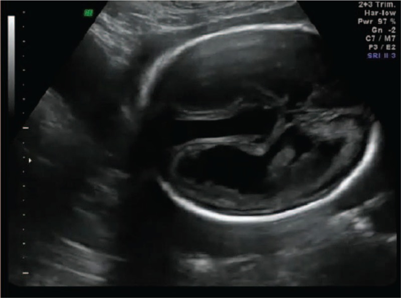
Vein of Galen aneurysmal malformation (VGAM) and hydrocephalus in gray scale (cranium axial plane).
We also noted expansion of the third ventricle, cisterna magna, and brain parenchyma and cerebellum was hypoplastic (Figs. 2–5).
Figure 2.
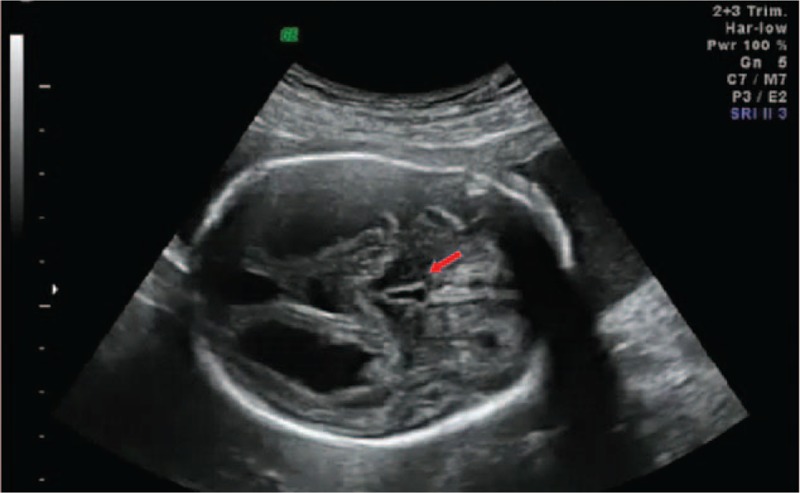
Dilatation of third ventricle in gray scale.
Figure 5.
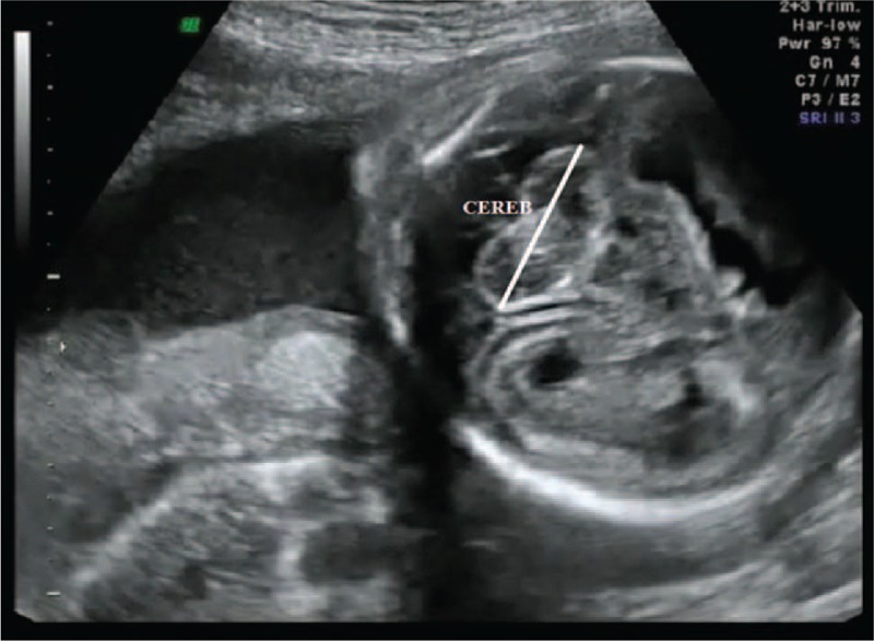
Cerebellar hypoplasia in gray scale.
Figure 3.
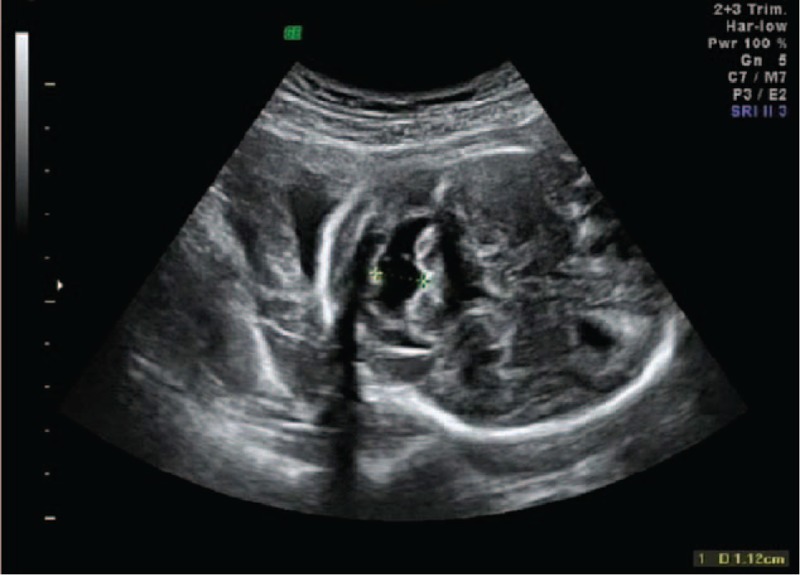
Dilatation of cisterna magna in gray scale.
Figure 4.
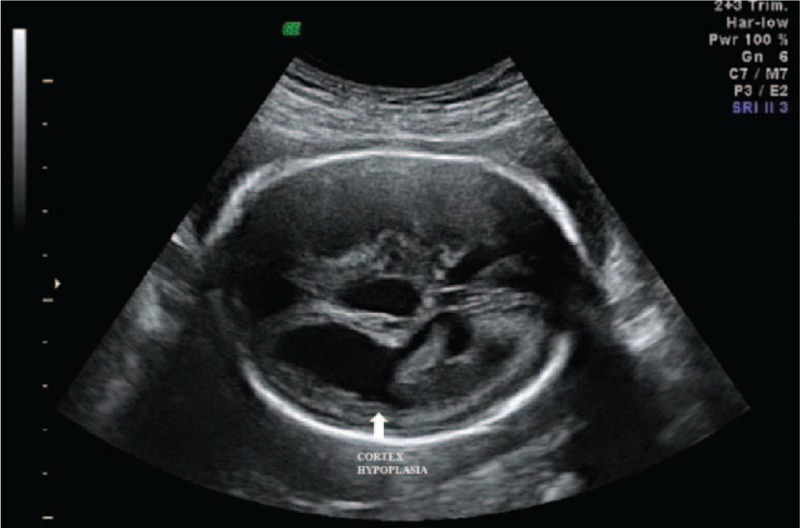
Cortex hypoplasia in gray scale.
Color Doppler examination revealed an arteriovenous pattern with significant arteriovenous expansion at the midline (Fig. 6).
Figure 6.
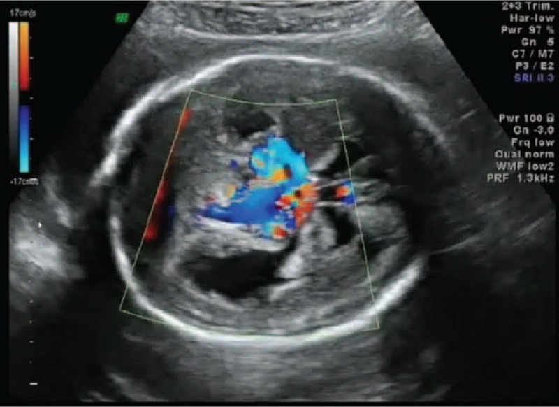
Color Doppler of VGAM.
On pulsed Doppler examination the waves indicated vascular hyperkinetic syndrome in the circle of Willis with a decline in vascular resistance and an increase in peak systolic velocity, as well as an arterial pattern in the sagittal sinus (Fig. 7). These are all characteristic of the arteriovenous shunt from the aneurysmal malformation of the vein of Galen.
Figure 7.
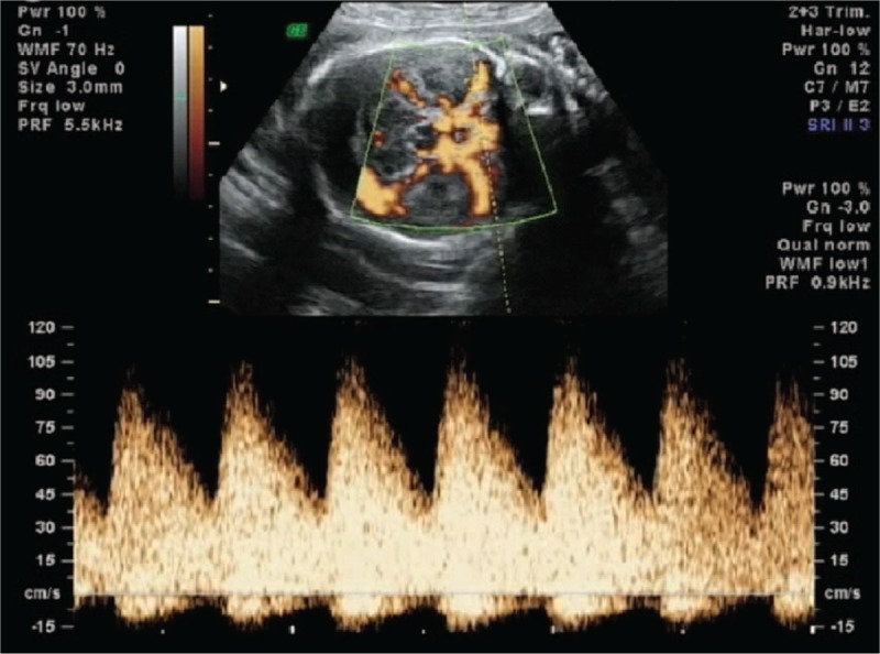
Pulsed Doppler of VGAM.
Three-dimensional ultrasound was performed with a Voluson 730 Pro machine (General Electric Healthcare, Zipf, Austria) with a convex volumetric transducer (R-4-8) and it revealed the expansion of the cerebral venous sinuses and the connections among them (Figs. 8 and 9).
Figure 8.
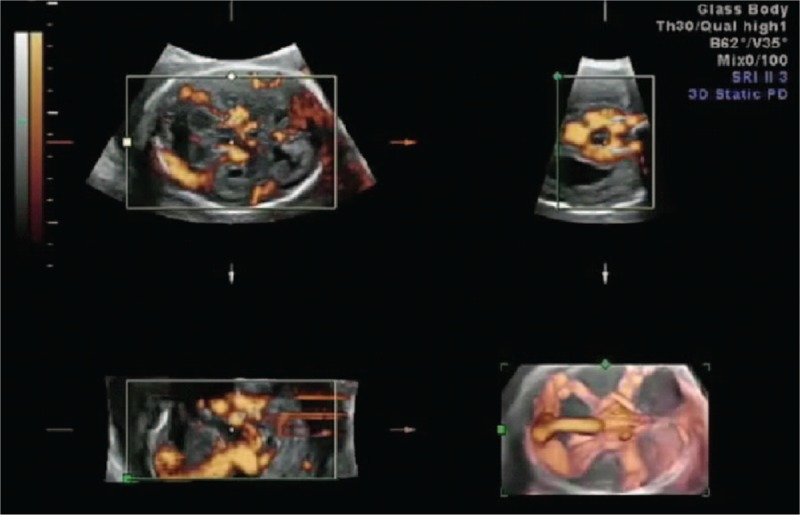
Reconstruction 3D power Doppler of VGAM.
Figure 9.
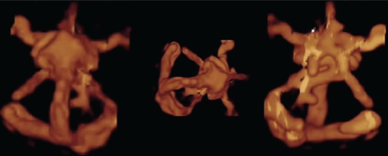
Reconstruction 3D power Doppler arteriovenous shunts.
Examination of the cervical region showed jugular vein, superior vena cava distention. Distally cardiomegaly with left heart overload without mitral or tricuspid regurgitation was seen (Fig. 10).
Figure 10.
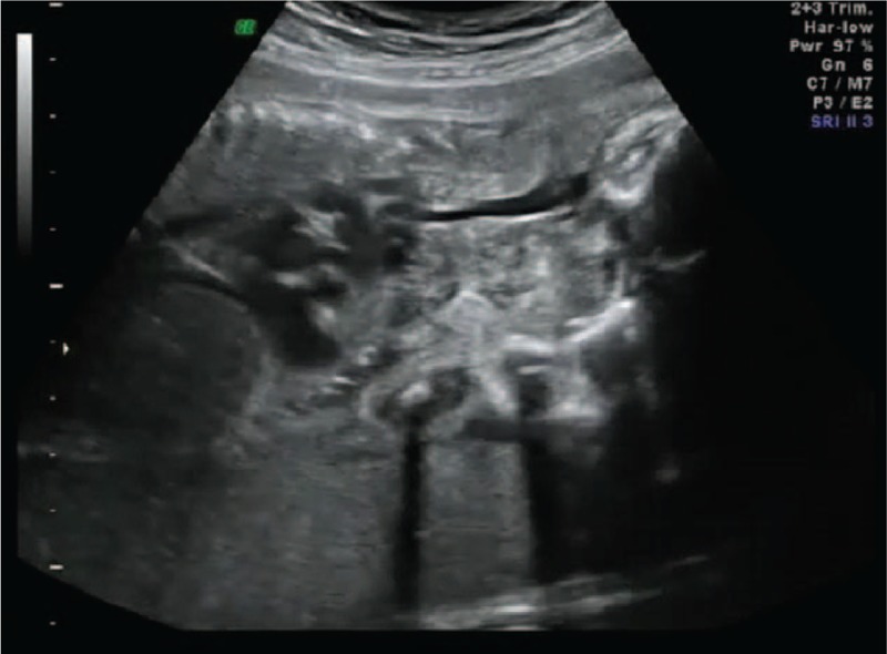
Jugular vein dilatation.
No polyhydramnios or any other fetal abnormalities associated therewith were seen, but the placenta was found in a ventral position covering the internal cervical orifice.
The parents were informed about the diagnosis of VGAM by a multidisciplinary team consisting of a neurosurgeon, pediatric neurologist, and neonatologist specialized in maternal–fetal medicine. The immediate fetal and neonatal risks were presented: hydrocephalus, heart failure associated with hydrops fetalis, cerebral atrophy, encephalomalacia, spasms, and cortical calcification in the neonatal stage. MRI investigation was suggested to point out the anatomical and vascular details of the abnormality, given the advance stage of the pregnancy. At first, the parents chose to terminate the pregnancy therapeutically after taking note of the negative prognostic factors. However, 24 hours after the hospitalization, the patient experienced uterine contractions followed by severe vaginal bleeding which called for an emergency cesarean section. A male infant weighing of 700 g was delivered. The newborn was admitted to the Neonatal Intensive Care Unit with full respiratory and medical support but the sustained cardiac failure resulted in neonatal death within the first hour after birth. The anatomopathological examination confirmed the diagnosis of VGAM.
3. Discussion
VGAM is a severe and complex vascular malformation characterized by multiple arteriovenous shunts between the vein of Galen and the choroidal arteries. Because of these shunts , vascular steal occurs and this determines the increase in blood returning to the heart at the level of cerebral cortex, resulting in overload of the right heart and a progressive heart failure.[3,4] Fetal heart failure leads to progressive hydrops. The secondary cerebral effects of the malformation are caused by both the mass effect and the phenomenon of cerebral vascular steal which could lead to hydrocephalus, cerebral infarcts, and leukomalacia.[8,9] The prenatal diagnosis of this abnormality is made means of 2D ultrasonography, color mapping, pulsed Doppler, 3D power Doppler, and MRI.[9,10] Postnatal confirmation is made by means of angiography, MRI, computed tomography, and transfontanellar ultrasound.[11,12] Identification of the turbulent vascular flow inside a supratentorial cystic mass, on the midline, when using color or pulsed Doppler provides a diagnosis and can distinguish between an aneurysm in the vein of Galen and an arachnoid or porencephalic cyst, or a Dandy Walker malformation. So, using color Doppler is essential for differential diagnose of VGAM with other cystic anomalies of the midline of brain. Also prenatal MRI can be used to rule out differential diagnosis with: choroid plexus cysts, porencephalic cysts, arachnoid cysts, pineal tumors.[9,10]
Due to the rarity of the malformation, the existing literature mostly involves case reports.[2,5,6,13] Some case series have presented prognostic factors and the therapeutic approach. The negative prognostic factors are determined by the correlation of the vascular malformation with severe cardiomegaly, tricuspid regurgitation, right atrial enlargement and dilatation of the superior vena cava and brain injury.[14–16] The prognosis of VGAM is poor when cerebral defects or cardiac dysfunction where detected prenatally, so a good outcome can be expected if no neurological or cardiac defects are present and the prognosis is poor if such defects are present.[11] The mural type angioarchitecture of a VGAM can be evaluated prenatally using 3-dimensional color power Doppler or MRI, but is most often diagnosed postnatally. The angioarchitecture of the VGAM appears to be an indicator of poor prognosis, the mural type malformation has a better prognosis than the choroidal type malformation.[12,13] There is no literature on the optimal methods for delivering these infants. Nevertheless, it is recommended that delivery should take place at a tertiary care center with doctors specialized in pediatric neuroradiology, neurosurgery, and cardiology.[14] In a retrospective study, the elective cesarean section was performed in all 10 cases while in another study, 12 of 17 were delivered vaginally while 5 were delivered by C-section.[11,16]
Little has been reported regarding the postpartum development of the infants over time. In the series published by Rodesch et al,[15] 13 of 17 infants survived during the neonatal period and 12 underwent brain aneurysm embolization, which is the elective therapeutic method at present.
In conclusion, we presented a case of VGAM, an extremely rare abnormality associated with a centrally inserted placenta. Color and pulsed Doppler ultrasound played a defining role in making the diagnosis and 3D power Doppler ultrasound accurately identified the anatomical links and the presence of negative prognostic factors.
Footnotes
Abbreviations: CT = computed tomography, MRI = magnetic resonance imaging, VGAM = vein of Galen aneurysmal malformation.
CI and SV contributed equally to this article.
The authors have no conflicts of interest to disclose.
References
- [1].Long DM, Seljeskog EL, Chou SN, et al. Giant arteriovenous malformations of infancy and childhood. J Neurosurg 1974;40:304–11. [DOI] [PubMed] [Google Scholar]
- [2].Beucher G, Fossey C, Belloy F, et al. Antenatal diagnosis and management of vein of Galen aneurysm: review illustrated by a case report. J Gynecol Obstet Biol Reprod (Paris) 2005;34:613–9. [DOI] [PubMed] [Google Scholar]
- [3].Raybaud CA, Strother CM, Hald JK. Aneurysms of the vein of Galen: embryonic considerations and anatomical features relating to the pathogenesis of the malformation. Neuroradiology 1989;31:109–28. [DOI] [PubMed] [Google Scholar]
- [4].Philippe GM, Declan P, O’Riordan D, et al. Diagnosis and management of vein of Galen aneurysmal malformations. J Perinatol 2005;25:242–51. [DOI] [PubMed] [Google Scholar]
- [5].Santo S, Pinto L, Clode N, et al. Prenatal ultrasonographic diagnosis of vein of Galen aneurysms—report of two cases. J Matern Fetal Neonatal Med 2008;21:209–11. [DOI] [PubMed] [Google Scholar]
- [6].Ruano R, Benachi A, Aubry MC, et al. Perinatal three dimensional color power Doppler ultrasonography of vein of Galen aneurysms. J Ultrasound Med 2003;22:1357–62. [DOI] [PubMed] [Google Scholar]
- [7].Ahmet E, Yeniel AÖ, Akdemir A, et al. Role of 3D power Doppler sonography in early prenatal diagnosis of Galen vein aneurysm. J Turkish German Gynecol Assoc 2013;14:178–81. [DOI] [PMC free article] [PubMed] [Google Scholar]
- [8].Rosenfeld JV, Fabinyi GC. Acute hydrocephalus in an elderly woman with an aneurysm of the vein of Galen. Neurosurgery 1984;15:852–4. [PubMed] [Google Scholar]
- [9].Wagner MW, Vaught AJ, Poretti A, et al. Vein of Galen aneurysmal malformation: prognostic factors depicted on fetal MRI. Neuroradiol J 2015;28:72–5. [DOI] [PMC free article] [PubMed] [Google Scholar]
- [10].Zhou LX, Dong SZ, Zhang MF. Diagnosis of Vein of Galen aneurysmal malformation using fetal MRI. Magn Reson Imaging 2016;doi:10.1002/jmri.25478. [DOI] [PubMed] [Google Scholar]
- [11].Doren M, Tercanli S, Holzgreve W. Prenatal sonographic diagnosis of a vein of Galen aneurysm: relevance of associated malformations for timing and mode of delivery. Ultrasound Obstet Gynecol 1995;6:287–9. [DOI] [PubMed] [Google Scholar]
- [12].Mai R, Rempen A, Kristen P. Prenatal diagnosis and prognosis of a vein of Galen aneurysm assessed by pulsed and color Doppler sonography. Ultrasound Obstet Gynecol 1996;7:228–9. [DOI] [PubMed] [Google Scholar]
- [13].Darji PJ, Gandhi VS, Banker H, et al. Antenatal diagnosis of aneurysmal malformation of the vein Galen—case report. BMJ Case Rep 2015;doi: 10.1136/bcr-2015-213785. [DOI] [PMC free article] [PubMed] [Google Scholar]
- [14].Deloissson B, Chalaoui E, Sonigo P. Hidden mortality of prenatally diagnosed vein of Galen aneurysmal malformation: retrospective study and review of the literature. Ultrasound Obstet Gynecol 2012;40:652–6. [DOI] [PubMed] [Google Scholar]
- [15].Rodesch G, Hui F, Alvarez H, et al. Prognosis of antenatally diagnosed vein of Galen aneurysmal malformations. Childs Nerv Syst 1994;10:79–83. [DOI] [PubMed] [Google Scholar]
- [16].Geibprasert S, Krings T, Armstrong D, et al. Predicting factors for the follow-up outcome and management decisions in vein of Galen aneurysmal malformations. Childs Nerv Syst 2010;26:35–46. [DOI] [PubMed] [Google Scholar]


