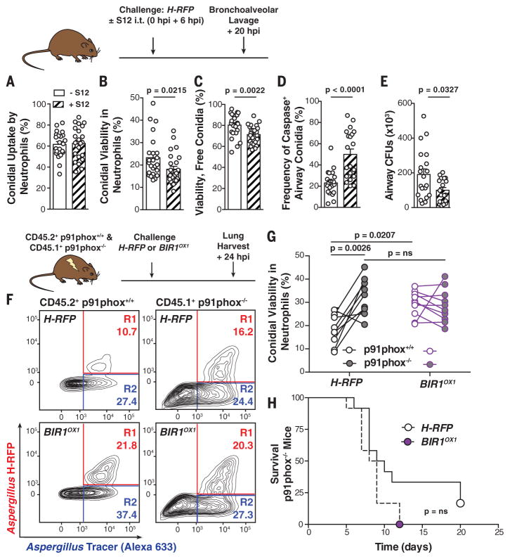
Fig. 4. NADPH oxidase induces conidial A-PCD.
(A to E) Mice were challenged with 3 × 10⁷ AF633+ H-RFP conidia with 7.5 mg per kg of weight of S12 or vehicle control, as indicated. The scatter plots show mean + SEM for (A) conidial uptake, (B) viability in airway neutrophils, (C) free airway conidial viability, (D) fungal caspase activity in free and leukocyte-engulfed airway conidia, and (E) fungal airway CFUs at 20 hpi (n = 20 per group). (F) Representative plots of neutrophils isolated from mixed bone marrow chimeric mice, as indicated above, and analyzed for conidial fluorescence. (G) H-RFP (black circle outline) and BIR1OX1 (purple circle) conidial viability in p91phox+/+ (white center) and p91phox−/− (gray center) neutrophils. The lines indicate paired data from a single mouse. (H) Survival of p91phox−/− mice challenged with 5 × 10⁴ H-RFP or BIROX1 conidia (n = 12 per group). All data were pooled from two independent experiments, with each circle denoting one mouse. Statistical analysis: [(A) to (G)] Mann-Whitney; (H) Log-rank (Mantel-Cox).

