Abstract
Plant sex chromosomes can be vastly different from those of the few historical animal model organisms from which most of our understanding of sex chromosome evolution is derived. Recently, we have seen several advancements from studies on green algae, brown algae, and land plants that are providing a broader understanding of the variable ways in which sex chromosomes can evolve in distant eukaryotic groups. Plant sex-determining genes are being identified and, as expected, are completely different from those in animals. Species with varying levels of differentiation between the X and Y have been found in plants, and these are hypothesized to be representing different stages of sex chromosome evolution. However, we are also finding that sex chromosomes can remain morphologically unchanged over extended periods of time. Where degeneration of the Y occurs, it appears to proceed similarly in plants and animals. Dosage compensation (a phenomenon that compensates for the consequent loss of expression from the Y) has now been documented in a plant system, its mechanism, however, remains unknown. Research has also begun on the role of sex chromosomes in sexual conflict resolution, and it appears that sex-biased genes evolve similarly in plants and animals, although the functions of these genes remain poorly studied. Because the difficulty in obtaining sex chromosome sequences is increasingly being overcome by methodological developments, there is great potential for further discovery within the field of plant sex chromosome evolution.
Keywords: Y degeneration, dioecy, sex chromosome turnover, sex-biased expression, sex chromosome sequencing
Introduction
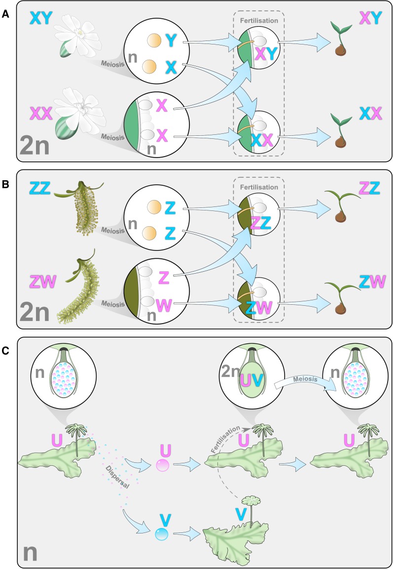
Sex chromosomes are unique in that each member of the chromosome pair has genetic material that differs partially from the other, giving a mechanism by which the sex of individuals can be determined. There are three systems by which this happens (fig. 1). One is the female heterogametic system, where females have two distinct sex chromosomes (ZW) and males are homogametic (ZZ), as can be observed in some willows, for example Salix suchowensis (Hou et al. 2015). Another is the male heterogametic system, such as that found in Silene latifolia (Bernasconi et al. 2009), where males have two distinct sex chromosomes (XY) and females are homogametic (XX). In species with an independent haploid phase in their life cycle, sex can be determined in the haploid gametophytes by a UV system (females U, males V) and the diploid sporophyte is then heterogametic (UV), as observed for example in Marchantia polymorpha (Yamato et al. 2007).
Fig. 1.—
Examples of sex chromosome systems in plants. (a) XY: male heterogamety (Silene latifolia), (b) ZW: female heterogamety (Salix suchowensis) and (c) UV: haplo-diploid system (Marchantia polymorpha), showing maternal (pink) and paternal (blue) sex chromosomes.
Sequence comparison within and between species of well-studied animal models suggests that sex chromosomes derive from an ordinary pair of autosomes that have evolved sex-determining genes and stopped recombining (Bachtrog 2013). For example, in placental mammals, Drosophila, and birds, most of the Y (or W) chromosome has stopped recombining (Bachtrog 2013; Ellegren 2011). As a result, this nonrecombining region degrades; the human Y chromosome, for example, has lost most of its original genes (Ross et al. 2005). The resulting imbalance of X genes (one copy of X genes in males vs. two in females) appears to have been countered by the evolution of dosage compensation in some animals, a mechanism that theoretically enables equivalent X gene expression in both sexes (Graves 2016).
Most of this knowledge on sex chromosome evolution comes from a few animal models, but a broader phylogenetic perspective is essential to test whether it can be generalized to other eukaryotes. Outside animals, sex chromosomes have mainly been studied in land plants, and to a lesser extent in green and brown algae. Consequently, this review will mainly focus on plant sex chromosomes and will mention data on algae whenever available.
Specifically, this review will cover the recent advances from studies on nonanimal systems that are giving us a more global perspective on the range of ways in which sex chromosomes evolve. It will explore the particularities of plant sex chromosomes regarding their origins and sex-determining genes, and discuss what they teach us about the causes of recombination suppression and the consequent degeneration of sex chromosomes. What follows is a presentation of why in some species, recombination suppression does not spread outside of the small region containing the sex-determining genes. The fields of plant dosage compensation and sexual conflict resolution are also explored, two topics that have not yet been tackled by recent reviews on plant sex chromosome evolution (Charlesworth 2016, 2015; Vyskot and Hobza 2015). Finally, with methodological advances making it easier than ever to obtain sex chromosome sequence data, there is great potential for new findings in this field, and suggestions for future research directions are provided.
The Origin of Sex-Determining Genes and Sex Chromosomes in Plants
Very few (∼40) sex chromosomes have so far been identified in plants (Ming et al. 2011). This contrasts dramatically with the number of dioecious plants (species with male and female individuals, i.e., species that are likely to carry sex chromosomes). Indeed, 75% of liverworts (6,000 species), 50% of leafy mosses (7,250 species), 36% of gymnosperms (381 species), and 5–6% of angiosperms (15,600 species) are dioecious (Ming et al. 2011; Renner 2014). Estimates of the frequency of dioecy in brown and green algae are currently missing. In angiosperms, hermaphroditism is supposedly the ancestral breeding system (Endress and Doyle 2015; Sauquet, seminar communication), and some families are entirely dioecious (Kafer et al. 2014; Renner 2016) such as Salicaceae (Manchester et al. 2006), Caricaceae (Carvalho and Renner 2012), and Ebenacaeae (Akagi et al. 2014), which suggests that dioecy is ancient in these families. However, dioecious and hermaphroditic species are often assigned to the same angiosperm genus, which suggests that dioecy is often of recent origin. Homomorphic sex chromosomes (where both sex chromosomes are the same size) can therefore be expected in many dioecious angiosperms, because not enough time has passed for them to differentiate morphologically (see section on degeneration below). As the sex chromosomes are often expected to be homomorhpic, genetic markers would be required for their detection (because cytology can only identify heteromorphic sex chromosomes) and this could explain why so few of them have been identified. In angiosperms, dioecy evolved independently from hermaphroditism somewhere between 871 and 5000 times (Renner 2014). Therefore, it is likely that many sex chromosome systems are yet to be discovered in plants, along with the mechanism for their origin.
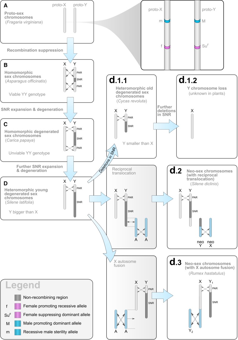
There is evidence that plant sex chromosomes derive from a pair of autosomes (Nicolas et al. 2005; Filatov 2005; Bergero and Charlesworth 2009). Theory predicts that at least two closely linked sex-determining genes are necessary for the birth of sex chromosomes; the so-called “two-gene model” (Charlesworth and Charlesworth 1978; see fig. 2): a male sterility mutation (recessive in X/Y systems, dominant in Z/W systems) and a female sterility mutation (dominant in X/Y systems, recessive in Z/W systems). In UV systems, the dominance of mutations does not matter as sex is expressed in the haploid phase (fig. 1c).
Fig. 2.—
Example progression of XY sex chromosome evolution. Note that this is only one potential evolutionary pathway, not all stages are obligatory and each stage of the pathway is not necessarily associated with the age of the system. In (a), the YY genotype is viable and only sex-determining genes differ (as shown on the zoom). Recombination can be suppressed in the immediate area around the sex-determining genes (b) or further suppressed along flanking regions (c), this can lead to the accumulation of repeated elements and a consequent increase in size of the Y (d). The Y chromosome can also become smaller than the X chromosome through deletions in the SNR (d1.1) (Segawa et al. 1971). Neo-sex chromosomes can evolve with reciprocal translocation (d2) (Howell et al. 2009) or with X autosome fusion (d3), (Smith 1964). Example organisms exhibiting each stage are given in parentheses. Recombining regions are indicated with crossed double-ended arrows.
Some studies have revealed the possibility of the “two-gene model” occurring in plants. For example, in the subdioecious plant Fragaria virginiana, the male function region and the female function region have been mapped to the same linkage group, separated by approximately 6 cM and recombination between the two loci was shown to lead to hermaphrodites and neuters (both male and female sterile) in cross progenies (Spigler et al. 2008). The name given to sex chromosomes at this first stage of their evolution is “proto-sex chromosomes” (fig. 2a). In Fragaria virginiana, because the mutation causing male sterility is dominant and the one causing female sterility is recessive, the system consists of proto-ZW sex chromosomes. Another example is the papaya, where hermaphrodites are determined by a Yh-specific region that recently evolved (around 4000 years ago) from a much older Y specific region (that evolved around 9 million years, hereafter My, ago), possibly after the loss of the dominant female sterility mutation (VanBuren et al. 2015). However, further genetic analyses will be necessary to confirm the two-gene model in this species. In Silene latifolia, the X/Y chromosomes are heteromorphic and mutants with deletions on the Y have revealed that there are two to three main sex-determining regions (at least one female-suppressing and at least one male-promoting), as expected under the (at-least) “two-gene model” (Zluvova et al. 2007; Bergero et al. 2008; Kazama et al. 2016). The genes, however, still remain to be identified.
So far, only two plant sex-determining genes have been identified as being located on sex chromosomes. One (called MID) functions in the green algae Volvox carteri to govern gametic differentiation and is only present in the minus haplotype (Ferris and Goodenough 1997). Another sex-determining gene is OGI, a small RNA-encoded on the Y specific region of the crop species Diospyros lotus (persimmon). OGI has male-specific expression and was found to down-regulate the expression of MeGi; a gene that represses male function (Akagi et al. 2014). However, MeGi is autosomal and therefore the interplay between MeGi and OGI does not fit well with the “two-gene model,” unless another yet unknown Y gene has a female-sterility mutation (Renner 2016). For other species, candidate sex-determining genes have been proposed but their location on sex chromosomes has yet to be assessed. For instance in Asparagus officinalis, pollen development aborts late in females at the microspore maturation stage, and Harkess et al. (2015) showed that the AMS (Aborted Microspores) gene has male-biased expression. This makes this species a good candidate for finding a male sterility gene, as this same gene is known to be involved in microspore maturation in another plant (Arabidopsis thaliana), but further analyses are required to evaluate whether this gene is indeed located on the Y chromosome for Asparagus officinalis.
There are other known sex-determining genes in plants, but they all come from monoecious cucurbits (Boualem et al. 2008, 2009, 2015; Martin et al. 2009); monoecious meaning that male and female flowers are on a single plant that does not have sex chromosomes. In these plants the WIP1 gene inhibits female function when active, but WIP1 can be inhibited by another gene (ACS11). By fixing a recessive loss-of-function mutation in ACS11, and maintaining WIP1 as polymorphic with a functional and a nonfunctional allele, Boualem et al. (2015) created an artificial dioecious population of melons (Cucumis melo) and maintained it over time. This experiment demonstrated that dioecy can be determined by a single gene, because only the WIP1 gene determines the sex of individuals once a loss-of-function mutation is fixed in ACS11. It remains unknown however, whether these mutations have been recruited in closely related dioecious species with sex chromosomes. If they have been, this would not fit the “two-gene model.”
The “two-gene model” and the evolutionary pathway leading to dioecy through gynodioecy (the coexistence of females and hermaphrodites within a species) could explain why in angiosperms, XY systems seem to be more frequent than ZW (Ming et al. 2011). However, as the above examples indicate, it is unclear how widespread the “two-gene model” is in reality for angiosperms and alternative models could exist in dioecious plants (Renner 2016). Also, given that there are few angiosperms with an identified sex chromosome system, the prevalence of XY systems could be due to a sampling bias.
The Differentiation of Sex Chromosomes
Once proto-sex chromosomes have established, they can continue to differentiate into sex chromosomes through a number of different mechanisms. Several species of plants and algae have sex chromosomes that show varying degrees of differentiation, each of which could represent different stages of sex chromosome evolution (fig. 2). Fragaria virginiana could be seen as representing the first stage of differentiation. Recombination events between the two sex-determining loci of their proto-Z and proto-W generate neuters and hermaphrodites (Spigler et al. 2008). In this species, neuters are selected against as they cannot reproduce, and hermaphrodites are expected to be selected against if they have a lower fitness compared with males and females. Theory predicts that this scenario would select for recombination suppression between the two sex-determining loci and the proto-sex chromosomes would be transformed into actual sex chromosomes. As a consequence, the male-specific region of the Y chromosome would stop recombining and become a sex-specific nonrecombining region (hereafter SNR). Genes located in the SNR and/or its X-homologous region are called sex-linked genes. The PAR (pseudoautosomal region) surrounding the SNR would continue to recombine in both males and females, but the SNR-homologous region on the X would continue to recombine only in females. This early stage of sex chromosome evolution after recombination suppression between the two sex-determining genes can be observed in the plant Asparagus officinalis (fig. 2b;Telgmann-Rauber et al. 2007). The process unfolds in the same way for ZW systems, the only difference being that the female-specific region on the W does not recombine. In UV systems, both male-specific and female-specific regions do not recombine as the sporophyte is always heterogametic.
Unlike Asparagus officinalis, many species with sex chromosomes have much larger SNRs than the expected small region containing the two closely linked sex-determining genes, which suggests that sex chromosomes undergo further recombination suppression events. This has been confirmed at the molecular level by the study of X-Y, Z-W, or U-V divergence, whenever sequences are available (see Box 1 for sex chromosome sequencing methods). Indeed, after recombination suppression, SNRs accumulate substitutions separately and, using a molecular clock approach, it is possible to estimate the time when recombination stopped. Because sex-linked genes with similar X-Y, Z-W, or U-V divergence levels tend to be located in similar genomic regions (called strata), unless these regions have later been rearranged, one can infer that for each strata, recombination was suppressed at relatively different points in time. For example, in Silene latifolia, there have been at least two recombination suppression events (Nicolas et al. 2005) as genes fall into at least two categories of X-Y divergence. Recombination suppression also seems to be associated with chromosomal rearrangements, in particular with inversions. For example, in papaya two large inversion events appear to define the two strata present in this species (Wang et al. 2012). Several additional studies have revealed much variation in the characteristics and abundance of strata in plant and algal species (reviewed in Table 1).
Box 1 .
Obtaining Sex Chromosome Sequence Data
The amount of repetitive DNA present in SNRs makes their sequencing virtually impossible with whole-genome shotgun approaches, especially when using short-read NGS technologies. The few fully sequenced and assembled SNRs in plants are rather small regions (Table 1) that were obtained through sequencing of BAC (Bacterial Artificial Chromosome) clones organized in a physical map. The single-haplotype iterative mapping and sequencing (SHIMS; Hughes and Rozen 2012) in particular has provided high-quality assemblies of the mammalian Y chromosome (Skaletsky et al. 2003; Hughes et al. 2010, 2012). However, these strategies are labor-demanding and costly, which explains why only a handful of SNRs have been fully sequenced and assembled in the past.
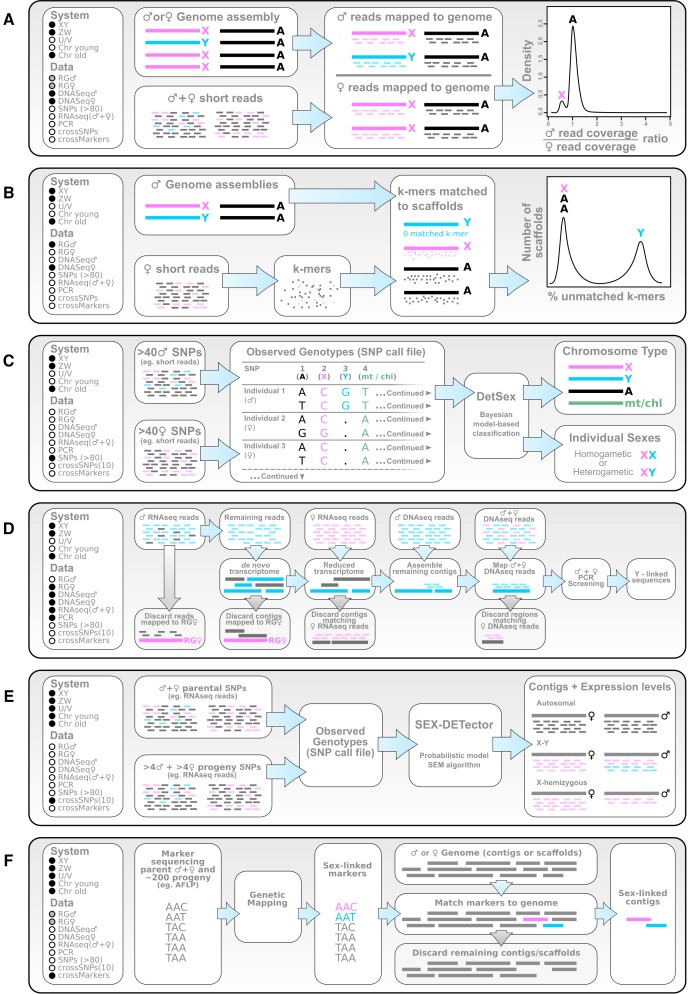
Producing high-quality assembly is not always necessary, and alternative, less expensive strategies have now been developed for identifying sex chromosome sequences based on NGS data. A first category of approaches relies on the comparison of one male and one female genome. Identifying X-linked scaffolds can be done by studying the genomic male over female read coverage ratio along the genome. This ratio is used as a proxy for ploidy: autosomal contigs will have a ratio of 1 whereas X-linked ones will have a ratio of 0.5 (fig. 3a;Vicoso and Bachtrog 2011; Vicoso, Kaiser, et al. 2013; Vicoso, Emerson, et al. 2013). The Y scaffolds can be identified by a subtraction approach between the male and female genomes as they are exclusively present in the male genome. Alternatively, one can also determine Y-linked sequences using an assembled genome and short reads from a female, because the reads are only expected to match autosomal or X-derived contigs/scaffolds, provided that repeats shared by the Y and the female genome are excluded prior to the analysis (fig. 3b;Carvalho and Clark 2013; Akagi et al. 2014). If the model species is well studied, it may be possible to use a combination of short reads from RNA-seq and genome data from male and female individuals alongside a reference genome to increase the number of known Y-linked genes (fig. 3d;Cortez et al. 2014). Segregation types can also be determined if one has access to genotyping data from many (at least 80) individuals, using a Bayesian method that ascertains the segregation type of a marker based on the inferred ploidy: autosomes are diploid in males and females, the X chromosome is diploid in females and haploid in males, the Y chromosome is specific to males and haploid and mitochondria are haploid in males and females (fig. 3c;Gautier 2014). All of these methods cited thus far assume that reads from one sex chromosome do not map to the other chromosome, so they are only adapted to old and diverged sex chromosomes. Another approach relies on studying how markers segregate among sexes. For example, one can use markers in genetic maps to differentiate scaffolds of sex chromosomes from others of an assembled genome (fig. 3f;Hou et al. 2015). Alternatively, one can infer sex markers using polymorphism data from males and females (without needing a cross or a genetic map), and use them to detect sex-linked scaffolds in a genome assembly (Al-Dous et al. 2011; Picq et al. 2014).
However, one rarely has access to a well assembled reference genome in the studied species or even a close relative, particularly when the genome is large (see Table 1 for available reference genomes). In this case, complexity and size can be reduced by using transcriptomes instead of complete genomes. For example, using RNA-seq data from male and female individuals of an inbred population, Muyle et al. (2012) used a SNP analysis to identify hundreds of sex-linked genes. The use of a cross (parents and their progeny of each sex) sequenced by RNA-seq is particularly promising as it is inexpensive and gives direct access to sex-linked genes and their expression levels. This approach has been applied to Silene latifolia (Chibalina and Filatov 2011; Bergero and Charlesworth 2011), Rumex hastatulus (Hough et al. 2014) and Rumex acetosa (Michalovova et al. 2015). More recently, a model-based probabilistic method was developed for this strategy (fig. 3e;Muyle et al. 2016).
Table 1.
Review of Plant and Algal Sex Chromosome Sequence Data
| Species (Taxon) | Reference Genome | System | Age | Strata X-Y/Z-W/U-V Divergence | Y/W/U/V Degeneration | Sequence Data |
|---|---|---|---|---|---|---|
| Asparagus officinalis (Asparagaceae, angiosperm) | — | XY (dominant Y) | — | — | Homomorphic, viable YY genotype, accumulation of repetitive sequences, low gene density | Four BAC clones (Telgmann-Rauber et al. 2007) |
| Carica papaya (Caricaceae, angiosperm) | Ming et al. (2008) | XX females, XY males and XYh hermaphrodites (dominant Y and Yh) | ∼9 My | 2 strata | Homomorphic, lethal YY, YhY and YhYh genotypes, high repeat density, 25% Yh and 14% X gene loss | 3.5 Mb X and 8.1 Mb Y and Yh sex specific complete regions (Wang et al. 2012; Gschwend et al. 2012; VanBuren et al. 2015) |
| Diospyros lotus (Ebenaceae, angiosperm) | Akagi et al. (2014) | XY | — | — | — | ∼1 Mb male specific region (Akagi et al. 2014) |
| Phoenix dactylifera (Arecaceae, angiosperm) | Al-Dous et al. (2011); Al-Mssallem et al. (2013) | XY (dominant Y) | ∼50 My | — | Heteromorphic (unusual smaller Y than X), low nucleotide diversity, long introns | Genetic markers of sex (Cherif et al. 2013), 4 non recombining Y scaffolds (Al-Dous et al. 2011) and X genetic map (Mathew et al. 2014) |
| Rumex hastatulus (Polygonaceae, angiosperm) | — | Females XX, males ancestrally XY but derived XY1Y2 (X-autosome dose) | 15–16 My (Navajas-Pérez et al. 2005) | — | Heteromorphic. 28% gene loss in ancestral Y, 8% in derived Y. High dN/dS. Less optimized codon usage. Reduced expression | RNA-seq data (Hough et al. 2014) |
| Silene latifolia (Caryophyllaceae, angiosperm) | Papadopulos et al. (2015) | XY (dominant Y) | ∼5 My (Rautenberg et al. 2010) | 1 PAR, 2–3 strata 5–25% synonymous divergence (Bergero et al. 2007, 2013) | Heteromorphic (giant Y). Lethal YY genotype. 48% Y genes with 10-fold reduced expression, 23% Y genes interrupted by stop codons. High dN/dS. Low nucleotide diversity. Increased intron length. High repeat density. Low gene density | RNA-seq data (Bergero and Charlesworth 2011; Chibalina and Filatov 2011; Muyle et al. 2012; Papadopulos et al. 2015), BAC clones (Blavet et al. 2015) and other genes referenced in (Bergero et al. 2013; Papadopulos et al. 2015) |
| Vitis vinifera L. subsp sylvestris (Vitaceae, angiosperm) | Jaillon et al. (2007) | XY | — | — | Homomorphic. Low nucleotide diversity | Partial 154.8kb genomic sequence (Picq et al. 2014) |
| Populus trichocarpa (Salicaceae, angiosperm) | Tuskan et al. (2006) | Unclear (XY or ZW) | — | — | Homomorphic, high dN/dS, reduced nucleotide diversity | ∼100 kbp Y specific region (Geraldes et al. 2015), ∼1 Mb W specific region(Yin et al. 2008; Tuskan et al. 2012) |
| Salix suchowensis (Salicaceae, angiosperm) | Dai et al. (2014) | ZW | — | — | Homomorphic | Sex linked scaffolds (Hou et al. 2015) |
| Ceratodon purpureus (Ditrichaceae, bryophyte) | — | UV | ∼6 My | Possibly three strata | Heteromorphic, reduced nucleotide diversity, premature stop codon | Nine genes (McDaniel et al. 2013, 2007) |
| Ectocarpus sp. (Ectocarpaceae, brown algae) | Cock et al. (2010) | UV (dominant V) | >70 My | No strata | Low gene density, high repeated DNA density, underrepresentation of optimal codons, lower expression level, longer introns | Complete 1 Mb genomic sequences (Ahmed et al. 2014) |
| Marchantia polymorpha (Hepaticeae, bryophyte) | — | UV | — | 70.6–93.6% nucleotide identity | Heteromorphic, low gene density | Complete 10 Mb V specific region (Yamato et al. 2007) |
| Volvox carteri (Volvocaceae, green algae) | Prochnik et al. (2010) | UV | — | Possibly 2 strata | Low gene density, high repeat density, reduced codon usage bias | Complete genomic sequences (Ferris et al. 2010) |
Note.—The number of plant species with sequence data contrasts dramatically with the number of plants with known sex chromosomes (~40; Ming et al. 2011), as well as the number of dioecious plants possibly having sex chromosomes (>30,000, see first section on sex determinism).
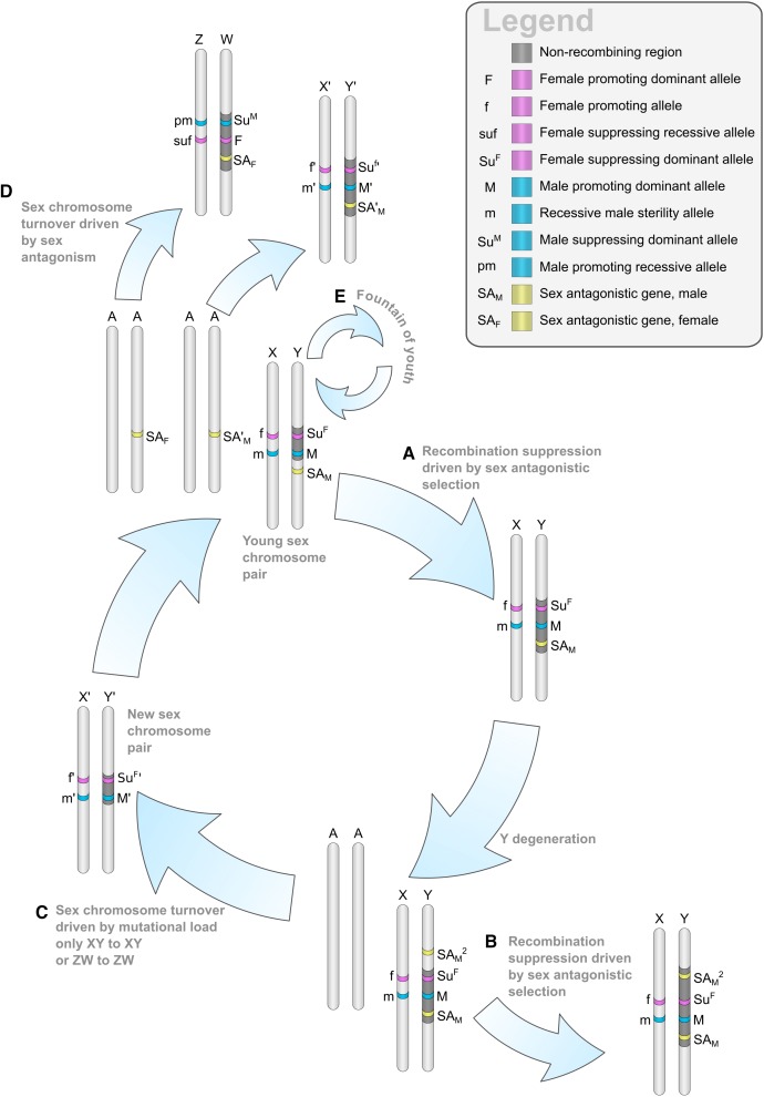
But what is the evolutionary force driving these additional events of recombination suppression on sex chromosomes? Theory predicts that sex-antagonistic mutations (advantageous for one sex and deleterious for the other) will be selected to be linked to the SNR, this way providing advantage to one sex without damaging the other (Charlesworth and Charlesworth 1980; Jordan and Charlesworth 2012; Rice 1984, 1987). This would lead to addition of genetic material on the SNR, either through spread into the flanking PAR (if the PAR contains sex-antagonistic genes, see fig. 4a and b), or through chromosomal rearrangements with autosomes (if sex-antagonistic genes are located on autosomes), generating neo-sex chromosomes (fig. 2d2 and d3).
Fig. 4.—
Mechanisms of sex chromosome turnover and absence/presence of recombination suppression: sex-antagonistic genes can induce recombination suppression if located on the PAR (a), resulting in larger SNRs and heteromorphic sex chromosomes ( here is a male beneficial sex-antagonistic gene that induces further recombination suppression (b) of the Y SNR). The accumulation of deleterious mutations on SNRs following recombination suppression can induce a sex chromosome turnover (c), possibly with endless cycles (but neither from an XY pair to a ZW pair nor from ZW to XY). Sex chromosomes can also be replaced by a new pair (d) if sex-antagonistic genes are located on autosomes (with this mechanism a change of system is possible, from ZW to XY and conversely, but the new pair can only replace a very young sex chromosome pair). The fountain of youth (e) could maintain sex chromosomes at a homomorphic state through X-Y (or Z-W) recombination in sex-reverted individuals.
As yet, no firm evidence definitively connects sex-antagonistic alleles to the evolution of reduced recombination of Y, W, U, or V sex chromosomes. If sex-antagonistic polymorphisms are present on the PAR, they could be maintained as polymorphic for a long evolutionary time because of partial linkage with the SNR, and this partial linkage should be visible through linkage disequilibrium (nonrandom association of the SNR with specific PAR alleles). Qiu et al. (2013) observed a high genetic diversity in the PAR in Silene latifolia, but found evidence of linkage disequilibrium between the PAR and the SNR for only one gene. The observed high diversity in the PAR could be due to sexually antagonistic polymorphism, but it is not clear yet whether a neutral model of partial linkage would be sufficient to explain the high diversity level.
Chromosomal rearrangements involving sex chromosomes and autosomes are common and have been observed for example in Silene latifolia where autosomal regions have been translocated to the sex chromosomes and are now nonrecombining (Bergero et al. 2013). Another example comes from the plant Silene diclinis, where a reciprocal translocation between an autosome and the ancestral Y chromosome led to two Xs and two Ys with a chain quadrivalent at meiosis metaphase I (fig. 2d2; Howell et al. 2009). In Rumex hastatulus some populations have a XY1Y2 system due to an X-autosome fusion (fig. 2d3; Smith 1964). These chromosomal rearrangements make it possible for genes that were autosomal to become sex-linked. If these events are selected for because of sex antagonism, they should be more frequent than rearrangements that do not involve sex chromosomes, but data to test this hypothesis are currently missing.
Fig. 3.—
Available methods for sequencing sex chromosomes, see Box 1 for more details. (a) Vicoso and Bachtrog (2011); Vicoso, Kaiser, et al. (2013); Vicoso, Emerson, et al. (2013), (b) Carvalho and Clark (2013), (c) Gautier (2014), (d) Cortez et al. (2014), (e) Muyle et al. (2016), (f) Hou et al. (2015). Sex chromosome system and required data are indicated with black filled circles when applicable and necessary, respectively, empty circles when not applicable/not necessary, or grey filled circles where only one of two options is required. Chr = Chromosome, RG = Reference Genome, >80 = more than 80 individuals, and “cross” refers to the requirement of parental and offspring data.
When comparing sex chromosomes between species, there are characteristics that indirectly point towards a role of sex antagonism in driving recombination suppression. A comparison of the two green algae Chlamydomonas reinhardtii and Volvox carteri suggests that the UV sex chromosomes of Volvox carteri derive from a mating type locus homologous to the one of Chlamydomonas reinhardtii, after the addition of genetic material to the SNRs (Ferris et al. 2010). The newly acquired genes of the SNRs show sex-specific patterns of expression and could be linked to the strong differentiation in gamete size (oogamy) observed in Volvox carteri, as compared with Chlamydomonas reinhardtii where male and female gametes have similar sizes (isogamy). This observation could be coincidental and more similar cases would be needed for a decisive conclusion, but it supports the link between sexual dimorphism and recombination suppression, possibly through sexually antagonistic selection. However, a more recent study showed that the sex-determining gene MID alone is able to change the sex of Volvox carteri transgenic individuals (Geng et al. 2014), suggesting sex dimorphism evolved through changes that are intrinsic to the MID gene product, rather than from the recruitment of other genes to U and V SNRs. Evidence of a link between sexual dimorphism and sexually antagonistic selection can also be found from Ectocarpus sp. that has U and V SNRs that are surprisingly small (under 1 Mb) given the old age of the system (>70 My). This could be related to the low level of sexual dimorphism in this species, and therefore, the low level of sexually antagonistic selection (Ahmed et al. 2014), especially because male and female gametes have different behavior (females do not swim but instead attract swimming males with pheromones, a process called physiological anisogamy), but differ very little in size (Lipinska et al. 2015).
So it is recombination suppression that drives the divergence of sex chromosomes and sometimes the formation of additional strata. However, the role of sex antagonism in recombination suppression is supported only by indirect evidence and remains to be assessed more thoroughly. One consequence of recombination suppression is that it causes SNRs to degenerate, and this will be explored in the next section.
The Degeneration of SNRs in Plants
Theory predicts that in SNRs, selective interference among genetically linked loci (called Hill-Robertson effects) will reduce effective population size and the efficacy of selection (Bachtrog 2006, 2013; Charlesworth and Charlesworth 2000). This, in turn, has a number of consequences to the way SNRs will evolve compared with other parts of the genome.
One expected consequence of recombination suppression that appears to be occurring in plants is the accumulation of repetitive sequences in the SNR. For instance, 79.3% of the hermaphrodite papaya SNR harbors repetitive sequences, compared with 52% genome-wide (Wang et al. 2012). Silene latifolia also appears to be accumulating repetitive sequences (fig. 2d); having a 570 Mb-Y chromosome that is much larger than the 420 Mb-X (Matsunaga et al. 1994). Another example is Coccinia grandis that, due to the accumulation of transposable elements and organellar DNA (Sousa et al. 2016), has a Y chromosome twice the size of any of its other chromosomes (Sousa et al. 2013). Repetitive sequence accumulation also occurs in SNRs of species with two Y chromosomes; Steflova et al. (2013) observed that specific transposable elements and satellites accumulate on the two Ys compared with other chromosomes of Rumex acetosa. In most of these cases involving transposable elements, the SNR can theoretically be further degraded, because transposable elements are hypothesized to favor chromosomal rearrangements through ectopic recombination and to promote recombination suppression if heterozygous in the PAR (Dooner and He 2008).
Although there are many examples of repetitive sequence accumulation in the SNRs of plants, it is unclear whether this is an obligatory step in Y degeneration, and whether this step is later followed by a shrinkage of the SNRs after extensive gene loss makes deletions possible (Ming et al. 2011). This shrinkage of the SNR has been observed in Cycas revoluta where the Y is smaller than the X (fig. 2d1.1). However, known sex chromosome sizes and ages in plants contradict this view that Y chromosomes would first expand and then shrink (Sousa et al. 2013). Note that the conclusions of this study are based on only a few species, and in order to understand whether shrinkage of the SNR occurs after expansion, the age and size of sex chromosomes should be assessed in different species.
There is at least one species (papaya), in which the X region homologous to the SNR shows a high repetitive content (65.4%) compared with the genome-wide average, although to a lesser extent than the SNR, suggesting that the efficacy of selection is also reduced on the X (Gschwend et al. 2012). This is consistent with the fact that the effective population size of the X chromosome is expected to be ¾ that of autosomes.
Additionally, the reduced efficacy of selection should also lead to increased gene loss. In plants, increased gene loss does not appear to be restricted to the Y, with there being at least one species (papaya) having both X and Yh with similar numbers of lost genes (Gschwend et al. 2012; Wang et al. 2012). Other species (e.g., Silene latifolia) have a higher rate of gene loss only on the Y (Bergero and Charlesworth 2011; Chibalina and Filatov 2011; Bergero et al. 2015; Papadopulos et al. 2015). In either case, the loss of genes and consequent degeneration of SNRs may cause YY and WW genotypes to be lethal in old and moderately old systems (Table 1). Despite this, it has been proposed that plants should lose genes on their SNRs at a slower pace compared with animals because of haploid selection acting on pollen (Charlesworth 2008; Chibalina and Filatov 2011). Indeed, in a diploid cell the Y/W nonrecombining region is sheltered by its homologous X/Z region, but in a haploid cell the loss of a Y/W gene would be a lot more deleterious and counter-selected. In support of this hypothesis, many genes are expressed and therefore likely to be under selection in haploid pollen tubes (Honys and Twell 2004), unlike haploid spermatozoa in animals (Schäfer et al. 1995). However, not all SNR genes are expressed in pollen tubes, which could limit the effect of haploid selection on SNR gene loss in plants. Theoretically, a similar idea applies to UV systems where gametophytes are haploid individuals and haploid selection should slow down U and V degeneration. Data on U/V and W gene loss rates are currently lacking, but there have been some initial studies on Y gene loss. In Silene latifolia for example, haploid selection was suggested to slow down the rate of Y gene loss compared with animal systems (Chibalina and Filatov 2011; Bergero and Charlesworth 2011). However, it has since been highlighted that this did not account for generation time when comparing rates across species (Bachtrog 2011). Since then, Papadopulos et al. (2015) used genomic data to show that 40% of Silene latifolia Y genes have been lost. Using this data, and taking into account the generation time of Silene latifolia and the age of its sex chromosomes, the rate of Y gene loss can be considered to be four to eight percent, per million generations (Table 2). This rate is very similar to the rate of neo-Y gene loss in Drosophila miranda (Table 2), which suggests that there is no major effect of haploid selection on the pace of Y degeneration. In contrast, Catarrhini (Old World monkeys and apes) have a much faster rate of Y gene loss (Table 2), with Hughes et al. (Hughes et al. 2012) showing that 75% of the initial Y genes on strata 4 and 5 of Catarrhini were lost after 5 My (using data from Human, Chimpanzee, and Macaque). This could be due to a reduced effective population size, which is expected to accelerate SNR degeneration. With results from only three lineages (Silene latifolia, Drosophila miranda, and Catarrhini), the above conclusions regarding the effect of haploid selection and effective population size on SNR gene loss rate are supported only weakly. Measurements of SNR gene loss are currently scarce because in order to estimate SNR ancestral gene content and infer gene loss, multiple, closely related species must be examined.
Table 2.
Comparison of Y Gene Loss Rates between Plants and Animals
| Strata 4 and 5 of Catarrhini Y | Silene latifolia Y | Drosophila miranda neo-Y | |
|---|---|---|---|
| % Y gene loss | 75 | 40 | 34 |
| Age (in My) | 5 | 5–10 | 1 |
| Generation time (in number of generation per year) | 0.25* | 1 | 10–20 |
| Rate of Y gene loss (in % per million generations) | 60 | 4–8 | 1.7–3.4 |
| Effective population size (Charlesworth 2009) | Low | Moderate | High |
| References | Hughes and Rozen (2012) | Papadopulos et al. (2015); Bernasconi et al. (2009); Rautenberg et al. (2010) | Zhou and Bachtrog (2012); Bachtrog (2013) |
Note.—Available estimations of SNR gene loss rates are shown for Silene latifolia, Drosophila miranda and Catarrhini (using data from humans, chimpanzees, and macaques). Silene latifolia and Drosophila miranda have a similar Y gene loss rate, which suggests that there is no effect of haploid pollen selection in plants. Meanwhile, Catarrhini have a very high Y gene loss rate, which could be linked to their small effective population size. See Supplementary Material online, for how generation time was estimated in Catarrhini.
Due to gene loss and accumulation of repeats, SNRs are also expected to have a lower gene density. This appears to be the case in several plants (Table 1), for instance there is a 4-fold decrease in gene density on the Y compared with the X in Silene latifolia (Blavet et al. 2015). Introns tend to be longer in SNRs compared with the rest of the genome due to the accumulation of repeats (Table 1).
Other expected consequences of SNR degeneration include a lower nucleotide diversity (due to a lower effective population size), lower SNR gene expression (possibly due to mutations in promoter regions impairing the fixation of transcription factors, or due to the silencing of repetitive elements which would spread to nearby genes), a higher dN/dS in SNR genes (due to an accumulation of deleterious nonsynonymous substitutions), and less optimized codon usage (Table 1). The results from studies on plant and algal sex chromosome evolution appear to be congruent with these theories predicting the degeneration of SNRs (Table 1) and suggest that plant and animal SNRs degenerate in a similar way.
The Forces Opposing Sex Chromosome Differentiation
Interestingly, some SNRs do not expand at all, and PARs can remain very large in some organisms. One theory that provides an explanation for this observation is called the “fountain of youth,” as it prevents the degeneration of SNRs (fig. 4e;Perrin 2009). There is evidence for this in some animals, where recombination patterns depend on phenotypic, rather than genotypic sex. Therefore, SNRs are expected to recombine in sex-reversed XY females (or sex-reversed ZW males) if the SNR is not too diverged. A similar process could occur in plants where the expression of dioecy can be labile; inconstant ZW females of Populus trichocarpa can be bisexual in some years (Stettler 1971), possibly allowing for Z-W recombination in pollen mother cells and causing sex chromosomes to be homomorphic. However, whether recombination patterns depend on phenotypic sex rather than genotypic sex (as in animals) still needs to be tested in plants.
Sex chromosome turnover can also prevent the accumulation of recombination suppression events. Theory predicts that a sex chromosome pair can be replaced by a new pair through two mechanisms (Blaser et al. 2014). The first mechanism (van Doorn and Kirkpatrick 2007, 2010) involves sex-antagonistic selection: a male-benefiting, female-deleterious mutation appearing on an autosome selects for a masculinizing mutation in its vicinity (and conversely for a female-benefiting mutation), possibly overtaking the previous masculinizing mutation on the old Y chromosome and leading to the evolution of a new sex chromosome pair (fig. 4d). In this first mechanism, transitions can change the system (from XY to ZW and conversely). The second mechanism (Blaser et al. 2013) involves the accumulation of deleterious loss-of-function mutations in SNRs that are expected to lower survival in the heterogametic sex. Transitions (from XY to XY or ZW to ZW) should occur as soon as this survival cost exceeds the benefits of having sex-antagonistic mutations linked to the sex-determining genes. In this second mechanism, transitions cannot change the heterogametic sex as transitions from XY to ZW are expected to fix the Y homologue as an autosomal pair, which is of course detrimental if transitions are precisely triggered by the mutational load accumulating on these Y chromosomes (fig. 4c).
In terms of documented evidence of these processes, a sex chromosome turnover event has been documented in Silene otites from XY to ZW (Slancarova et al. 2013). Additionally, many sex chromosome turnovers could have theoretically occurred in the Salicaceae family. The entire Salicaceae family is dioecious, which suggests that the ancestor was dioecious and that Salicaceae species could carry old sex chromosomes (45 My; Manchester et al. 2006). However, sex-specific markers can be mapped to different loci depending on which species of Salicaceae is observed. For example, sex-specific markers were mapped to chromosome XV in two species of the genus Salix: Salix viminalis (Pucholt et al. 2015) and Salix suchowensis (Hou et al. 2015; Chen et al. 2016) with a ZW system in both cases. In Populus (another genus of Salicaceae), sex-specific markers mapped to different positions on chromosome XIX depending on the species (Tuskan et al. 2012), with some species having an XY system (Populus nigra, Populus tremuloides, and Populus tremula) and others a ZW system (Populus alba and Populus deltoides). For Populus trichocarpa, two contradictory papers reported a ZW (Yin et al. 2008) and an XY system (Geraldes et al. 2015). These observations suggest that there have been high rates of sex chromosome turnover in the Salicaceae family, but the mechanisms that caused these turnovers are yet unknown.
The Evolution of Dosage Compensation in Nonanimal Taxa
With SNR degeneration, the heterogametic sex in ZW and XY systems has lower levels of expression than the homogametic sex, most of all after SNR gene losses that make the heterogametic sex partially aneuploid, which can be deleterious. A mechanism called dosage compensation has evolved in some species that allows for similar male and female expression levels (Charlesworth 1996), but more importantly, similar expression levels between sex chromosomes and their ancestral autosomal pair (Mank 2013).
Three dosage compensation mechanisms have so far been described in Drosophila, placental mammals and Caenorhabditis elegans (Disteche 2012; Ercan 2015). In Drosophila, the single X in males is hyperexpressed. In placental mammals, X chromosomes are hyperexpressed in both males and females compared with autosomes but one X is inactivated in females. In Caenorhabditis elegans, X chromosomes are also hyperexpressed in males, whereas the two X chromosomes in hermaphrodites are both down-regulated and hyperexpressed through different pathways. Several studies have revealed that the two canonical dosage compensation mechanisms of Caenorhabditis elegans and placental mammals only hold for some genes on the X. In placental mammals, genes involved in protein complexes, where stoichiometric balance is important, show the expected 2-fold increase in gene expression compared with autosomes (Pessia et al. 2012), but this is not observed when taking all X genes together at the transcript (Xiong et al. 2010) or protein level (Chen and Zhang 2015). Similarly, most X genes are not upregulated in Caenorhabditis elegans (Albritton et al. 2014). This suggests that only some genes are dosage-sensitive and dosage compensation evolves only for these genes (Pessia et al. 2014; Veitia et al. 2015). In humans, an alternative strategy involves the down-regulation of autosomal genes that are within the same protein interaction network as down-regulated sex-linked genes (Julien et al. 2012). Yet another strategy in humans is the duplication and relocation of sex-linked genes to autosomes (Hurst et al. 2015). In Caenorhabditis elegans, the orthologs of yeast haploinsufficient genes (genes causing deleterious effects when in a haploid state) are depleted from the X chromosome (Albritton et al. 2014), also suggesting that the strategy is to move a dosage-sensitive gene to an autosome. Therefore, Ohno’s hypothesis of X 2-fold upregulation (Ohno 1967, p. 19) is one of many different mechanisms that counteracted the potential haploinsufficiency of individual X-linked genes in males (Ercan 2015). Also, the study of dosage compensation in other taxa has shown that chromosome-wide dosage compensation has not evolved in all sex chromosomes, in particular it is more common in XY than ZW systems (Mank 2013; Mullon et al. 2015).
It was only relatively recently that the study of dosage compensation began to involve nonanimal systems. Indeed, the increasing availability of sex-linked gene sequences (Box 1) is now making this possible. For example, Chibalina and Filatov (2011) tested for dosage compensation in Silene latifolia by comparing male and female expression levels for X-hemizygous genes (sex-linked genes for which the Y copy is not expressed or was lost). They found that male and female expression levels significantly differed for these genes and concluded that there was no dosage compensation in this species. However, another study that focused on sex-linked genes with a preserved Y copy observed that males and females maintained similar expression levels even when Y expression was reduced due to degeneration (Muyle et al. 2012). The X-hemizygous gene SlWUS1 in Silene latifolia was shown (using qRT-PCR) to have lower expression levels in males than in females and to have both X copies expressed in females (Kazama et al. 2012), therefore, the study concluded that there was no evidence that this gene was dosage compensated or X-inactivated. However, without an outgroup reference, it could not be ruled out that expression in female buds had been increased, due perhaps to a specialization of that gene in the female function. Bergero et al. (2015) found that male expression was globally halved compared with females for 99 X-hemizygous genes, as expected under no dosage compensation (however a few genes individually showed patterns of dosage compensation). The authors also tested for dosage compensation in 99 XY gene pairs with high expression (over 100 Y reads), and degenerated Y copies (Y over X expression ratios below 0.75), and observed that the male over female expression level ratio was globally higher than the value expected without dosage compensation (both in bud and leaf transcriptomes). They concluded that partial dosage compensation for these XY genes could not be excluded, but that results could also be produced by male-biased expression. Unfortunately, male-biased genes were not identified in the data set, which could have made the two hypotheses distinguishable. From these four studies (Bergero et al. 2015; Chibalina and Filatov 2011; Kazama et al. 2012; Muyle et al. 2012), it seemed clear that X-hemizygous genes are mainly not dosage compensated in Silene latifolia, however the status of XY gene pairs was unclear and the possibility for partial dosage compensation remained (Toups et al. 2015). Another analysis of sex-linked genes in Silene latifolia compared male and female expression levels for the X chromosome (Papadopulos et al. 2015). Two clusters of X-hemizygous genes were identified: one cluster had a ratio of male X expression over female XX expression close to 1, suggesting dosage compensation, whereas another cluster of X-hemizygous genes had a ratio of around 0.5, suggesting no dosage compensation. The study thus confirmed complete compensation for some genes in Silene latifolia, even for X-hemizygous genes.
Only one other plant has been studied for dosage compensation to date: heartwing sorrel (Rumex hastatulus; Hough et al. 2014). In this study, the authors focused on 119 X-hemizygous genes and found that 79% were expressed at significantly lower levels in males than in females, which led them to conclude that there was no dosage compensation in Rumex hastatulus. However, the fact that genes with a preserved but degenerated Y copy maintained similar male and female expression levels (fig. 3 in Hough et al. 2014) actually does suggest dosage compensation.
Most studies thus far have focused on X-hemizygous genes to test for dosage compensation because, for these genes, the complete absence of Y expression would be most deleterious and selection for dosage compensation is expected to be strongest, especially in haploid tissues (Bergero et al. 2015). However, this theoretical expectation has not been tested, and the opposite could easily be argued: X-hemizygous genes could have lost their Y copy because dosage was not important for them and selection neither slowed down the loss of the Y copy nor selected for dosage compensation when degeneration inevitably occurred. The analysis by Papadopulos et al. (2015) showed that not all genes were affected by dosage compensation in Silene latifolia, similarly to placental mammals and Caenorhabditis elegans. This suggests that dosage compensation evolved only for dosage-sensitive genes and that some genes are insensitive to changes in dosage.
From these studies on plants, we can say that there is dosage compensation in other eukaryotes than just animals. Further studies on Silene latifolia and other plants are required to understand the underlying mechanisms of dosage compensation more clearly. There is also much room for methodological improvement in terms of how dosage compensation is quantified in plants. For example, a gene-by-gene statistical test for dosage compensation could be performed, where for X-hemizygous genes, male and female X expression could be compared in order to reject the null hypothesis (of no dosage compensation) that expression in males is half that of females. Additionally, for X/Y genes, X + Y expression in males could be compared with X + X expression in females. However, this type of statistical test could be complicated by the possible existence of buffering effects. Indeed, when only one copy of a gene is present with the same amount of expression machinery in cells, it has been shown to be expressed at a higher level than if two copies of the gene were present and shared the expression machinery among themselves (Malone et al. 2012). Therefore, X-hemizygous genes in males could be expressed at a higher level than half that of females, without involving the evolution of a specific dosage compensation mechanism. Thus, a threshold higher than 50% could be used in statistical tests to address this issue. Another problem is the reference mapping bias in RNA-seq analyses: reads that carry the same alleles as the reference tend to map more than others due to the mismatch cut-off of mapping programs (Wang and Clark 2014). This could be problematic in studies of dosage compensation if the reference genome or transcriptome tends to carry the X allele, which is expected if the reference is assembled from female data or a mix of male and female data. A solution is to build a new reference that carries all of the alternative alleles and to average read counts between both references (Wang and Clark 2014). Studies addressing dosage compensation in plants so far have also been using tools that do not deal with differential allele expression in RNA-seq data. This is especially important when SNR genes are less expressed due to degeneration. The SEX-DETector pipeline (fig. 3e) offers a solution with the use of the reads2snp genotyper that allows for allele-specific expression (Muyle et al. 2016). Finally, normalization of expression levels can be problematic for sex-linked genes due to the difference in the number of X chromosomes between males and females. Using an outgroup species without sex chromosomes to normalize expression levels could offer a solution to this bias. Also, having an outgroup without sex chromosomes allows one to infer the direction of expression changes and how X expression levels evolved in males and females.
The Resolution of Sexual Conflicts in the Genomes of Dioecious Plants
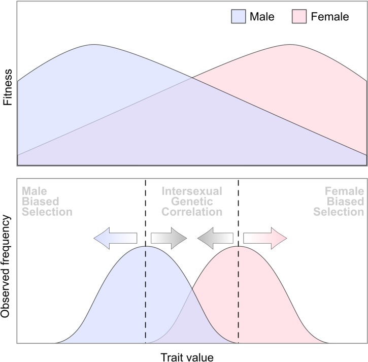
The evolution of sex chromosomes and separate sexes from hermaphroditism should trigger a burst of adaptation throughout the genome. This is because trade-offs between male and female reproductive functions can be resolved by expressing genes at different levels in males and females (Ellegren and Parsch 2007; and see fig. 5), but also through evolving male- and female-specific sequences with SNRs. Other mechanisms of this sexual conflict resolution are described in animal species. These involve gene duplication followed by neofunctionalization and alternative splicing (Betrán et al. 2004).
Fig. 5.—
Sex antagonism and evolution of gene expression level (made after Barrett and Hough 2013): A hypothetical scenario in which females (red) and males (blue) have different optima for the same trait, causing sex-biased selection (blue and red arrows). A shared genetic architecture can constrain the sexes from evolving toward their respective trait optima (grey arrows). However, sexual dimorphism can still evolve when such trade-offs exist, and this can involve sex-limited gene expression and the breakdown of strong intersexual genetic correlations, possibly facilitated by the evolution of SNRs.
In plants, little is known about sex-biased genes and the involvement of SNRs in their evolution (Barrett and Hough 2013) but several studies have given some insight. In Populus yunnanensis and Populus tremula, sex-biased genes do not appear to be enriched in the SNR (Jiang et al. 2012; Robinson et al. 2014). One of these species (Populus yunnanensis) had an enrichment of sex-biased genes in autosomes, whereas the other (Populus tremula) had barely any sex-biased genes. This establishes that in plants it is possible to resolve sexual conflicts without involving the SNR. In an algae, however, the SNR appears to be at least partially involved; Ectocarpus sp. has a PAR enriched with female-biased genes relative to autosomes, suggesting a role of partial linkage with the SNR in the evolution of sex-biased expression (Lipinska et al. 2015). For other species (Fucus vesiculosus and Asparagus officinalis), although sex-biased genes were identified, the involvement of the SNR is unknown (Martins et al. 2013; Harkess et al. 2015) and these are good candidate models for further exploration of this problem. In Silene latifolia, sex-biased genes were found to be significantly enriched among sex-linked genes compared with autosomal genes (Zemp et al. 2016), which is the first evidence of the role of SNR in sexual conflict resolution. Surprisingly, sexual conflicts in this species were mainly solved through changes in expression levels in females rather than males. Some species appear to use mechanisms other than sex-biased gene expression in order to solve sexual conflicts. One example is sex-specific alternative splicing; in Volvox, there is a SNR gene (retinoblastoma tumor suppressor homolog MAT3) that displays sexually regulated alternative splicing and evidence of gender-specific selection (Ferris et al. 2010). Another example is the resolution of sexual conflict through having a sex-specific gene (located in the SNR and therefore present in only one sex). Evidence for this was found in Marchantia polymorpha, where a V-specific repeated gene was found to be expressed specifically in the male sexual organ, similarly to human Y palindromes (Okada et al. 2001).
Conclusions and Perspectives
Plants and algae are convenient systems to study the evolution of sex chromosomes. Their sex chromosomes are often of moderately recent origin (Table 1) and closely related species without sex chromosomes allow for comparative analyses that reveal the direction of the sex chromosome pair’s evolution, the precise inference of gene loss on SNRs and the study of how expression levels evolved for sex-linked genes. This is however not specific to plants because some animal systems also have these characteristics (e.g., some fish and frogs).
The evolution of sex chromosomes in plants continues to bear similarities to that of animals. In recent years, this field has seen the discovery of recombination suppression, degeneration of SNRs, sex chromosome turnovers, homomorphic sex chromosomes in old systems, and dosage compensation in plants. Although these findings are congruent with our understanding of animal sex chromosome evolution, several theoretical differences still need to be investigated. It is still unclear whether the rate of SNR gene loss is lower in plants compared with animals (especially in UV systems) due to haploid selection. There is a great but untapped potential in the study of sex determination in plants. With so many pathways leading to dioecy, and there being only two sex-determining genes described so far for plant and algal sex chromosomes, plants and algae promise a diversity of genetic mechanisms for sex determination, possibly greater than that of animals.
Some aspects of sex chromosome evolution remain unexplained (even in animals), and plants could be useful in providing answers. For instance, the fountain of youth and sex chromosome turnover have been proposed to explain why the relationship between sex chromosome age and degeneration can be so blurry, but these ideas remain theoretical. It is also still unknown whether sex-antagonistic selection drives recombination suppression. Finally, further tests for dosage compensation in other plant and algal species should allow us to understand whether this is a broad-scale solution to the theoretical loss of genes in heterogametic sexes, or if there are more unique mechanisms that are yet to be discovered.
Supplementary Material
Supplementary material is available at Genome Biology and Evolution online.
Authors’ Contributions
Aline Muyle and Gabriel Marais conceived the outline of the review. Aline Muyle wrote a first version of the manuscript. Rylan Shearn prepared the figures. All authors contributed to the intermediate and final stages of the manuscript.
Supplementary Material
Acknowledgments
We thank two anonymous referees, Susanne Renner and editor Kateryna Makova for their helpful comments, Jeff Lemaitre for helpful discussions about generation times, and Benjamin Herran for preparing a previous version of figure 3. This work was supported by ANR (grant numbers: ANR-12-BSV7-0002-04 and ANR-14-CE19-0021-01).
Literature Cited
- Ahmed S, et al. 2014. A haploid system of sex determination in the brown alga Ectocarpus sp. Curr Biol. 24:1945–1957. [DOI] [PubMed] [Google Scholar]
- Akagi T, Henry IM, Tao R, Comai L. 2014. Plant genetics. A Y-chromosome-encoded small RNA acts as a sex determinant in persimmons. Science 346:646–650. [DOI] [PubMed] [Google Scholar]
- Albritton SE, et al. 2014. Sex-biased gene expression and evolution of the X chromosome in nematodes. Genetics 197:865–883. [DOI] [PMC free article] [PubMed] [Google Scholar]
- Al-Dous EK, et al. 2011. De novo genome sequencing and comparative genomics of date palm (Phoenix dactylifera). Nat Biotechnol. 29:521–527. [DOI] [PubMed] [Google Scholar]
- Al-Mssallem IS, et al. 2013. Genome sequence of the date palm Phoenix dactylifera L. Nat Commun. 4:2274. [DOI] [PMC free article] [PubMed]
- Bachtrog D. 2006. A dynamic view of sex chromosome evolution. Curr Opin Genet Dev. 16:578–585. [DOI] [PubMed] [Google Scholar]
- Bachtrog D. 2011. Plant sex chromosomes: a non-degenerated Y?. Curr Biol. 21:R685–R688. [DOI] [PubMed] [Google Scholar]
- Bachtrog D. 2013. Y-chromosome evolution: emerging insights into processes of Y-chromosome degeneration. Nat Rev Genet. 14:113–124. [DOI] [PMC free article] [PubMed] [Google Scholar]
- Barrett SCH, Hough J. 2013. Sexual dimorphism in flowering plants. J Exp Bot. 64:67–82. [DOI] [PubMed] [Google Scholar]
- Bergero R, Charlesworth D. 2011. Preservation of the Y transcriptome in a 10-million-year-old plant sex chromosome system. Curr Biol. 21:1470–1474. [DOI] [PubMed] [Google Scholar]
- Bergero R, Charlesworth D. 2009. The evolution of restricted recombination in sex chromosomes. Trends Ecol Evol. 24:94–102. [DOI] [PubMed] [Google Scholar]
- Bergero R, Charlesworth D, Filatov DA, Moore RC. 2008. Defining regions and rearrangements of the Silene latifolia Y chromosome. Genetics 178:2045–2053. [DOI] [PMC free article] [PubMed] [Google Scholar]
- Bergero R, Forrest A, Kamau E, Charlesworth D. 2007. Evolutionary strata on the X chromosomes of the dioecious plant Silene latifolia: evidence from new sex-linked genes. Genetics 175:1945–1954. [DOI] [PMC free article] [PubMed] [Google Scholar]
- Bergero R, Qiu S, Charlesworth D. 2015. Gene loss from a plant sex chromosome system. Curr Biol. 25(9): 1234–1240. [DOI] [PubMed] [Google Scholar]
- Bergero R, Qiu S, Forrest A, Borthwick H, Charlesworth D. 2013. Expansion of the pseudo-autosomal region and ongoing recombination suppression in the Silene latifolia sex chromosomes. Genetics 194:673–686. [DOI] [PMC free article] [PubMed] [Google Scholar]
- Bernasconi G, et al. 2009. Silene as a model system in ecology and evolution. Heredity 103:5–14. [DOI] [PubMed] [Google Scholar]
- Betrán E, Emerson JJ, Kaessmann H, Long M. 2004. Sex chromosomes and male functions: where do new genes go?. Cell Cycle 3:873–875. [PubMed] [Google Scholar]
- Blaser O, Grossen C, Neuenschwander S, Perrin N. 2013. Sex-chromosome turnovers induced by deleterious mutation load. Evol Int J Org Evol. 67:635–645. [DOI] [PubMed] [Google Scholar]
- Blaser O, Neuenschwander S, Perrin N. 2014. Sex-chromosome turnovers: the hot-potato model. Am Nat. 183:140–146. [DOI] [PubMed] [Google Scholar]
- Blavet N, et al. 2015. Identifying new sex-linked genes through BAC sequencing in the dioecious plant Silene latifolia. BMC Genomics 16:546. [DOI] [PMC free article] [PubMed] [Google Scholar]
- Boualem A, et al. 2009. A conserved ethylene biosynthesis enzyme leads to andromonoecy in two cucumis species. PloS One 4:e6144. [DOI] [PMC free article] [PubMed] [Google Scholar]
- Boualem A, et al. 2015. A cucurbit androecy gene reveals how unisexual flowers develop and dioecy emerges. Science 350:688–691. [DOI] [PubMed] [Google Scholar]
- Boualem K, et al. 2008. Cloning and expression of genes involved in conidiation and surface properties of Penicillium camemberti grown in liquid and solid cultures. Res Microbiol. 159:110–117. [DOI] [PubMed] [Google Scholar]
- Carvalho AB, Clark AG. 2013. Efficient identification of Y chromosome sequences in the human and Drosophila genomes. Genome Res. 23:1894–1907. [DOI] [PMC free article] [PubMed] [Google Scholar]
- Carvalho FA, Renner SS. 2012. A dated phylogeny of the papaya family (Caricaceae) reveals the crop’s closest relatives and the family’s biogeographic history. Mol Phylogenet Evol. 65:46–53. [DOI] [PubMed] [Google Scholar]
- Charlesworth B. 2009. Fundamental concepts in genetics: effective population size and patterns of molecular evolution and variation. Nat Rev Genet. 10:195–205. [DOI] [PubMed] [Google Scholar]
- Charlesworth B. 1996. The evolution of chromosomal sex determination and dosage compensation. Curr Biol. 6:149–162. [DOI] [PubMed] [Google Scholar]
- Charlesworth B, Charlesworth D. 1978. A model for the evolution of dioecy and gynodioecy. Am Nat. 112:975–997. [Google Scholar]
- Charlesworth B, Charlesworth D. 2000. The degeneration of Y chromosomes. Philos Trans R Soc Lond B Biol Sci. 355:1563–1572. [DOI] [PMC free article] [PubMed] [Google Scholar]
- Charlesworth D. 2015. Plant contributions to our understanding of sex chromosome evolution. New Phytol. 208:52–65. [DOI] [PubMed] [Google Scholar]
- Charlesworth D. 2016. Plant sex chromosomes. Annu Rev Plant Biol. 67:397–420. [DOI] [PubMed] [Google Scholar]
- Charlesworth D. 2008. Plant sex chromosomes. Genome Dyn. 4:83–94. [DOI] [PubMed] [Google Scholar]
- Charlesworth D, Charlesworth B. 1980. Sex differences in fitness and selection for centric fusions between sex-chromosomes and autosomes. Genet Res. 35:205–214. [DOI] [PubMed] [Google Scholar]
- Chen X, Zhang J. 2015. No X-chromosome dosage compensation in human proteomes. Mol Biol Evol. 32(6):1456–1460. [DOI] [PMC free article] [PubMed] [Google Scholar]
- Chen Y, Wang T, Fang L, Li X, Yin T. 2016. Confirmation of single-locus sex determination and female heterogamety in willow based on linkage analysis. PloS One 11:e0147671. [DOI] [PMC free article] [PubMed] [Google Scholar]
- Cherif E, et al. 2013. Male-specific DNA markers provide genetic evidence of an XY chromosome system, a recombination arrest and allow the tracing of paternal lineages in date palm. New Phytol. 197:409–415. [DOI] [PubMed] [Google Scholar]
- Chibalina MV, Filatov DA. 2011. Plant Y chromosome degeneration is retarded by haploid purifying selection. Curr Biol. 21:1475–1479. [DOI] [PubMed] [Google Scholar]
- Cock JM, et al. 2010. The Ectocarpus genome and the independent evolution of multicellularity in brown algae. Nature 465:617–621. [DOI] [PubMed] [Google Scholar]
- Cortez D, et al. 2014. Origins and functional evolution of Y chromosomes across mammals. Nature 508:488–493. [DOI] [PubMed] [Google Scholar]
- Dai X, et al. 2014. The willow genome and divergent evolution from poplar after the common genome duplication. Cell Res. 24:1274–1277. [DOI] [PMC free article] [PubMed] [Google Scholar]
- Disteche CM. 2012. Dosage compensation of the sex chromosomes. Annu Rev Genet. 46:537–560. [DOI] [PMC free article] [PubMed] [Google Scholar]
- Dooner HK, He L. 2008. Maize genome structure variation: interplay between retrotransposon polymorphisms and genic recombination. Plant Cell 20:249–258. [DOI] [PMC free article] [PubMed] [Google Scholar]
- van Doorn GS, Kirkpatrick M. 2010. Transitions between male and female heterogamety caused by sex-antagonistic selection. Genetics 186:629–645. [DOI] [PMC free article] [PubMed] [Google Scholar]
- van Doorn GS, Kirkpatrick M. 2007. Turnover of sex chromosomes induced by sexual conflict. Nature 449:909–912. [DOI] [PubMed] [Google Scholar]
- Ellegren H. 2011. Sex-chromosome evolution: recent progress and the influence of male and female heterogamety. Nat Rev Genet. 12:157–166. [DOI] [PubMed] [Google Scholar]
- Ellegren H, Parsch J. 2007. The evolution of sex-biased genes and sex-biased gene expression. Nat Rev Genet. 8:689–698. [DOI] [PubMed] [Google Scholar]
- Endress PK, Doyle JA. 2015. Ancestral traits and specializations in the flowers of the basal grade of living angiosperms. Taxon 64:1093–1116. [Google Scholar]
- Ercan S. 2015. Mechanisms of X chromosome dosage compensation. J Genomics. 3:1–19. [DOI] [PMC free article] [PubMed] [Google Scholar]
- Ferris P, et al. 2010. Evolution of an expanded sex-determining locus in Volvox. Science 328:351–354. [DOI] [PMC free article] [PubMed] [Google Scholar]
- Ferris PJ, Goodenough UW. 1997. Mating type in Chlamydomonas is specified by mid, the minus-dominance gene. Genetics 146:859–869. [DOI] [PMC free article] [PubMed] [Google Scholar]
- Filatov DA. 2005. Substitution rates in a new Silene latifolia sex-linked gene, SlssX/Y. Mol Biol Evol. 22:402–408. [DOI] [PubMed] [Google Scholar]
- Gautier M. 2014. Using genotyping data to assign markers to their chromosome type and to infer the sex of individuals: a Bayesian model-based classifier. Mol Ecol Resour. 14:1141–1159. [DOI] [PubMed] [Google Scholar]
- Geng S, De Hoff P, Umen JG. 2014. Evolution of sexes from an ancestral mating-type specification pathway. PLoS Biol. 12:e1001904. [DOI] [PMC free article] [PubMed] [Google Scholar]
- Geraldes A, et al. 2015. Recent Y chromosome divergence despite ancient origin of dioecy in poplars (Populus). Mol Ecol.24(13):3243–3256. [DOI] [PubMed] [Google Scholar]
- Graves JAM. 2016. Evolution of vertebrate sex chromosomes and dosage compensation. Nat Rev Genet. 17:33–46. [DOI] [PubMed] [Google Scholar]
- Gschwend AR, et al. 2012. Rapid divergence and expansion of the X chromosome in papaya. Proc Natl Acad Sci U S A. 109:13716–13721. [DOI] [PMC free article] [PubMed] [Google Scholar]
- Harkess A, et al. 2015. Sex-biased gene expression in dioecious garden asparagus (Asparagus officinalis). New Phytol. 207(3):883–892. [DOI] [PubMed] [Google Scholar]
- Honys D, Twell D. 2004. Transcriptome analysis of haploid male gametophyte development in Arabidopsis. Genome Biol. 5:R85. [DOI] [PMC free article] [PubMed] [Google Scholar]
- Hou J, et al. 2015. Different autosomes evolved into sex chromosomes in the sister genera of Salix and Populus. Sci Rep. 5:9076. [DOI] [PMC free article] [PubMed] [Google Scholar]
- Hough J, Hollister JD, Wang W, Barrett SCH, Wright SI. 2014. Genetic degeneration of old and young Y chromosomes in the flowering plant Rumex hastatulus. Proc Natl Acad Sci U S A. 111:7713–7718. [DOI] [PMC free article] [PubMed] [Google Scholar]
- Howell EC, Armstrong SJ, Filatov DA. 2009. Evolution of neo-sex chromosomes in Silene diclinis. Genetics 182:1109–1115. [DOI] [PMC free article] [PubMed] [Google Scholar]
- Hughes JF, et al. 2010. Chimpanzee and human Y chromosomes are remarkably divergent in structure and gene content. Nature 463:536–539. [DOI] [PMC free article] [PubMed] [Google Scholar]
- Hughes JF, et al. 2012. Strict evolutionary conservation followed rapid gene loss on human and rhesus Y chromosomes. Nature 483:82–86. [DOI] [PMC free article] [PubMed] [Google Scholar]
- Hughes JF, Rozen S. 2012. Genomics and genetics of human and primate Y chromosomes. Annu Rev Genomics Hum Genet. 13:83–108. [DOI] [PubMed] [Google Scholar]
- Hurst LD, Ghanbarian AT, Forrest ARR, FANTOM consortium. Huminiecki L. 2015. The constrained maximal expression level owing to haploidy shapes gene content on the mammalian X chromosome. PLoS Biol. 13:e1002315. [DOI] [PMC free article] [PubMed] [Google Scholar]
- Jiang H, et al. 2012. Transcriptional profiling analysis in Populus yunnanensis provides insights into molecular mechanisms of sexual differences in salinity tolerance. J Exp Bot 63:3709–3726. [DOI] [PMC free article] [PubMed] [Google Scholar]
- Jordan CY, Charlesworth D. 2012. The potential for sexually antagonistic polymorphism in different genome regions. Evol Int J Org Evol. 66:505–516. [DOI] [PubMed] [Google Scholar]
- Julien P, et al. 2012. Mechanisms and evolutionary patterns of mammalian and avian dosage compensation. PLoS Biol. 10:e1001328. [DOI] [PMC free article] [PubMed] [Google Scholar]
- Kafer J, et al. 2014. Dioecy is associated with higher diversification rates in flowering plants. J Evol Biol. 27:1478–1490. [DOI] [PubMed] [Google Scholar]
- Kazama Y, et al. 2016. A new physical mapping approach refines the sex-determining gene positions on the Silene latifolia Y-chromosome. Sci Rep. 6:18917. [DOI] [PMC free article] [PubMed] [Google Scholar]
- Kazama Y, et al. 2012. SlWUS1: an X-linked gene having no homologous Y-linked copy in Silene latifolia. G3 (Bethesda) 2:1269–1278. [DOI] [PMC free article] [PubMed] [Google Scholar]
- Lipinska A, et al. 2015. Sexual dimorphism and the evolution of sex-biased gene expression in the brown alga ectocarpus. Mol Biol Evol. 32(6):1581–1597. [DOI] [PubMed] [Google Scholar]
- Malone JH, et al. 2012. Mediation of Drosophila autosomal dosage effects and compensation by network interactions. Genome Biol. 13(4):r28. [DOI] [PMC free article] [PubMed] [Google Scholar]
- Manchester SR, Judd WS, Handley B. 2006. Foliage and fruits of early poplars (Salicaceae: Populus) from the Eocene of Utah, Colorado, and Wyoming. Int J Plant Sci. 167:897–908. [Google Scholar]
- Mank JE. 2013. Sex chromosome dosage compensation: definitely not for everyone. Trends Genet. 29:677–683. [DOI] [PubMed] [Google Scholar]
- Martin A, et al. 2009. A transposon-induced epigenetic change leads to sex determination in melon. Nature 461:1135–1138. [DOI] [PubMed] [Google Scholar]
- Martins MJF, Mota CF, Pearson GA. 2013. Sex-biased gene expression in the brown alga Fucus vesiculosus. BMC Genomics 14:294. [DOI] [PMC free article] [PubMed] [Google Scholar]
- Mathew LS, et al. 2014. A first genetic map of date palm (Phoenix dactylifera) reveals long-range genome structure conservation in the palms. BMC Genomics 15:285. [DOI] [PMC free article] [PubMed] [Google Scholar]
- Matsunaga S, Hizume M, Kawano S, Kuroiwa T. 1994. Cytological analyses in Melandrium album: genome size, chromosome size and fluorescence in situ hybridization. Cytologia (Tokyo) 59:135–141. [Google Scholar]
- McDaniel SF, Neubig KM, Payton AC, Quatrano RS, Cove DJ. 2013. Recent gene-capture on the UV sex chromosomes of the moss Ceratodon purpureus. Evol Int J Org Evol. 67:2811–2822. [DOI] [PMC free article] [PubMed] [Google Scholar]
- McDaniel SF, Willis JH, Shaw AJ. 2007. A linkage map reveals a complex basis for segregation distortion in an interpopulation cross in the moss Ceratodon purpureus. Genetics 176:2489–2500. [DOI] [PMC free article] [PubMed] [Google Scholar]
- Michalovova M, Kubat Z, Hobza R, Vyskot B, Kejnovsky E. 2015. Fully automated pipeline for detection of sex linked genes using RNA-Seq data. BMC Bioinformatics 16:78. [DOI] [PMC free article] [PubMed] [Google Scholar]
- Ming R, et al. 2008. The draft genome of the transgenic tropical fruit tree papaya (Carica papaya Linnaeus). Nature 452:991–996. [DOI] [PMC free article] [PubMed] [Google Scholar]
- Ming R, Bendahmane A, Renner SS. 2011. Sex chromosomes in land plants. Annu Rev Plant Biol. 62:485–514. [DOI] [PubMed] [Google Scholar]
- Mullon C, Wright AE, Reuter M, Pomiankowski A, Mank JE. 2015. Evolution of dosage compensation under sexual selection differs between X and Z chromosomes. Nat Commun. 6:7720. [DOI] [PMC free article] [PubMed] [Google Scholar]
- Muyle A, et al. 2012. Rapid de novo evolution of X chromosome dosage compensation in Silene latifolia, a plant with young sex chromosomes. PLoS Biol. 10:e1001308. [DOI] [PMC free article] [PubMed] [Google Scholar]
- Muyle A, et al. 2016. SEX-DETector: a probabilistic approach to study sex chromosomes in non-model organisms. Genome Biol Evol. 8:2530–2543. [DOI] [PMC free article] [PubMed] [Google Scholar]
- Navajas-Pérez R, et al. 2005. The evolution of reproductive systems and sex-determining mechanisms within rumex (polygonaceae) inferred from nuclear and chloroplastidial sequence data. Mol Biol Evol. 22:1929–1939. [DOI] [PubMed] [Google Scholar]
- Nicolas M, et al. 2005. A gradual process of recombination restriction in the evolutionary history of the sex chromosomes in dioecious plants. PLoS Biol. 3:e4. [DOI] [PMC free article] [PubMed] [Google Scholar]
- Ohno S. 1967. Sex chromosomes and sex-linked genes. Vol. 1. Berlin, Heidelberg, NY: Springer-Verlag. p. 192.
- Okada S, et al. 2001. The Y chromosome in the liverwort Marchantia polymorpha has accumulated unique repeat sequences harboring a male-specific gene. Proc Natl Acad Sci U S A. 98:9454–9459. [DOI] [PMC free article] [PubMed] [Google Scholar]
- Papadopulos AST, Chester M, Ridout K, Filatov DA. 2015. Rapid Y degeneration and dosage compensation in plant sex chromosomes. Proc Natl Acad Sci U S A. 112:13021–13026. [DOI] [PMC free article] [PubMed] [Google Scholar]
- Perrin N. 2009. Sex reversal: a fountain of youth for sex chromosomes?. Evol Int J Org Evol. 63:3043–3049. [DOI] [PubMed] [Google Scholar]
- Pessia E, Engelstädter J, Marais GAB. 2014. The evolution of X chromosome inactivation in mammals: the demise of Ohno’s hypothesis? Cell Mol Life Sci. 71:1383–1394. [DOI] [PMC free article] [PubMed] [Google Scholar]
- Pessia E, Makino T, Bailly-Bechet M, McLysaght A, Marais GAB. 2012. Mammalian X chromosome inactivation evolved as a dosage-compensation mechanism for dosage-sensitive genes on the X chromosome. Proc Natl Acad Sci U S A. 109:5346–5351. [DOI] [PMC free article] [PubMed] [Google Scholar]
- Picq S, et al. 2014. A small XY chromosomal region explains sex determination in wild dioecious V. vinifera and the reversal to hermaphroditism in domesticated grapevines. BMC Plant Biol. 14:229. [DOI] [PMC free article] [PubMed] [Google Scholar]
- Prochnik SE, et al. 2010. Genomic analysis of organismal complexity in the multicellular green alga Volvox carteri. Science 329:223–226. [DOI] [PMC free article] [PubMed] [Google Scholar]
- Pucholt P, Rönnberg-Wästljung A-C, Berlin S. 2015. Single locus sex determination and female heterogamety in the basket willow (Salix viminalis L.). Heredity 114(6):575–583. [DOI] [PMC free article] [PubMed] [Google Scholar]
- Qiu S, Bergero R, Charlesworth D. 2013. Testing for the footprint of sexually antagonistic polymorphisms in the pseudoautosomal region of a plant sex chromosome pair. Genetics 194:663–672. [DOI] [PMC free article] [PubMed] [Google Scholar]
- Rautenberg A, Hathaway L, Oxelman B, Prentice HC. 2010. Geographic and phylogenetic patterns in Silene section Melandrium (Caryophyllaceae) as inferred from chloroplast and nuclear DNA sequences. Mol Phylogenet Evol. 57:978–991. [DOI] [PubMed] [Google Scholar]
- Renner SS. 2016. Pathways for making unisexual flowers and unisexual plants:Moving beyond the ‘two mutations linked on one chromosome’ model. Am J Bot. 103:587–589. [DOI] [PubMed] [Google Scholar]
- Renner SS. 2014. The relative and absolute frequencies of angiosperm sexual systems: dioecy, monoecy, gynodioecy, and an updated online database. Am J Bot. 101:1588–1596. [DOI] [PubMed] [Google Scholar]
- Rice W. 1984. Sex-chromosomes and the evolution of sexual dimorphism. Evolution 38:735–742. [DOI] [PubMed] [Google Scholar]
- Rice W. 1987. The accumulation of sexually antagonistic genes as a selective agent promoting the evolution of reduced recombination between primitive sex-chromosomes. Evolution 41:911–914. [DOI] [PubMed] [Google Scholar]
- Robinson KM, et al. 2014. Populus tremula (European aspen) shows no evidence of sexual dimorphism. BMC Plant Biol. 14:276. [DOI] [PMC free article] [PubMed] [Google Scholar]
- Ross MT, et al. 2005. The DNA sequence of the human X chromosome. Nature 434:325–337. [DOI] [PMC free article] [PubMed] [Google Scholar]
- Schäfer M, Nayernia K, Engel W, Schäfer U. 1995. Translational control in spermatogenesis. Dev Biol. 172:344–352. [DOI] [PubMed] [Google Scholar]
- Segawa M, Kishi S, Tatuno S. 1971. Sex chromosomes of Cycas revoluta. Jpn J Genet. 46:33. [Google Scholar]
- Skaletsky H, et al. 2003. The male-specific region of the human Y chromosome is a mosaic of discrete sequence classes. Nature 423:825–837. [DOI] [PubMed] [Google Scholar]
- Slancarova V, et al. 2013. Evolution of sex determination systems with heterogametic males and females in silene. Evol Int J Org Evol. 67:3669–3677. [DOI] [PubMed] [Google Scholar]
- Smith BW. 1964. The evolving karyotype of Rumex hastatulus. Evolution 18:93–104. [Google Scholar]
- Sousa A, Bellot S, Fuchs J, Houben A, Renner SS. 2016. Analysis of transposable elements and organellar DNA in male and female genomes of a species with a huge Y-chromosome reveals distinct Y-centromeres. Plant J. 88(3):387–396. [DOI] [PubMed] [Google Scholar]
- Sousa A, Fuchs J, Renner SS. 2013. Molecular cytogenetics (FISH, GISH) of Coccinia grandis : a ca. 3 myr-old species of cucurbitaceae with the largest Y/autosome divergence in flowering plants. Cytogenet Genome Res. 139:107–118. [DOI] [PubMed] [Google Scholar]
- Spigler RB, Lewers KS, Main DS, Ashman T-L. 2008. Genetic mapping of sex determination in a wild strawberry, Fragaria virginiana, reveals earliest form of sex chromosome. Heredity 101:507–517. [DOI] [PubMed] [Google Scholar]
- Steflova P, et al. 2013. Contrasting patterns of transposable element and satellite distribution on sex chromosomes (XY1Y2) in the dioecious plant Rumex acetosa. Genome Biol Evol. 5:769–782. [DOI] [PMC free article] [PubMed] [Google Scholar]
- Stettler R. 1971. Variation in sex expression of black cottonwood and related hybrids. Silvae Genet. 20:42–46. [Google Scholar]
- Telgmann-Rauber A, Jamsari A, Kinney MS, Pires JC, Jung C. 2007. Genetic and physical maps around the sex-determining M-locus of the dioecious plant asparagus. Mol Genet Genomics. 278:221–234. [DOI] [PubMed] [Google Scholar]
- Toups M, Veltsos P, Pannell JR. 2015. Plant sex chromosomes: lost genes with little compensation. Curr Biol. 25:R427–R430. [DOI] [PubMed] [Google Scholar]
- Tuskan GA, et al. 2012. The obscure events contributing to the evolution of an incipient sex chromosome in Populus: a retrospective working hypothesis. Tree Genet Genomes. 8:559–571. [Google Scholar]
- VanBuren R, et al. 2015. Origin and domestication of papaya Yh chromosome. Genome Res. 25:524–533. [DOI] [PMC free article] [PubMed] [Google Scholar]
- Veitia RA, Veyrunes F, Bottani S, Birchler JA. 2015. X chromosome inactivation and active X upregulation in therian mammals: facts, questions, and hypotheses. J Mol Cell Biol. 7:2–11. [DOI] [PubMed] [Google Scholar]
- Vicoso B, Bachtrog D. 2011. Lack of global dosage compensation in Schistosoma mansoni, a female-heterogametic parasite. Genome Biol Evol. 3:230–235. [DOI] [PMC free article] [PubMed] [Google Scholar]
- Vicoso B, Emerson JJ, Zektser Y, Mahajan S, Bachtrog D. 2013. Comparative sex chromosome genomics in snakes: differentiation, evolutionary strata, and lack of global dosage compensation. PLoS Biol. 11:e1001643. [DOI] [PMC free article] [PubMed] [Google Scholar]
- Vicoso B, Kaiser VB, Bachtrog D. 2013. Sex-biased gene expression at homomorphic sex chromosomes in emus and its implication for sex chromosome evolution. Proc Natl Acad Sci U S A. 110:6453–6458. [DOI] [PMC free article] [PubMed] [Google Scholar]
- Vyskot B, Hobza R. 2015. The genomics of plant sex chromosomes. Plant Sci Int J Exp Plant Biol. 236:126–135. [DOI] [PubMed] [Google Scholar]
- Wang J, et al. 2012. Sequencing papaya X and Yh chromosomes reveals molecular basis of incipient sex chromosome evolution. Proc Natl Acad Sci U S A. 109:13710–13715. [DOI] [PMC free article] [PubMed] [Google Scholar]
- Wang X, Clark AG. 2014. Using next-generation RNA sequencing to identify imprinted genes. Heredity 113:156–166. [DOI] [PMC free article] [PubMed] [Google Scholar]
- Xiong Y, et al. 2010. RNA sequencing shows no dosage compensation of the active X-chromosome. Nat Genet. 42:1043–1047. [DOI] [PubMed] [Google Scholar]
- Yamato KT, et al. 2007. Gene organization of the liverwort Y chromosome reveals distinct sex chromosome evolution in a haploid system. Proc Natl Acad Sci U S A. 104:6472–6477. [DOI] [PMC free article] [PubMed] [Google Scholar]
- Yin T, et al. 2008. Genome structure and emerging evidence of an incipient sex chromosome in Populus. Genome Res. 18:422–430. [DOI] [PMC free article] [PubMed] [Google Scholar]
- Zemp N, et al. 2016. Evolution of sex-biased gene expression in a dioecious plant. Nat Plants 2(11):16168. [DOI] [PubMed] [Google Scholar]
- Zhou Q, Bachtrog D. 2012. Sex-specific adaptation drives early sex chromosome evolution in Drosophila. Science 337:341–345. [DOI] [PMC free article] [PubMed] [Google Scholar]
- Zluvova J, et al. 2007. Early events in the evolution of the Silene latifolia Y chromosome: male specialization and recombination arrest. Genetics 177:375–386. [DOI] [PMC free article] [PubMed] [Google Scholar]
Associated Data
This section collects any data citations, data availability statements, or supplementary materials included in this article.


