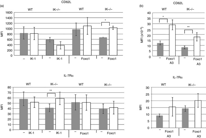
Figure 6.

Restoring Ikaros or Foxo1 to Ikaros null T cells differentially impacts the expression of CD62L and interleukin‐7 receptor α (IL‐7Rα). (a) Purified splenic CD4 T cells from wild‐type and Ikaros null (IK −/−) mice were transduced with retroviruses prepared using MSCV IRES H‐2Kk (−), MSCV IRES Ik‐1 H‐2Kk (IK‐1) and MSCV Foxo1 Thy‐1.1 (Foxo1) constructs. Results are presented as a compilation of mean fluorescence intensities for CD62L and IL‐7Rα expression from at least four independent experiments. Differences are not significant unless marked with asterisk(s) (*P = 0·0233, **P = 0·032). Error bars are representative of ± SEM. (b) Purified splenic CD4 T cells from wild‐type and Ikaros null (IK −/−) mice were transduced with retroviruses prepared using MSCV IRES Thy‐1.1 (−) or MSCV Foxo1‐A3 Thy‐1.1 (Foxo1‐A3) constructs. Data are a compilation of results from five independent experiments. Differences are not significant unless marked with asterisk(s) (*P = 0·0152, **P = 0·0353).
