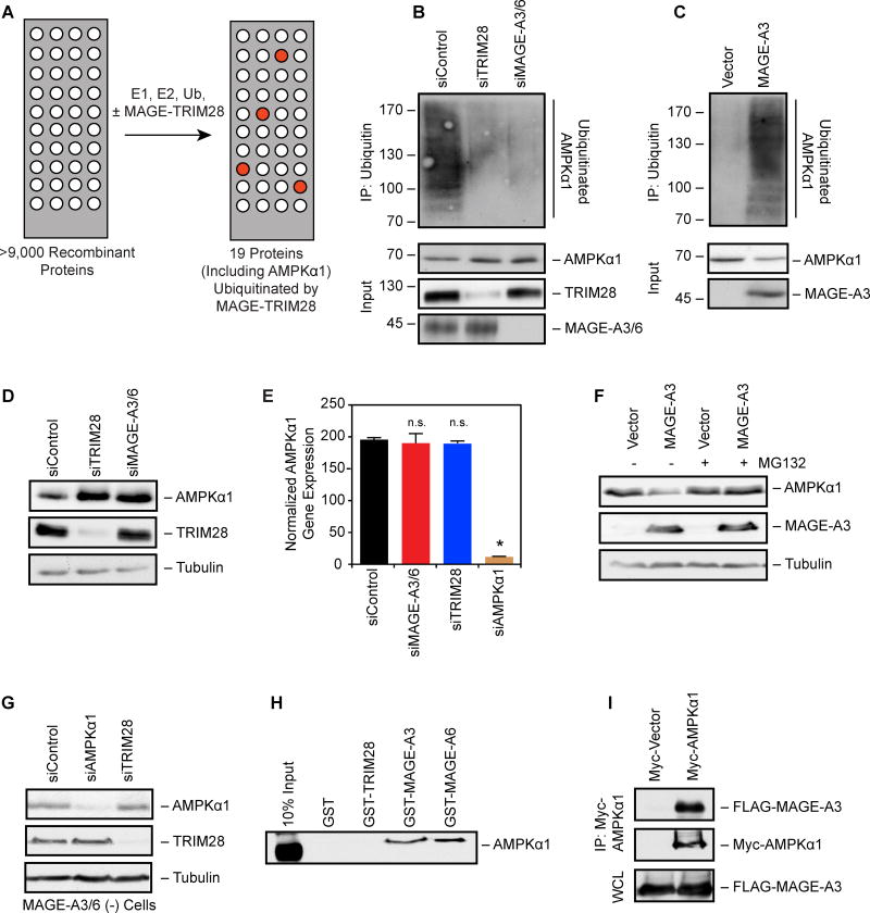
Figure 3. MAGE-A3/6-TRIM28 E3 ubiquitin ligase ubiquitinates and degrades AMPKα1.
(A) Schematic of in vitro screen for MAGE-TRIM28 ubiquitination substrates using protein arrays.
(B) AMPKα1 ubiquitination requires MAGE-A3/6-TRIM28. HeLa (MAGE-A3/6-positive) were treated with the indicated siRNAs for 24 hrs before transfection with Myc-tagged ubiquitin for 48 hrs before anti-Myc immunoprecipitation (IP) and immunoblotting was performed (n=3).
(C) Expression of MAGE-A3 promotes AMPKα1 ubiquitination. MAGE-A3/6-negative HEK293 cells stably expressing FLAG-MAGE-A3 were transfected with Myc-ubiquitin 48 hrs before anti-Myc IP and immunoblotting was performed (n=3).
(D) Knockdown of MAGE-A3/6-TRIM28 increases AMPKα1 protein levels. MAGE-A3/6-positive cells were treated with the indicated siRNAs for 72 hrs and then blotted for the indicated proteins (n > 3).
(E) Knockdown of MAGE-A3/6-TRIM28 does not affect AMPKα1 mRNA levels. MAGE-A3/6-positive cells were treated with the indicated siRNAs for 72 hrs and then AMPKα1 mRNA levels were determined by RT-QPCR (n=3). Data are represented as the mean + SD. Asterisks indicates p<0.05.
(F) MAGE-A3 promotes proteasome-dependent AMPKα1 degradation. MAGE-A3/6-negative cells expressing vector of MAGE-A3 were treated with 5 µM MG132 for 4 hrs before immunoblotting (n=3).
(G) TRIM28-mediated AMPKα1 degradation requires MAGE-A3/6. MAGE-A3/6-negative HEK293 cells were transfected with the indicated siRNA for 72 hrs before cell lysates were immnoblotted (n>3).
(H) GST pulldown assays reveal AMPKα1 directly binds to MAGE-A3 and MAGE-A6, but not TRIM28 (22–418) or GST (n=3).
(I) HeLa cells expressing FLAG-MAGE-A3 or FLAG-vector along with Myc-AMPKα1 were subjected to anti-Myc IP and immunoblotting. WCL represents whole cell lysate. Data representative of multiple experiments (n=2). See also Table S2.

