Abstract
Although many studies have discussed the association of abnormally expressed silent information regulator 1 (Sirt1) with the prognosis of patients with a variety of solid carcinomas, they failed to agree on whether excessive Sirt1 indicates a good or poor overall survival for the patients. We conducted the current meta-analysis to illustrate the prognostic value of Sirt1 in solid malignancies. Articles published before December 2016 were searched using Pubmed and Web of Science. The studies were selected for the meta-analysis based on certain criteria. A total of 7,369 cases from 37 studies were included, in which 48.6% of the patients overexpressed Sirt1. The overall survival (OS) and clinical features, such as age and TNM stage, were analyzed using RevMan 5.3 software. Sirt1 overexpression was significantly correlated with the OS (HR: 1.52, 95% CI: [1.23, 1.88], P = 0.0001), especially in liver cancer (HR: 1.78, 95% CI: [1.46, 2.18], P < 0.00001) and lung cancer (HR: 1.80, 95% CI: [1.06, 3.05], P = 0.03), which suggested that the overexpression of Sirt1 indicates poor prognosis of patients with solid cancers.
Keywords: Sirt1, solid malignancy, prognosis, meta-analysis
INTRODUCTION
The increase in cancer prevalence and mortality and its impact on social economy have drawn enormous attention towards investigations into the occurrence, development and metastasis of cancer [1]. A large number of cell signaling pathways have been discovered and studied, and accumulating evidence has shown that epigenetic regulation of gene expression contributes significantly to solid malignancy [2]. Aberrant activation of key epigenetic pathways, including Sirt1 signaling, contributes to carcinogenesis in a variety of tumors, suggesting a potential therapeutic target for future treatments [3].
Sirt1, a proto member of the sirtuin family, is an NAD+-dependent histone deacetylase. Sirt1 modifies histones and non-histone proteins through deacetylation [4]. Sirt1 plays pivotal roles in a variety of physiological processes, such as cell metabolism, proliferation, senescence, apoptosis, and tumorigenesis [3, 4]. It exercises its functions through p53 [5], FoxO1 [6], NF-κB [7] and other signaling pathways. Sirt1, because of its tumorigenic characteristics, can be targeted for therapy, which may provide a longer lifespan and better quality of life for cancer patients. Interestingly, Sirt1 seems to have dual roles in cancer. Sirt1 promotes tumorigenesis by boosting cell survival under stress conditions but facilitates uncontrolled cell proliferation, and additionally, it can defend against carcinomas by increasing genomic stability and limiting cellular replicative lifespan [8]. Additionally, Sirt1 expression is increased in ovarian cancer [9] and gastric cancer [10], whereas it is reduced in liver cancer and breast cancer [11]. Therefore, it remains controversial whether Sirt1 overexpression indicates a good or poor prognosis. Although the majority of the evidence shows that overexpressed Sirt1 has a negative prognostic effect on cancer patients, Jung et al. [12] failed to draw the same conclusion, and their data suggested that higher Sirt1 expression resulted in a better survival status. Consequently, the clinical significance of SIRT1 in cancers is complex and requires further investigation. Therefore, in the present study, we conducted an exhaustive meta-analysis and subgroup analyses to understand the prognostic effects of Sirt1 overexpression in solid malignancies, with the aim to provide evidence for improved targeted regimens.
RESULTS
Eligible studies
Most of the studies found during the initial search were excluded based on the selection criteria such as inappropriate article type, replicated data or insufficient original information. Eventually, 37 qualified studies containing 7,369 cases were included for analyses. Figure 1 shows the selection workflow of eligible studies for our meta-analysis.
Figure 1. Flow chart of the selection for the meta-analysis.
Demographic characteristics of the included studies
Among the 37 studies, the majority (15) of them were conducted in China, followed by Korea (n = 13) and other countries. The majority of the studies were based on breast cancer (n = 8), followed by colorectal carcinoma, hepatocellular carcinoma, gastric cancer (n = 5, respectively), lung cancer (n = 4), and other types of solid carcinoma. The sample sizes ranged from 40 to 557, with a median of 144 patients. Among the 37 studies, 34 studies described the correlation between overall survival and Sirt1 expression, 9 trials demonstrated the relationship between disease-free survival and Sirt1 expression, 6 studies discussed relapse-free survival and Sirt1 expression, and 3 articles studied the correlation of cancer-specific survival and abnormal expression of Sirt1. Other details and features were recorded and are summarized in Table 1. All the eligible entries scored higher than six by NOS, suggesting a high methodological quality across all studies.
Table 1. Demographic information of included studies.
| Reference | Country | Cancer type | No. | Male/Femaleale | TNM Stagee | Sirt1 high (+) | Sirt1 low | Follow-up range months | NOS score |
|---|---|---|---|---|---|---|---|---|---|
| Zhang 2016 [13] | China | breast cancer | 149 | All female | I-III | 68 | 81 | NA | 7 |
| Chen 2014 [14] | China | colorectal adenocarcinoma | 102 | 56/46 | II-IV | 44 | 58 | NA | 7 |
| Jang 2012 [15] | South Korea | colorectal adenocarcinoma | 497 | 281/216 | I-IV | 208 | 289 | NA | 8 |
| Jung 2013 [12] | South Korea | colorectal adenocarcinoma | 349 | 208/141 | I-IV | 235 | 114 | NA | 8 |
| Cheng 2016 [16] | China | colorectal adenocarcinoma | 90 | 47/43 | I-IV | 37 | 53 | NA | 7 |
| He 2016 [17] | China | esophageal squamous cell carcinoma | 86 | 64/22 | I-III | 54 | 32 | NA | 7 |
| Chen 2014a [18] | China | esophageal squamous cell carcinoma | 206 | 152/54 | NA | 95 | 111 | 5–86 | 7 |
| Zhang 2013 [19] | China | gastroesophageal junction cancer | 90 | NA | NA | 46 | 44 | NA | 7 |
| Cha 2009 [10] | South Korea | gastric carcinoma | 177 | 135/42 | I-IV | 130 | 47 | NA | 7 |
| Cao 2014 [20] | China | breast cancer | 122 | All female | I-IV | 94 | 28 | 2–161 | 7 |
| Kang 2012 [21] | South Korea | gastric carcinoma | 452 | 309/143 | I-IV | 255 | 197 | NA | 7 |
| Feng 2011 [22] | China | gastric carcinoma | 90 | NA | NA | 46 | 44 | NA | 7 |
| Noguchi 2014 [23] | Japan | gastric carcinoma | 557 | 391/166 | I-IV | 345 | 212 | 6–142 | 8 |
| Hao 2014 [24] | China | hepatocellular carcinoma | 99 | 89/10 | I-IV | 76 | 23 | NA | 6 |
| Song 2014 [25] | China | hepatocellular carcinoma | 300 | 267/33 | I-IV | 145 | 155 | 3–83 | 7 |
| Jang 2012 [18] | South Korea | hepatocellular carcinoma | 154 | 132/22 | I-IV | 55 | 99 | NA | 7 |
| Li 2016 [26] | China | hepatocellular carcinoma | 72 | 65/7 | I-III | 41 | 31 | NA | 6 |
| Chen 2012 [27] | China | hepatocellular carcinoma | 172 | 142/30 | NA | 95 | 77 | 45–236 | 7 |
| Noguchi 2013 [28] | Japan | head and neck squamous cell carcinoma | 437 | 356/81 | NA | 348 | 89 | 1–174 | 8 |
| Chung 2015 [29] | South Korea | breast cancer | 427 | All female | NA | 227 | 150 | NA | 7 |
| Yu 2013 [30] | China | laryngeal and hypopharyngeal carcinomas | 46 | NA | NA | 17 | 29 | NA | 7 |
| Grbesa 2015 [31] | Spain | lung cancer | 105 | 93/12 | NA | 52 | 53 | NA | 7 |
| Noh 2013 [32] | South Korea | lung cancer | 144 | 82/62 | NA | 75 | 40–136 | 7 | |
| Li 2015 [33] | China | lung cancer | 75 | 39/36 | I-IV | 56 | 19 | NA | 7 |
| Shin 2016 [34] | South Korea | ovarian cancer | 45 | NA | NA | 16 | 29 | NA | 6 |
| Lee 2010 [35] | South Korea | breast cancer | 122 | All female | NA | 82 | 40 | NA | 7 |
| Mvunta 2016 [36] | Japan | ovarian cancer | 68 | All female | NA | 11 | 57 | NA | 7 |
| Stenzinger 2013 [37] | Germany | pancreatic cancer | 113 | NA | NA | 32 | 81 | NA | 7 |
| Noh 2013 [38] | South Korea | renal cell carcinoma | 200 | 140/60 | I-IV | 119 | 81 | NA | 7 |
| Batra 2016 [39] | India | retinoblastoma | 94 | 62/32 | NA | 49 | 45 | NA | 6 |
| Kim 2013 [40] | South Korea | soft tissue sarcomas | 104 | 59/45 | NA | 74 | 30 | NA | 7 |
| Benard 2015 [41] | Dutch | colorectal adenocarcinoma | 254 | 128/126 | I-III | NA | NA | NA | 6 |
| Chung 2016 [42] | South Korea | breast cancer | 344 | All female | NA | 146 | 198 | NA | 8 |
| Jin 2016 [43] | South Korea | breast cancer | 319 | All female | I-III | 107 | 212 | NA | 7 |
| Wu 2012 [44] | China | breast cancer | 134 | All female | NA | 72 | 62 | NA | 6 |
| Gharabaghi 2016 [45] | Iran | lung cancer | 40 | 23/17 | NA | 27 | 13 | NA | 6 |
| Derr 2014 [46] | Dutch | breast cancer | 460 | All female | I-III | NA | NA | 2–330 | 7 |
NOS: Newcastle-Ottawa Scale.
NA: not available.
Correlation of Sirt1 expression with the overall survival and subgroup analyses
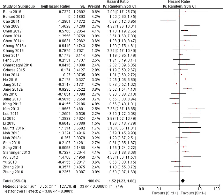
Thirty-four trials offered data on the correlation between the overall survival and Sirt1 expression. Our calculations showed that higher Sirt1 expression indicated an unfavorable overall survival for solid malignancies (HR: 1.52, 95% CI: [1.23, 1.88], P = 0.0001, Figure 2). Because of the significant heterogeneity (I2 = 74%), we applied a random-effects model for the statistical analysis. To determine possible sources of heterogeneity among studies, we grouped the original articles for subgroup analyses, based on several factors. In the cancer subgroup, Sirt1 overexpression was associated with a worse overall survival in liver cancer (n = 5, HR: 1.78, 95% CI: [1.46, 2.18], P < 0.00001, I2 = 0%, Figure 3) and lung cancer (n = 4, HR: 1.80, 95% CI: [1.06, 3.05], P = 0.03, I2 = 37%, Figure 4), whereas Sirt1 overexpression was not correlated with the overall survival in breast cancer (n = 7, HR: 1.25, 95% CI: [0.70, 2.22], P = 0.46, I2 = 75%, Supplementary Figure 1), colorectal carcinoma (n = 5, HR: 1.12, 95% CI: [0.66,1.89], P = 0.67, I2 = 81%, Supplementary Figure 2), and gastric carcinoma (n = 4, HR: 1.44, 95% CI: [0.60, 3.45], P = 0.41, I2 = 81%, Supplementary Figure 3). When the studies were sub grouped based on TNM clinical stages, we found several studies that discussed pre-terminal stages (TNM I-III) (n = 6, HR: 1.16, 95% CI: [0.98, 1.38], P = 0.09, I2 = 8%, Supplementary Figure 4), and only one study [14] that included advanced TNM stages (II-IV) with HR: 3.51, 95% CI: (1.68, 7.33). Moreover, in studies that covered all stages (TNM I-IV), overexpression of Sirt1 suggested a poorer overall outcome (n = 12, HR: 1.35, 95% CI: [0.87, 2.11], P = 0.18, I2 = 86%, Supplementary Figure 5). Two studies evaluated the nuclear and cytoplasmic Sirt1 expressions separately, with distinctive results. One article showed that overexpression of Sirt1 in the cytoplasm is indicative of a good prognosis (P < 0.005), whereas another suggested that increased expression of Sirt1 significantly correlated with poor patient survival (HR = 2.617, P = 0.019). The remaining studies detected the nuclear expression of Sirt1 expression (n = 17, HR: 1.56, 95% CI: [1.15, 2.12], P = 0.004, I2 = 80%).
Figure 2. The correlation between Sirt1 expression and overall survival in solid malignancies.
Figure 3. The correlation between Sirt1 expression and overall survival of liver cancer.
Figure 4. The correlation between Sirt1 expression and overall survival of lung cancer.
Correlation of Sirt1 expression with disease-free survival, relapse-free survival and cancer-specific survival
Unfortunately, our analysis failed to conclude the significance between increased Sirt1 expression and relapse-free survival (RFS, n = 6, HR: 1.58, 95% CI: [0.97, 2.60], P = 0.07, I2 = 84%, Supplementary Figure 6), disease-free survival (DFS, n = 9, HR: 1.23, 95% CI: [0.88, 1.73], P = 0.22, I2 = 71%, Supplementary Figure 7) or cancer-specific survival (CSS, n = 3, HR: 1.40, 95% CI: [0.60, 3.30], P = 0.44, I2 = 89%, Supplementary Figure 8) among solid malignancies.
Sensitivity analysis
Excluding studies about breast cancer (n = 27, HR: 1.60, 95% CI: [1.26, 2.04], P = 0.0001, I2 = 74%), colorectal cancer (n = 29, HR: 1.62, 95% CI: [1.29, 2.03], P < 0.0001, I2 = 70%), and gastric cancer (n = 30, HR: 1.54, 95% CI: [1.23, 1.92], P = 0.0001, I2 = 74%) had no substantial impact on the outcome of overall survival; however, a large heterogeneity was consistently observed.
Eliminating studies that scored 6 on the NOS scale did not alter the unfavorable prognostic effect of Sirt1 overexpression on the overall survival in patients with solid malignancies (n = 27, HR: 1.53, 95% CI: [1.20, 1.94], P = 0.0006, I2 = 78%).
Publication bias
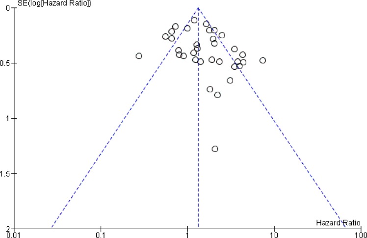
We used funnel plots to visualize publication bias. (Figure 5).
Figure 5. The funnel plots for this meta-analysis.
DISCUSSION
The dual nature of Sirt1 in cancer remains a controversy and could be a consequence of several factors including its different expression levels in various types of carcinoma, its subcellular location, and diverse downstream substrates. Earlier studies predominately reported nuclear Sirt1 expression levels, whereas more recent studies have included cytoplasmic Sirt1 expression levels. It has been proposed that subcellular localization may account for the dual roles of SIRT1 in normal versus cancer cells [47]. One study suggested that the cytoplasmic SIRT1 originates in the nucleus and plays a role in enhancing caspase-dependent apoptosis [48]. All these studies suggested that cytoplasmic expression of Sirt1 is a worse indicator of poor prognosis in cancer patients than the nuclear expression of Sirt1. However, of all the included studies, there were only two articles that discussed the relationship between Sirt1 cytoplasmic expression and patient survival, and regardless, the two studies failed to draw the same conclusion. This inconsistency could be because of different scoring criteria or differing protocols, and therefore, additional studies that precisely illuminate the impact of cytoplasmic Sirt1 expression are needed.
Although an overwhelming number of studies have established evidence that indicates an unfavorable impact of Sirt1 overexpression on patient prognosis in a wide range of carcinomas, several recent investigations revealed a superior survival duration in cases with abnormal expressions of Sirt1. The exact cause for this inconsistency is unknown. Thus, from a clinical perspective, the significance of Sirt1 in patient survival is unknown due to the lack of convincing evidence, and therefore, a comprehensive study is urgently needed.
Based on our knowledge, our study is the first and most versatile meta-analysis that systematically elucidates the prognostic role of Sirt1 overexpression in solid malignancies. Altogether, the results of our analyses strongly support the current mainstream point that Sirt1 redundancy was significantly correlated with patient overall survival in carcinomas. Furthermore, this unfavorable prognostic impact was independent of TNM stages. However, our quantitative analysis found that Sirt1 overexpression was not associated with patient survival in breast cancer, colorectal cancer, and gastric cancer, which was inconsistent with a majority of previous findings, and this contradiction could result from the data collection process. Some of the included study data were acquired from figures in the articles because of a lack of individual patient data. It is also worth mentioning that our subgroup analyses of the correlations between abnormal Sirt1 expression and breast cancer or gastric cancer did not lead to the same conclusions as the meta-analysis [49, 50]. These inconsistencies originated from the differences in literature selection criteria: we excluded data that were not published in English and those from geo database in cases of data duplication.
Apart from the interesting results, there are some limitations to this quantitative meta-analysis. First, the heterogeneity among the studies remained, despite the usage of a random-effects model and subgroup analyses. The heterogeneity could have resulted in outcome bias. Second, we barely explored the correlation between Sirt1 overexpression and patient survival in terms of clinical parameters. Other elements that may contribute to the heterogeneity, such as pathological grade, body mass index, and mean age, were not analyzed due to the lack of sufficient data. Finally, because of a shortage of original individual patient data, we performed a quantitative meta-analysis based mostly on secondary data, which could lead to inaccurate results.
In spite of the limitations mentioned above, there is plenty of pragmatic value in this full-scale, quantitative meta-analysis. First, Sirt1 was identified as a biomarker of overall survival in solid malignancies, especially in liver cancer and lung cancer. Second, we proposed that cytoplasmic rather than nuclear Sirt1 expression deserves more attention. Additional and more in-depth clinical studies are needed because current studies indicate that Sirt1 can serve as a more accurate prognostic predictor in carcinomas.
MATERIALS AND METHODS
Search strategy
We performed a thorough electronic search for relevant studies using PubMed and Web of Science that were published before December 2016. The search terms “Sirt1 AND (cancer OR neoplasm OR carcinoma OR malignancy)” were applied, and we initially identified 3474 studies for further examination. Both abstracts and full texts were elaborately screened to exclude irrelevant articles. Additionally, we reviewed the citation lists of the retrieved articles to guarantee the sensitivity of the search process. This procedure was carried out by two authors separately, and discrepancies were resolved by mutual discussions.
Selection criteria
Studies that met the following criteria were considered eligible and were included in our quantitative meta-analysis: (1) articles written in English and published before Dec. 2016; (2) studies discussing the correlation between Sirt1 expression and patient prognosis in human solid malignancies; (3) a minimal follow-up duration of 3 years; (4) a minimal sample-size of 10 participants; and (5) the diagnosis of solid malignancy was histologically and pathologically confirmed. Studies were excluded on the basis of the following criteria: (1) duplicate or overlapping populations; (2) lack of enough statistical data for further quantification analyses; (3) review articles or case reports; (4) animal studies; and (5) articles based on the Geo database. All evaluations were independently conducted by two authors to ensure the accuracy of the selection process.
Data extraction
General information, overall survival, cancer-specific survival, disease-free survival, and recurrence-free survival were extracted from qualified studies independently by two investigators. The original survival data for both comparative groups were calculated from the text, tables or Kaplan-Meier curves. The survival information from Kaplan-Meier curves were digitized and extracted using Enguage Digitizer 4.1. Any disagreements were resolved by mutual discussions. All extraction procedures were performed with the aid of predefined standardized extraction forms.
Methodological quality assessment
Newcastle-Ottawa Scale (NOS) [51] was applied for the quality evaluation of each selected article because all the eligible studies were observational studies. Certain adaptive modifications were made to revise the scale to match the practical needs of the analysis. The scale contained three categories including selection, comparability and outcome, and the maximum score was nine. Methodological high quality studies were those that scored more than six on this scale. The assessment process was conducted independently by two authors.
Statistical analysis
All quantitative calculations were performed using Review Manager 5.3 (Cochrane Collaboration, Oxford, England). The hazard ratio (HR) at a 95% confidential interval (CI) was used to measure the correlation between Sirt1 expression and patient survival. The data, including the general survival analyses and sub-group comparisons, calculated from the articles were included in the form of generic inverse variables. Heterogeneity among studies was calculated using both the I2 test and Q-test, and I2 > 25% or P < 0.05 was defined as significant heterogeneity; therefore, a random-effects model (the DerSimonian and Laird method) was used. In all other cases, the fixed-effects model (Mantel-Haenszel method) was used. Additionally, we conducted a sensitivity analysis to test the consistency of the selected studies. Publication bias was determined using funnel plots, and P < 0.05 signified a statistically significant publication bias. All P values were 2-tailed.
SUPPLEMENTARY MATERIALS FIGURES
Acknowledgments
We sincerely appreciate our lab members for providing methodological support for this publication.
Abbreviations
- Sirt1
silent information regulator 1
- OS
overall survival
- DFS
disease-free survival
- RFS
relapse-free survival
- CSS
cancer-specific survival
Authors’ contributions
C.W. and S.R. contributed to the design of the study. C.W. contributed to the manuscript writing. C.W., W.Y., F.D., Y.G. and J.T. contributed to data extraction and analysis of the study. T.H. and S.R. contributed to the financial support and revision of the manuscript.
CONFLICTS OF INTEREST
None.
FUNDING
This work was supported by the National Natural Science Foundation of China (grant no. 81001171) and the Key Technologies R&D Program of Hubei Province (grant no. 2007AA302B07).
REFERENCES
- 1.Torre LA, Bray F, Siegel RL, Ferlay J, Lortet-Tieulent J, Jemal A. Global cancer statistics, 2012. CA Cancer J Clin. 2015;65:87–108. doi: 10.3322/caac.21262. [DOI] [PubMed] [Google Scholar]
- 2.Shen H, Laird PW. Interplay between the cancer genome and epigenome. Cell. 2013;153:38–55. doi: 10.1016/j.cell.2013.03.008. [DOI] [PMC free article] [PubMed] [Google Scholar]
- 3.Saunders LR, Verdin E. Sirtuins: critical regulators at the crossroads between cancer and aging. Oncogene. 2007;26:5489–504. doi: 10.1038/sj.onc.1210616. [DOI] [PubMed] [Google Scholar]
- 4.Blander G, Guarente L. The Sir2 family of protein deacetylases. Annu Rev Biochem. 2004;73:417–35. doi: 10.1146/annurev.biochem.73.011303.073651. [DOI] [PubMed] [Google Scholar]
- 5.Yi YW, Kang HJ, Kim HJ, Kong Y, Brown ML, Bae I. Targeting mutant p53 by a SIRT1 activator YK-3-237 inhibits the proliferation of triple-negative breast cancer cells. Oncotarget. 2013;4:984–94. doi: 10.18632/oncotarget.1070. [DOI] [PMC free article] [PubMed] [Google Scholar]
- 6.Choi HK, Cho KB, Phuong NT, Han CY, Han HK, Hien TT, Choi HS, Kang KW. SIRT1-mediated FoxO1 deacetylation is essential for multidrug resistance-associated protein 2 expression in tamoxifen-resistant breast cancer cells. Mol Pharm. 2013;10:2517–27. doi: 10.1021/mp400287p. [DOI] [PubMed] [Google Scholar]
- 7.Yeung F, Hoberg JE, Ramsey CS, Keller MD, Jones DR, Frye RA, Mayo MW. Modulation of NF-kappaB-dependent transcription and cell survival by the SIRT1 deacetylase. EMBO J. 2004;23:2369–80. doi: 10.1038/sj.emboj.7600244. [DOI] [PMC free article] [PubMed] [Google Scholar]
- 8.Taylor DM, Maxwell MM, Luthi-Carter R, Kazantsev AG. Biological and potential therapeutic roles of sirtuin deacetylases. Cell Mol Life Sci. 2008;65:4000–18. doi: 10.1007/s00018-008-8357-y. [DOI] [PMC free article] [PubMed] [Google Scholar]
- 9.Jang KY, Kim KS, Hwang SH, Kwon KS, Kim KR, Park HS, Park BH, Chung MJ, Kang MJ, Lee DG, Moon WS. Expression and prognostic significance of SIRT1 in ovarian epithelial tumours. Pathology. 2009;41:366–71. doi: 10.1080/00313020902884451. [DOI] [PubMed] [Google Scholar]
- 10.Cha EJ, Noh SJ, Kwon KS, Kim CY, Park BH, Park HS, Lee H, Chung MJ, Kang MJ, Lee DG, Moon WS, Jang KY. Expression of DBC1 and SIRT1 is associated with poor prognosis of gastric carcinoma. Clin Cancer Res. 2009;15:4453–9. doi: 10.1158/1078-0432.CCR-08-3329. [DOI] [PubMed] [Google Scholar]
- 11.Wu M, Wei W, Xiao X, Guo J, Xie X, Li L, Kong Y, Lv N, Jia W, Zhang Y, Xie X. Expression of SIRT1 is associated with lymph node metastasis and poor prognosis in both operable triple-negative and non-triple-negative breast cancer. Med Oncol. 2012;29:3240–9. doi: 10.1007/s12032-012-0260-6. [DOI] [PubMed] [Google Scholar]
- 12.Jung W, Hong KD, Jung WY, Lee E, Shin BK, Kim HK, Kim A, Kim BH. SIRT1 Expression Is Associated with Good Prognosis in Colorectal Cancer. Korean J Pathol. 2013;47:332–9. doi: 10.4132/KoreanJPathol.2013.47.4.332. [DOI] [PMC free article] [PubMed] [Google Scholar]
- 13.Zhang WW, Luo JY, Yang F, Wang YC, Yin YM, Strom A, Gustafsson JA, Guan XX. BRCA1 inhibits AR-mediated proliferation of breast cancer cells through the activation of SIRT1. Scientific Reports. 2016:6. doi: 10.1038/srep22034. [DOI] [PMC free article] [PubMed] [Google Scholar]
- 14.Chen XJ, Sun K, Jiao SF, Cai N, Zhao X, Zou HB, Xie YX, Wang ZS, Zhong M, Wei LX. High levels of SIRT1 expression enhance tumorigenesis and associate with a poor prognosis of colorectal carcinoma patients. Scientific Reports. 2014:4. doi: 10.1038/srep07481. [DOI] [PMC free article] [PubMed] [Google Scholar]
- 15.Jang SH, Min KW, Paik SS, Jang KS. Loss of SIRT1 histone deacetylase expression associates with tumour progression in colorectal adenocarcinoma. J Clin Pathol. 2012;65:735–9. doi: 10.1136/jclinpath-2012-200685. [DOI] [PubMed] [Google Scholar]
- 16.Cheng F, Su L, Yao C, Liu L, Shen J, Liu C, Chen X, Luo Y, Jiang L, Shan J, Chen J, Zhu W, Shao J, et al. SIRT1 promotes epithelial-mesenchymal transition and metastasis in colorectal cancer by regulating Fra-1 expression. Cancer Lett. 2016;375:274–83. doi: 10.1016/j.canlet.2016.03.010. [DOI] [PubMed] [Google Scholar]
- 17.He Z, Yi J, Jin L, Pan B, Chen L, Song H. Overexpression of Sirtuin-1 is associated with poor clinical outcome in esophageal squamous cell carcinoma. Tumour Biol. 2016;37:7139–48. doi: 10.1007/s13277-015-4459-y. [DOI] [PubMed] [Google Scholar]
- 18.Jang KY, Noh SJ, Lehwald N, Tao GZ, Bellovin DI, Park HS, Moon WS, Felsher DW, Sylvester KG. SIRT1 and c-Myc promote liver tumor cell survival and predict poor survival of human hepatocellular carcinomas. PLoS One. 2012;7:e45119. doi: 10.1371/journal.pone.0045119. [DOI] [PMC free article] [PubMed] [Google Scholar]
- 19.Zhang LH, Huang Q, Fan XS, Wu HY, Yang J, Feng AN. Clinicopathological significance of SIRT1 and p300/CBP expression in gastroesophageal junction (GEJ) cancer and the correlation with E-cadherin and MLH1. Pathol Res Pract. 2013;209:611–7. doi: 10.1016/j.prp.2013.03.012. [DOI] [PubMed] [Google Scholar]
- 20.Cao YW, Li WQ, Wan GX, Li YX, Du XM, Li YC, Li F. Correlation and prognostic value of SIRT1 and Notch1 signaling in breast cancer. J Exp Clin Cancer Res. 2014;33:97. doi: 10.1186/s13046-014-0097-2. [DOI] [PMC free article] [PubMed] [Google Scholar]
- 21.Kang Y, Jung WY, Lee H, Lee E, Kim A, Kim BH. Expression of SIRT1 and DBC1 in Gastric Adenocarcinoma. Korean J Pathol. 2012;46:523–31. doi: 10.4132/KoreanJPathol.2012.46.6.523. [DOI] [PMC free article] [PubMed] [Google Scholar]
- 22.Feng AN, Zhang LH, Fan XS, Huang Q, Ye Q, Wu HY, Yang J. Expression of SIRT1 in gastric cardiac cancer and its clinicopathologic significance. Int J Surg Pathol. 2011;19:743–50. doi: 10.1177/1066896911412181. [DOI] [PubMed] [Google Scholar]
- 23.Noguchi A, Kikuchi K, Zheng H, Takahashi H, Miyagi Y, Aoki I, Takano Y. SIRT1 expression is associated with a poor prognosis, whereas DBC1 is associated with favorable outcomes in gastric cancer. Cancer Med. 2014;3:1553–61. doi: 10.1016/j.molcel.2014.07.011. doi: 10.1016/j.molcel.2014.07.011. [DOI] [PMC free article] [PubMed] [Google Scholar]
- 24.Hao C, Zhu PX, Yang X, Han ZP, Jiang JH, Zong C, Zhang XG, Liu WT, Zhao QD, Fan TT, Zhang L, Wei LX. Overexpression of SIRT1 promotes metastasis through epithelial-mesenchymal transition in hepatocellular carcinoma. BMC Cancer. 2014;14:978. doi: 10.1186/1471-2407-14-978. [DOI] [PMC free article] [PubMed] [Google Scholar]
- 25.Song S, Luo M, Song Y, Liu T, Zhang H, Xie Z. Prognostic role of SIRT1 in hepatocellular carcinoma. J Coll Physicians Surg Pak. 2014;24:849–54. doi: 11.2014/jcpsp.849854. [PubMed] [Google Scholar]
- 26.Li YM, Xu SC, Li J, Zheng L, Feng M, Wang XY, Han KQ, Pi HF, Li M, Huang XB, You N, Tian YW, Zuo GH, et al. SIRT1 facilitates hepatocellular carcinoma metastasis by promoting PGC-1 alpha-mediated mitochondrial biogenesis. Oncotarget. 2016;7:29255–74. doi: 10.18632/oncotarget.8711. [DOI] [PMC free article] [PubMed] [Google Scholar]
- 27.Chen HC, Jeng YM, Yuan RH, Hsu HC, Chen YL. SIRT1 promotes tumorigenesis and resistance to chemotherapy in hepatocellular carcinoma and its expression predicts poor prognosis. Ann Surg Oncol. 2012;19:2011–9. doi: 10.1245/s10434-011-2159-4. [DOI] [PubMed] [Google Scholar]
- 28.Noguchi A, Li X, Kubota A, Kikuchi K, Kameda Y, Zheng H, Miyagi Y, Aoki I, Takano Y. SIRT1 expression is associated with good prognosis for head and neck squamous cell carcinoma patients. Oral Surg Oral Med Oral Pathol Oral Radiol. 2013;115:385–92. doi: 10.1016/j.oooo.2012.12.013. [DOI] [PubMed] [Google Scholar]
- 29.Chung YR, Kim H, Park SY, Park IA, Jang JJ, Choe JY, Jung YY, Im SA, Moon HG, Lee KH, Suh KJ, Kim TY, Noh DY, et al. Distinctive role of SIRT1 expression on tumor invasion and metastasis in breast cancer by molecular subtype. Hum Pathol. 2015;46:1027–35. doi: 10.3978/j.issn.2304-3881.2014.08.06. doi: 10.3978/j.issn.2304-3881.2014.08.06. [DOI] [PubMed] [Google Scholar]
- 30.Yu XM, Liu Y, Jin T, Liu J, Wang J, Ma C, Pan XL. The Expression of SIRT1 and DBC1 in Laryngeal and Hypopharyngeal Carcinomas. Plos One. 2013:8. doi: 10.1371/journal.pone.0066975. [DOI] [PMC free article] [PubMed] [Google Scholar]
- 31.Grbesa I, Pajares MJ, Martinez-Terroba E, Agorreta J, Mikecin AM, Larrayoz M, Idoate MA, Gall-Troselj K, Pio R, Montuenga LM. Expression of sirtuin 1 and 2 is associated with poor prognosis in non-small cell lung cancer patients. PLoS One. 2015;10:e0124670. doi: 10.1371/journal.pone.0124670. [DOI] [PMC free article] [PubMed] [Google Scholar]
- 32.Noh SJ, Baek HA, Park HS, Jang KY, Moon WS, Kang MJ, Lee DG, Kim MH, Lee JH, Chung MJ. Expression of SIRT1 and cortactin is associated with progression of non-small cell lung cancer. Pathol Res Pract. 2013;209:365–70. doi: 10.1016/j.prp.2013.03.011. [DOI] [PubMed] [Google Scholar]
- 33.Li C, Wang LL, Zheng L, Zhan XH, Xu B, Jiang JT, Wu CP. SIRT1 expression is associated with poor prognosis of lung adenocarcinoma. Oncotargets and Therapy. 2015;8:977–84. doi: 10.2147/ott.s82378. [DOI] [PMC free article] [PubMed] [Google Scholar]
- 34.Shin DH, Choi YJ, Jin P, Yoon H, Chun YS, Shin HW, Kim JE, Park JW. Distinct effects of SIRT1 in cancer and stromal cells on tumor promotion. Oncotarget. 2016;7:23975–87. doi: 10.18632/oncotarget.8073. [DOI] [PMC free article] [PubMed] [Google Scholar]
- 35.Lee H, Kim KR, Noh SJ, Park HS, Kwon KS, Park BH, Jung SH, Youn HJ, Lee BK, Chung MJ, Koh DH, Moon WS, Jang KY. Expression of DBC1 and SIRT1 is associated with poor prognosis for breast carcinoma. Hum Pathol. 2011;42:204–13. doi: 10.1016/j.humpath.2010.05.023. [DOI] [PubMed] [Google Scholar]
- 36.Mvunta DH, Miyamoto T, Asaka R, Yamada Y, Ando H, Higuchi S, Ida K, Kashima H, Shiozawa T. Overexpression of SIRT1 is Associated With Poor Outcomes in Patients With Ovarian Carcinoma. Appl Immunohistochem Mol Morphol. 2016;25:415–421. doi: 10.1097/pai.0000000000000316. [DOI] [PubMed] [Google Scholar]
- 37.Stenzinger A, Endris V, Klauschen F, Sinn B, Lorenz K, Warth A, Goeppert B, Ehemann V, Muckenhuber A, Kamphues C, Bahra M, Neuhaus P, Weichert W. High SIRT1 expression is a negative prognosticator in pancreatic ductal adenocarcinoma. BMC Cancer. 2013;13:450. doi: 10.1186/1471-2407-13-450. [DOI] [PMC free article] [PubMed] [Google Scholar]
- 38.Noh SJ, Kang MJ, Kim KM, Bae JS, Park HS, Moon WS, Chung MJ, Lee H, Lee DG, Jang KY. Acetylation status of P53 and the expression of DBC1, SIRT1, and androgen receptor are associated with survival in clear cell renal cell carcinoma patients. Pathology. 2013;45:574–80. doi: 10.1097/PAT.0b013e3283652c7a. [DOI] [PubMed] [Google Scholar]
- 39.Batra A, Kashyap S, Singh L, Bakhshi S. Sirtuin1 Expression and Correlation with Histopathological Features in Retinoblastoma. Ocular Oncology and Pathology. 2016;2:86–90. doi: 10.1159/000439594. [DOI] [PMC free article] [PubMed] [Google Scholar]
- 40.Kim JR, Moon YJ, Kwon KS, Bae JS, Wagle S, Yu TK, Kim KM, Park HS, Lee JH, Moon WS, Lee H, Chung MJ, Jang KY. Expression of SIRT1 and DBC1 is associated with poor prognosis of soft tissue sarcomas. PLoS One. 2013;8:e74738. doi: 10.1371/journal.pone.0074738. [DOI] [PMC free article] [PubMed] [Google Scholar]
- 41.Benard A, Goossens-Beumer IJ, van Hoesel AQ, Horati H, de Graaf W, Putter H, Zeestraten EC, Liefers GJ, van de Velde CJ, Kuppen PJ. Nuclear expression of histone deacetylases and their histone modifications predicts clinical outcome in colorectal cancer. Histopathology. 2015;66:270–82. doi: 10.1111/his.12534. [DOI] [PubMed] [Google Scholar]
- 42.Chung SY, Jung YY, Park IA, Kim H, Chung YR, Kim JY, Park SY, Im SA, Lee KH, Moon HG, Noh DY, Han W, Lee C, et al. Oncogenic role of SIRT1 associated with tumor invasion, lymph node metastasis, and poor disease-free survival in triple negative breast cancer. Clin Exp Metastasis. 2016;33:179–85. doi: 10.1007/s10585-015-9767-5. [DOI] [PubMed] [Google Scholar]
- 43.Jin MS, Hyun CL, Park IA, Kim JY, Chung YR, Im SA, Lee KH, Moon HG, Ryu HS. SIRT1 induces tumor invasion by targeting epithelial mesenchymal transition-related pathway and is a prognostic marker in triple negative breast cancer. Chem Biol Drug Des. 2016;37:4743–53. doi: 10.1111/cbdd.12680. doi: 10.1111/cbdd.12680. [DOI] [PubMed] [Google Scholar]
- 44.Wu MQ, Wei WD, Xiao XS, Guo JL, Xie XH, Li LS, Kong YN, Lv N, Jia WH, Zhang Y, Xie XM. Expression of SIRT1 is associated with lymph node metastasis and poor prognosis in both operable triple-negative and non-triple-negative breast cancer. Medical Oncology. 2012;29:3240–9. doi: 10.1007/s12032-012-0260-6. [DOI] [PubMed] [Google Scholar]
- 45.Gharabaghi MA. Diagnostic investigation of BIRC6 and SIRT1 protein expression level as potential prognostic biomarkers in patients with non-small cell lung cancer. Clin Respir J. 2016 Oct 21 doi: 10.1111/crj.12572. [Epub ahead of print] [DOI] [PubMed] [Google Scholar]
- 46.Derr RS, van Hoesel AQ, Benard A, Goossens-Beumer IJ, Sajet A, Dekker-Ensink NG, de Kruijf EM, Bastiaannet E, Smit VT, van de Velde CJ, Kuppen PJ. High nuclear expression levels of histone-modifying enzymes LSD1, HDAC2 and SIRT1 in tumor cells correlate with decreased survival and increased relapse in breast cancer patients. BMC Cancer. 2014;14:604. doi: 10.1242/dev.110627. doi: 10.1242/dev.110627. [DOI] [PMC free article] [PubMed] [Google Scholar]
- 47.Song NY, Surh YJ. Janus-faced role of SIRT1 in tumorigenesis. Ann N Y Acad Sci. 2012;1271:10–9. doi: 10.1111/j.1749-6632.2012.06762.x. [DOI] [PMC free article] [PubMed] [Google Scholar]
- 48.Tanno M, Sakamoto J, Miura T, Shimamoto K, Horio Y. Nucleocytoplasmic shuttling of the NAD+-dependent histone deacetylase SIRT1. J Biol Chem. 2007;282:6823–32. doi: 10.1074/jbc.M609554200. [DOI] [PubMed] [Google Scholar]
- 49.Cao YW, Li YC, Wan GX, Du XM, Li F. Clinicopathological and prognostic role of SIRT1 in breast cancer patients: a meta-analysis. Int J Clin Exp Med. 2015;8:616–24. [PMC free article] [PubMed] [Google Scholar]
- 50.Jiang B, Chen JH, Yuan WZ, Ji JT, Liu ZY, Wu L, Tang Q, Shu XG. Prognostic and clinical value of Sirt1 expression in gastric cancer: A systematic meta-analysis. J Huazhong Univ Sci Technolog Med Sci. 2016;36:278–84. doi: 10.1007/s11596-016-1580-0. [DOI] [PubMed] [Google Scholar]
- 51.Stang A. Critical evaluation of the Newcastle-Ottawa scale for the assessment of the quality of nonrandomized studies in meta-analyses. Eur J Epidemiol. 2010;25:603–5. doi: 10.1007/s10654-010-9491-z. [DOI] [PubMed] [Google Scholar]
Associated Data
This section collects any data citations, data availability statements, or supplementary materials included in this article.


