Abstract
Pectus carinatum is a chest deformity characterized by a protrusion of the sternum and ribs (usually 3–7 ribs). The treatment of these patients varies in relation to age. In younger patients we prefer to use a custom-made brace, surgery is the elective treatment for older patients. The minimally-invasive technique (Abramson procedure) is used rarely and for mild defects, whereas open surgery is still preferred by many surgeons to repair major deformities. In our institution we use a modified Ravitch approach trough a vertical incision, which is performed on top of the most prominent part of the defect. The first step is the mobilisation of the pectoralis muscle to allow the exposure of the skeletal structure of the sternum and of the deformed costal cartilages. The second step is to perform multiple parasternal rib cartilage resections to shorten the overabundant length that causes the deformity, avoiding damaging the perichondrium. The third step consists of a wedge osteotomy at the level of the most prominent point of the sternum. The last step is the remodelling and the stabilization of the chest wall. The sternum stabilization is obtained through the placement of one titanium bar and with the filling of the space created at the osteotomy line with fragments of cartilages or with demineralized bone tissue. The perichondrial sheats of the ribs are sutured to the sternum with absorbable sutures. The postoperative pain management should be a priority in order to avoid further complications. In our institution we use a patient-controlled analgesia (PCA) with morphine on the day of the surgery. On the first postoperative day we remove the PCA and start an oral therapy with the combination of opioids and non-steroidal anti-inflammatory drugs. Early mobilisation is also a milestone in the postoperative management of these patients.
Keywords: Pectus carinatum, modified Ravitch procedure, titanium bars
Introduction
Pectus carinatum is a chest deformity which appears to be caused by overgrowth of the costal cartilages and undergrowth of ribs, which results in prominent ribs (usually 3–7 ribs) and it is normally associated with the protrusion of the sternum (1,2). The deformity can be present bilaterally or unilaterally; in this case the sternum is usually rotated to the side of the defect. For both pectus excavatum and carinatum the pathogenesis is largely unknown although various hypotheses exist. The presence of this deformity can be isolated or in the context of a genetic syndrome, most frequently Marfan syndrome and Noonan syndrome (3).
The minimally-invasive technique [Abramson procedure (4)] is used rarely and for mild defects, whereas open surgery is still preferred by many surgeons to repair major deformities. The classic open surgery procedure is the Ravitch technique (5), but open surgery nowadays has many modifications, from the techniques of resection of the deformed cartilages to the stabilization techniques of the sternum.
Patients’ selection
We believe that the age of the patients and the flexibility of the chest wall are very important in deciding the best correction method. In younger patients we prefer to use a custom-made brace (Medicalex type) (6). Surgery is for older patients as they tend to have a lower compliance with the brace. Moreover the amount of pressure needed to achieve correction is likely to cause skin breakdown. Moderate to severe asymmetry is also a criteria for open surgery.
Surgical technique (modified Ravitch technique)
The patient is in supine position. A vertical incision is performed on top of the most prominent part of the defect. Some surgeons advocate the use of a submammary incisions (especially in women) claiming that this affords better cosmetic results. In our opinion such incision is by necessity quite large and requires extensive dissection of soft tissues, potentially leading to infections and seromas.
The length of the incision is around 10 cm (Figure 1), the skin is then retracted to reach to upper and lower most part of the defect. The pectoralis muscle should be mobilized to allow the exposure of the skeletal structure of the sternum and of the deformed costal cartilages. We prefer to mobilize the muscle with the cut setting of the diathermy rather than coagulation. This allows a much better exposure of the underlying structures without charring the perichondrium. Such manoeuvre greatly facilitates the subsequent exposure of the perichondrium.
Figure 1.
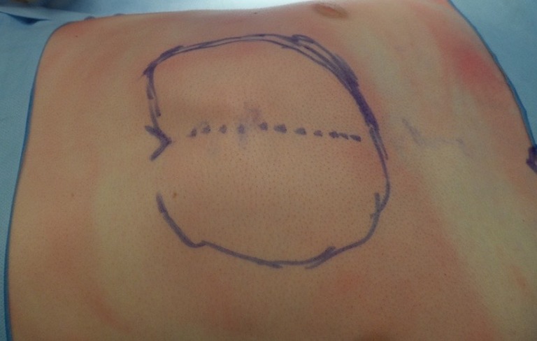
Position and length of the incision.
The next step consists of multiple parasternal rib cartilage resections to shorten the overabundant length that causes the deformity. Paramount attention should be paid to avoid damaging the perichondrium, otherwise there is a significant risk of chondrodystrophy. A 4–5-cm incision is made on top of the perichondrium of the deformed cartilages. We prefer a door-trap incision to allow circumferential lifting of the perichondrium. This manoeuvre facilitates subsequent reapproximation with PDS sutures. The incision should be made with a blade rather than with diathermy as the latter will melt the anatomical plane making exposure of the costal cartilage very difficult (Figure 2). Four or five of the most deformed costal cartilages are then resected from each side using periosteum elevators (Figure 3). This is applied circumferentially paying attention to stay within the perichondrium to avoid damaging the intercostal vessels. The costal cartilages should be preserved and used for further restabilization subsequently. Using this approach, the lesion of the pleura and intercostal nerves and vessels is kept to a minimum.
Figure 2.
Exposure of the perichondrium (7). Available online: http://www.asvide.com/articles/856
Figure 3.
Removal of the cartilage (8). Available online: http://www.asvide.com/articles/857
After resection of the cartilages, a wedge osteotomy should be performed with an oscillator blade at the level of the most prominent point (Figure 4). In symmetric defects this is made horizontally, in asymmetric defects is necessary sometime to perform a derotational osteotomy. Usually only the outer table of the bone is incised, than the sternum is compressed posteriorly to create a space in correspondence of the osteotomy line. One of the resected cartilages is cut to form a triangular prism and placed in the space created at the osteotomy line to keep the sternum depressed. Further reinforcement can be achieved with demineralized bone tissue (DBX® Demineralized Bone Matrix, Synthes, West Chester, PA, USA), which promotes bone growth.
Figure 4.
Wedge osteotomy using an oscillator blade (9). Available online: http://www.asvide.com/articles/858
Then the last step is the remodelling and stabilization of the chest wall. The sternum stabilization is obtained through the placement of one titanium bar (SternaLock® Blu, Biomet Microfixation Inc., Jacksonville, FL, USA) over the sternal body at the osteotomy line (Figure 5). The plate is used to provide stability for the first three months. It can be removed in case of problems, but we tend to leave it in permanently. There are reports of polymer plates used to stabilize the sternum (11). Whilst the big advantage is that they are reabsorbable, we do not believe they provide, in adult population, enough rigidity. Moreover they tend to cause seromas during the reabsorption process. Sometimes it is necessary to perform a second wedge osteotomy at the level of the least depressed part of the sternum, usually just above the xiphoid process. This manoeuvre allows perfect alignment of the sternum. The wedge osteotomy is reinforced with number 1 absorbable sutures.
Figure 5.
Fixation of the titanium bar on the sternum (10). Available online: http://www.asvide.com/articles/859
Lastly the perichondrial sheats of the ribs are then sutured to the sternum with absorbable sutures and shortened by plication if needed (Figure 6). A Redivac drain is finally place over the sternum and the pectoral muscle is refixed at the midline of the sternum, followed by the closure of the skin.
Figure 6.
Fixation of the perichondrial sheats of the ribs with the sternum (12). Available online: http://www.asvide.com/articles/860
Postoperative management
The pain management should be a priority in order to avoid further complications. In our institution we use a patient-controlled analgesia (PCA) with morphine on the day of the surgery. On the first postoperative day we remove the PCA and start an oral therapy with the combination of opioids (oxycodone) and non-steroidal anti-inflammatory drugs (usually paracetamol and diclofenac). All patients are mobilized right after surgery and continue having intense physiotherapy until they are discharged, usually between the second and the third postoperative day.
Tips and tricks
There are some tips and tricks that can help to achieve good results with minimal complications. For example, the Aquamantys device is particularly helpful in reducing blood loss and risk of infected hematoma. We recommend using small sharp instruments to achieve a good dissection of the perichondrium. Another advice is always try to achieve a balance between perfect correction and chest wall instability. Therefore we advise not to remove the first rib to avoid instability of the thoracic outlet and arm dysfunction.
Acknowledgements
None.
Footnotes
Conflicts of Interest: The authors have no conflicts of interest to declare.
References
- 1.Robicsek F, Sanger PW, Taylor FH, et al. The surgical treatment of chondrosternal prominence (pectus carinatum). J Thorac Cardiovasc Surg 1963;45:691-701. [PubMed] [Google Scholar]
- 2.Park CH, Kim TH, Haam SJ, et al. The etiology of pectus carinatum involves overgrowth of costal cartilage and undergrowth of ribs. J Pediatr Surg 2014;49:1252-8. 10.1016/j.jpedsurg.2014.02.044 [DOI] [PubMed] [Google Scholar]
- 3.Cobben JM, Oostra RJ, van Dijk FS. Pectus excavatum and carinatum. Eur J Med Genet 2014;57:414-7. 10.1016/j.ejmg.2014.04.017 [DOI] [PubMed] [Google Scholar]
- 4.Abramson H, D'Agostino J, Wuscovi S. A 5-year experience with a minimally invasive technique for pectus carinatum repair. J Pediatr Surg 2009;44:118-23; discussion 123-4. 10.1016/j.jpedsurg.2008.10.020 [DOI] [PubMed] [Google Scholar]
- 5.Ravitch MM. Operative Correction of Pectus Carinatum (Pigeon Breast). Ann Surg 1960;151:705-14. 10.1097/00000658-196005000-00011 [DOI] [PMC free article] [PubMed] [Google Scholar]
- 6.Colozza S, Bütter A. Bracing in pediatric patients with pectus carinatum is effective and improves quality of life. J Pediatr Surg 2013;48:1055-9. 10.1016/j.jpedsurg.2013.02.028 [DOI] [PubMed] [Google Scholar]
- 7.Scarci M, Bertolaccini L, Bedetti B, et al. Exposure of the perichondrium. Asvide 2016;3:102. Available online: http://www.asvide.com/articles/856
- 8.Scarci M, Bertolaccini L, Bedetti B, et al. Removal of the cartilage. Asvide 2016;3:103. Available online: http://www.asvide.com/articles/857
- 9.Scarci M, Bertolaccini L, Bedetti B, et al. Wedge osteotomy using an oscillator blade. Asvide 2016;3:104. Available online: http://www.asvide.com/articles/858
- 10.Scarci M, Bertolaccini L, Bedetti B, et al. Fixation of the titanium bar on the sternum. Asvide 2016;3:105. Available online: http://www.asvide.com/articles/859
- 11.Yuksel M, Bostanci K, Eldem B. Stabilizing the sternum using an absorbable copolymer plate after open surgery for pectus deformities: New techniques to stabilize the anterior chest wall after open surgery for pectus excavatum. Multimed Man Cardiothorac Surg 2011;2011:mmcts.2010.004879. [DOI] [PubMed]
- 12.Scarci M, Bertolaccini L, Bedetti B, et al. Fixation of the perichondrial sheats of the ribs with the sternum. Asvide 2016;3:106. Available online: http://www.asvide.com/articles/860


