Abstract
Background
Minimally invasive techniques for non-oncologic lung resections especially fungal infections are not widely employed. Through this video we share our experience of one such case of a robotic resection of pulmonary aspergilloma.
Methods
A 55-year-old male with recurrent hemoptysis underwent surgical resection of post tuberculosis aspergilloma of right upper lobe using a 4-arm DaVinci Robot.
Results
He received antituberculous drugs for 6 weeks pre-operatively. Systemic antifungals were given 2 weeks prior and continued for 3 months postoperatively. The operative time was 188 minutes and blood loss was 560 mL. Postoperative Chest X-rays showed complete lung expansion.
Conclusions
Robotic resection of lung is technically possible with good clinical outcomes even in infective pathologies. Robotic technique allows excellent 3D visualisation and good dexterity for easier and safe dissection of adhesions, as well as effective and precise anatomical lung resections for pulmonary aspergilloma
Keywords: Aspergilloma, robotic, resection
Introduction
Medical treatment of pulmonary aspergilloma is usually difficult and ineffective. Surgical resection of aspergilloma is likely to result in a complete cure, however it is technically challenging owing to dense adhesions, indurated hilar structures and high chances of intraoperative and early postoperative bleeding. The mortality following surgery is also significant. There has been a constant evolution in search of the appropriate surgical approach for pulmonary aspergilloma. Minimally invasive thoracic surgery for such cases has been reported in literature with many centres around the world now adopting video-assisted thoracic surgery (VATS) pulmonary resections in selected cases. However robotics for management of aspergilloma of lung is unheard of. Through this video we will share with you our experience of a robotic assisted thoracoscopic right upper lobectomy in a 55-year-old male with an aspergilloma in a post-tubercular fibrocavitary lesion in the right upper lobe causing recurrent hemoptysis.
Operative technique
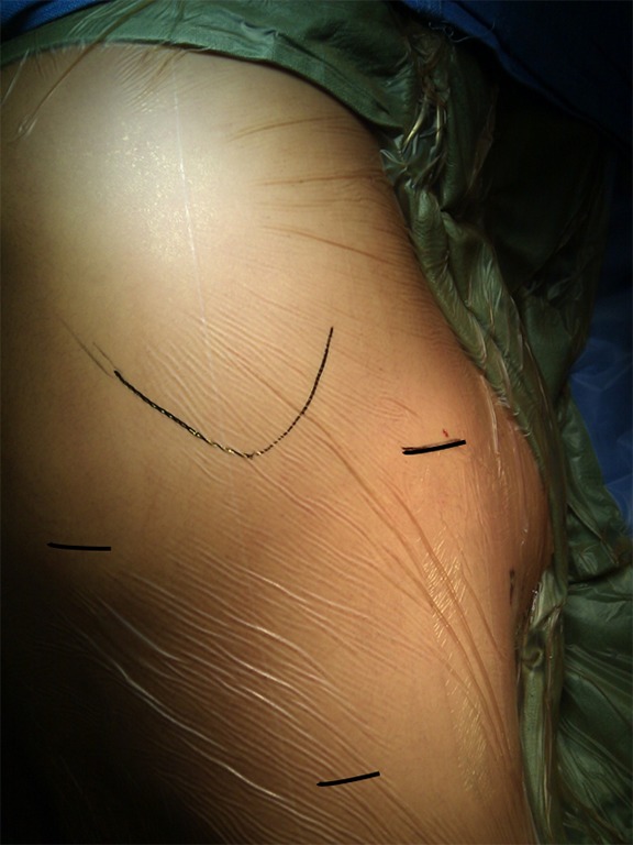
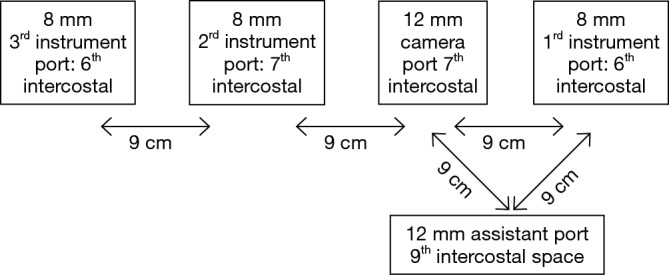
The patient was operated in left lateral decubitus position under general anesthesia with single lung ventilation and using extra long ventilator tubings and lines. Double lumen intubation was done in the lateral position and a Fogarty catheter was placed on the non-affected side to prevent spillage of fungus to the normal lung. Incisions and ports were placed as shown in Figures 1,2.
Figure 1.

Pre-operative marking of incisions after placing the patient in lateral position.
Figure 2.

Descriptive diagram of port placement for robotic right upper lobectomy.
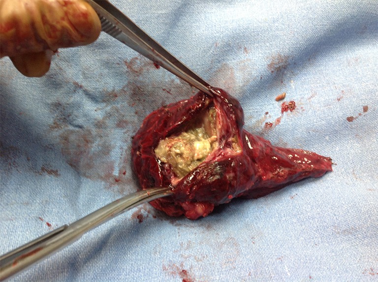
Following the incisions and port placement a 4-arm DaVinci Robot was docked to the patient and adhesiolysis started (Figure 3). The superior pulmonary vein was first dissected and divided using an endoscopic stapler, which was followed by the division of upper lobe bronchus. All arterial branches to the affected lobe were carefully dissected & divided later. Dense adhesions of the apex to the upper lobe were then divided. They were purposely not taken down initially for two reasons. Firstly they act as a good retraction for the lobe and secondly as they are highly vascular, lysing them early on in the procedure leads to blood loss and obscured vision. The robot was then undocked and subsequently one of the incisions was extended to facilitate the removal of the specimen from the chest, which was extracted out in an endobag (Figure 4).
Figure 3.

Surgical video of a robotic right upper lobectomy (1). Available online: http://www.asvide.com/articles/862
Figure 4.

Cut-open section of right upper lobe showing the fungal ball.
A number 28 intercostal chest drain was placed after ensuring hemostasis and connected to a digital suction device.
Comments
Traditionally surgical approach for aspergilloma is through a liberal thoracotomy. However the last decade has seen a rise in the number of VATS pulmonary resections performed for aspergilloma.
Gossot et al. have shared their experience of 15 patients with invasive pulmonary aspergillosis (IPA), and they showed that full thoracoscopic resection (including lobectomies) of these fungal infections is possible with less postoperative morbidity (2). Whitson et al. have described a successful thoracoscopic lingulectomy for IPA (3). A recent retrospective study (4) compared VATS in the treatment of simple mycetoma and complex mycetoma. They concluded that VATS can be safely applied to simple aspergilloma and complex aspergilloma without infiltration of the hilum and allows for an early postoperative recovery.
With the advent of robotics in thoracic surgery, case volumes for robotic pulmonary resections has increased significantly during the last few years, and thoracic surgeons have been able to adopt the robotic approach safely.
Multiple published articles have shown the efficacy and safety of robotic pulmonary resection including lobectomy, segmentectomy, and even several reports of pneumonectomy (5-8).
Recently Kent et al. made a comparison in outcomes between robotic, open, and VATS procedures, using the State Inpatient Databases (SID). They advocate that robotic lung resection appears to be an appropriate alternative to VATS and is associated with improved outcomes compared with open thoracotomy (9).
However no data exists in literature where non-oncologic lung resections, especially of infective etiologies have been carried out with robotics. Encouraging results of thoracoscopic surgery or VATS in management of pulmonary aspergilloma and our growing comfort with the robot for pulmonary resections led us to start our series of robotic resections for pulmonary aspergilloma
Robotic technique allows excellent 3D visualisation and good dexterity for easier and safe dissection of adhesions. The 360 degree freedom of movement of the endowrist offers effective and precise anatomical lung resections for pulmonary aspergilloma because of easier dissection around the broncho-vascular structures. It appears to be superior over conventional techniques in terms of pinpoint intra-operative visualization, filtering of tremors around crucial structures, less bleeding and less post-operative air leaks and hence early removal of chest drain, less postoperative pain, minimal or no wound infections, reduced hospitalisation and excellent cosmesis. Disadvantages of the robot are the initial long learning curve, availability and cost.
It is extremely important to not hesitate and quickly convert to a VATS or thoracotomy whenever facing intraoperative difficulty.
The safety and efficacy of robotic lung resections for pulmonary aspergilloma however needs to be established through a study with large number of patients.
Acknowledgements
None.
Informed Consent: Written informed consent was obtained from the patient. A copy of the written consent is available for review by the Editor-in-Chief of this journal.
Footnotes
Conflicts of Interest: The authors have no conflicts of interest to declare.
References
- 1.Khan AZ, Ali K, Khandelwal S, et al. Surgical video of a robotic right upper lobectomy. Asvide 2016;3:108. Available online: http://www.asvide.com/articles/862
- 2.Gossot D, Validire P, Vaillancourt R, et al. Full thoracoscopic approach for surgical management of invasive pulmonary aspergillosis. Ann Thorac Surg 2002;73:240-4. 10.1016/S0003-4975(01)03280-5 [DOI] [PubMed] [Google Scholar]
- 3.Whitson BA, Maddaus MA, Andrade RS. Thoracoscopic lingulectomy for invasive pulmonary aspergillosis. Am Surg 2007;73:279-80. [PubMed] [Google Scholar]
- 4.Ichinose J, Kohno T, Fujimori S. Video-assisted thoracic surgery for pulmonary aspergilloma. Interact Cardiovasc Thorac Surg 2010;10:927-30. 10.1510/icvts.2009.232066 [DOI] [PubMed] [Google Scholar]
- 5.Park BJ, Melfi F, Mussi A, et al. Robotic lobectomy for non-small cell lung cancer (NSCLC): long-term oncologic results. J Thorac Cardiovasc Surg 2012;143:383-9. 10.1016/j.jtcvs.2011.10.055 [DOI] [PubMed] [Google Scholar]
- 6.Cerfolio RJ, Bryant AS, Skylizard L, et al. Initial consecutive experience of completely portal robotic pulmonary resection with 4 arms. J Thorac Cardiovasc Surg 2011;142:740-6. 10.1016/j.jtcvs.2011.07.022 [DOI] [PubMed] [Google Scholar]
- 7.Veronesi G, Galetta D, Maisonneuve P, et al. Four-arm robotic lobectomy for the treatment of early-stage lung cancer. J Thorac Cardiovasc Surg 2010;140:19-25. 10.1016/j.jtcvs.2009.10.025 [DOI] [PubMed] [Google Scholar]
- 8.Melfi FM, Mussi A. Robotically assisted lobectomy: learning curve and complications. Thorac Surg Clin 2008;18:289-95, vi-vii. 10.1016/j.thorsurg.2008.06.001 [DOI] [PubMed] [Google Scholar]
- 9.Kent M, Wang T, Whyte R, et al. Open, video-assisted thoracic surgery, and robotic lobectomy: review of a national database. Ann Thorac Surg 2014;97:236-42; discussion 242-4. 10.1016/j.athoracsur.2013.07.117 [DOI] [PubMed] [Google Scholar]


