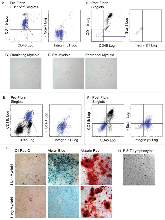
Figure 5.
Myeloid cells from other tissues are incapable of complete mesenchymal differentiation after fibrin culture. Myeloid (CD45POS/CD11bPOS) cells were purified from the circulation (A, B & C), bone marrow (BM) and peritoneal fluid, (D), liver and lung (E, F & G), and B and T lymphocytes (H). Cells from n = 3 male and female animals were pooled experimented; independent experiments conducted in duplicate. The cells were cultured in fibrin clots for 5 days, recovered by plasmin hydrolysis of the clots and plated on plastic surfaces in MesenCult medium with SCSS. A) Flow analysis of pre-fibrin circulating myeloid cells. B) Flow cytometry analysis of post-fibrin “myeloid” cells. C) Post fibrin circulating cells or D) BM or peritoneal cells exhibited minimal plastic adherence and failed to proliferate. Flow cytometry analysis of E) pre-fibrin or F) post-fibrin liver “myeloid” cells. G) Post-fibrin cells from liver or lung were plastic adherent and proliferated. At confluence they were exposed to adipogenic, chondrogenic or osteogenic conditions for 14–21 d and stained with Oil Red O, Alcian Blue or Alizarin Red to assess adipogenesis, chondrogenesis or osteogenesis, respectively. H) Post-fibrin lymphocytes exhibited limited plastic adherence and failed to proliferate in SCSS medium. All images 20x magnification.

