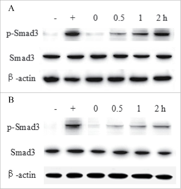
Figure 9.

The Smad3 activation analysis of HSC-T6 cells. Cells were incubated with 30 nM eTGFBR2 (A) or HSA-eTGFBR2 (B) for 0, 0.5, 1, 2 h, and then exposed to 0.4 nM TGF-β1 for 30 min. -: DMEM medium for negative control; +: 0.4 nM TGF-β1 for positive control.
