Abstract
Background:
The obstructive hydronephrosis is a term that implicates the structural and functional changes of the kidneys as a result of difficulties in the flow of urine. Hydronephrosis specifically describes dilation and swelling of the kidney. Hydronephrosis is a condition that typically occurs when the kidney swells due to the failure of normal drainage of urine from the kidney to the bladder. Our aim was to evaluate the degree of hydronephrosis, causes and diagnostic method. Material and
Methods:
This is a study of 136 patients that have been treated at the Department of Urology, University Clinical Centre of Kosovo, Prishtina. For diagnosis of hydronephrosis in our patient, we used as equipment the Color Doppler ultrasound, with resolution of 3.5 MHz–8 MHz.
Results:
Out of 136 participants in the study, 91 (66.9%) were males and 45 (33.1%) females, with significant difference (P=0.000). The average age for males was 49 years old, whereas for females was 33. This study included patients with a diagnosis of symptomatic hydronephrosis with various causes and degrees. All patients were presented with hydronephrosis. The hydronephrosis grade varied from the stage I up to the IV. In our study we have difference grade of hydronephrosis, X2 test, P= 0.114. The most common causes of hydronephrosis in our study were; kidney stone, ureteral stones, neck stenosis PU, pregnancy, infiltrating bladder cancer, bladder neck contracture, prostate adenoma, infiltrating prostatic carcinoma etc. In this study we have indentified different causes, of which stones dominate as the most usual causes of hydronephrosis P= 0.0001.
Conclusion:
The Ultrasound is an easy method to be applied, non invasive, and a fast one to help and diagnose the obstructive hydronephrosis. The ultrasound has a high sensitivity and should be used as a screening method followed by other methods, as necessary. Hydronphrosis is most commonly presented to men with an average age about fifties. We came to the conclusion that the main causes of hydronephrosis are kidney stone, followed by ureteral stones, in which, in a larger percentage, they appear with the II degree of hydronephrosis.
Keywords: Hydronephrosis, grade, causes
1. INTRODUCTION
Hydronephrosis describes the situation where the urine collecting system of the kidney is dilated. Hydronephrosis specifically describes dilation and swelling of the kidney, while the term hydroureter is used to describe swelling of the ureter. The presence of hydronephrosis or hydroureter can be physiologic or pathologic (1, 2). The obstructive hydronephrosis is a term that implicates the structural and functional changes of the kidneys as a result of difficulties in the flow of urine (difficulties in urinating). The uropathy can be caused by the difficulties or interruption of the flow of urine in the kidney, urethra, bladder, prostate, changes of retro-peritoneum or vascular system (3, 4).
The Ultrasound is a suitable method of early detection of obstructive uropathies especially in cases of hydronephrosis.
Hydronephrosis may be unilateral involving just one kidney or bilateral involving both. Also, it may be acute or chronic. The etiology of hydronephrosis and/or hydroureter is calculi in young adults, while prostatic hypertrophy or carcinoma, retroperitoneal or pelvic neoplasms, and calculi in older patients (5, 6, 7).
Dilatation of the ureters and renal pelvis is seen to the pregnant women. Dilatation can come as a result of progesterone effects and mechanical compression of the ureters at the pelvic brim. This hydronephrosis is a normal finding in pregnant women (8, 9).
Common symptoms of hydronephrosis include: flank pain, abdominal mass, nausea and vomiting, urinary tract infection, fever, painful urination (dysuria), increased urinary frequency, increased urinary urgency, etc (10, 11, 12).
Basic exams and tests for the diagnosis of hydronephrosis include; ultrasound of the kidneys or abdomen, MRI of the abdomen, CT-scan of the kidneys or abdomen, intravenous pyelogram (IVP), (12, 13).
2. MATERIALS AND METHODS
This study was approved by the Ethical Committee of the Faculty of Medicine, University of Prishtina, and the research was conducted in accordance with the Declaration of Helsinki guidelines. Written informed consent was obtained from all study participants before inclusion in the study.
This is a study of 136 patients that have been treated at the Department of Urology, University Clinical Centre of Kosovo, Prishtina. The equipment used Color Doppler ultrasound, with resolution 3.5 Mhz-8 Mhz.
This study included patients with a diagnosis of symptomatic hydronephrosis with various causes and degrees. Prior to study, hemogram, blood urea, serum creatinine, urine examination, urine culture sensitivity, was done for participating patients.
Statistical analysis
Processing of date is done with the statistical package InStat 3. The obtained data has been presented through tables and figures. Data are presented as proportions and 95% Cl. Testing of the data was done with X2 test. The difference statistically significant is if P< 0.05.
3. RESULTS
This study included patients with a diagnosis of symptomatic hydronephrosis with various causes and degrees.
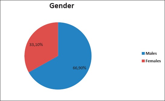
Out of 136 participants in the study, 91 (66.9%) were males and 45 (33.1%) females, with significant diference P=0.000. The average age for males was 49 years old, whereas for females was 33 (Figure 1).
Figure 1.

Gender distribution X2 test; P= 0.000.
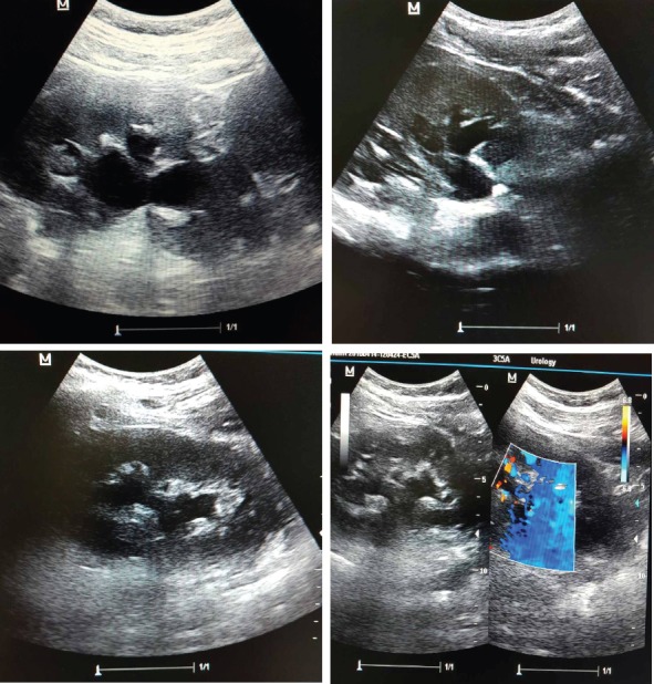
Figure 2.

Cases with hydronephrosis (with ultrasound)
All patients were presented with hydronephrosis. The hydronephrosis grade varied from the stage I up to the IV. In our study we have difference grade of hydronephrosis, X2 test, P=0.114. Stage I hydronephrosis was 22.8% of cases, P-Value (16.5-30.5); Stage II hydronephrosis was: 48%, P value (40.3-56.9); Stage III was: 16.2 %, P Value (10.9-23.3); Stage IV: was 12.5%, P value (8.0-19.1) (Table 1).
Table 1.
Hydronephrosis grade and percentage, X2 test, P= 0.114

The most common causes of hydronephrosis in our study were; kidney stone, ureteral stones, neck stenosis PU, pregnancy, infiltrating bladder cancer, bladder neck contracture, prostate adenoma, infiltrating prostatic carcinoma etc.
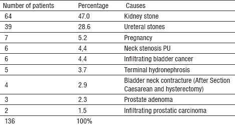
In Table 2 we have presented different causes of hydronephrosis, of which stones dominate as the most usual causes, P= 0.0001.
Table 2.
Causes of hydronephrosis and percentage. X2–test, Stones/Other causes P= 0.0001

Out of 136 patients, 64 (47.0%) patients had kidney stones, 7 of them had a coral-form stones (5 cases of complete coral-form stones and 2 of them in complete coral-form stones). 39 (28.6 %) of cases with hydronephrosis had bladder stones. 7 (5.2 %) of cases with hydronephrosis were pregnant. 6 (4.4 %) of cases with hydronephrosis had neck stenosis PU (which were verified by U.I.V, CT urography and retrograde urethropielography).
There were 6 (4.4 %) cases of hydronephrosis caused by infiltrating bladder cancer. 5 (3.7 %) cases of terminal hydronephrosis (a function-scintigram and radiorenogram). 4 (2.9 %) of cases with hydronephrosis were women with bladder neck contracture (After Sectio caesarean and hysterectomy; ligatura ureteris) 3 (2.3 %) cases of hydronephrosis caused by prostate adenoma, and 2 (1.5%) cases of hydronephrosis caused by infiltrating prostatic carcinoma (Table 2).
4. DISCUSSION
The obstructive hydronephrosis is a term that implicates the structural and functional changes of the kidneys as a result of difficulties in the flow of urine. The uropathy can be caused by the difficulties or interruption of the flow of urine in the kidney, urethra, bladder, prostate, changes of retro-peritoneum or vascular system (3, 4).
The Ultrasound is a suitable method of early detection of obstructive uropathies especially in cases of hydronephrosis.
Hydronephrosis may be unilateral involving just one kidney or bilateral involving both. Also, it may be acute or chronic. The etiology of hydronephrosis and/or hydroureter is calculi in young adults, while prostatic hypertrophy or carcinoma, retroperitoneal or pelvic neoplasms, and calculi in older patients (5, 6).
Dilatation of the ureters and renal pelvis is seen of pregnant women. Dilatation can come as a result of progesterone effects and mechanical compression of the ureters at the pelvic brim. This hydronephrosis is a normal finding in pregnant women (7, 8).
Common symptoms of hydronephrosis include: flank pain, abdominal mass, nausea and vomiting, urinary tract infection, fever, painful urination (dysuria), increased urinary frequency, increased urinary urgency (9-12).
In our study, of 136 participants in the study, 66.9% of which were males and 33.1% females, with significant difference P=0.000. The average age for males was 49 years old, whereas for females was 33 (Figure 1).
All patients were presented with hydronephrosis. The hydronephrosis grade varied from the stage I up to the IV, (Table 1), X2 test, P=0.114.
Stage I hydronephrosis was in 22.8% of cases, P-Value (16.5-30.5); stage II hydronephrosis was 48%, P value (40.3-56.9); Stage III was 16.2%, P Value (10.9-3.3); stage IV was 12.5%, P value (8.0-19.1).
In Table 2 we have presented causes of hydronephrosis, we have done testing by X2 test stones/other causes P=0.0001. 47.0% of the patients had kidney stones; 28.6 % of cases with hydronephrosis had uretral stones; 5.2 % of cases with hydronephrosis were pregnant women; 4.4 % of cases with hydronephrosis had neck stenosis PU; 4.4 % of cases of hydronephrosis caused by infiltrating bladder cancer; 3.7 % cases of terminal hydronephrosis (a function-scintigram and radiorenogram); 2.9 % of cases with hydronephrosis were women with; bladder neck contracture After Sectio caesarean and hysterectomy; ligature ureteris; 2.3 % cases of hydronephrosis caused by prostate adenoma, and 1.5% cases of hydronephrosis caused by infiltrating prostatic carcinoma.
Hydronephrosis is not a primary disease. It is a secondary condition that results from some other underlying disease. It is a structural condition that is the result of a blockage or obstruction in the urinary tract.
Ultrasound adds a functional evaluation of the urinary tract when combined with clinical findings allows performing the appropriate management (11, 12, 13).
5. CONCLUSION
Ultrasound is an easy method to be applied, non invasive and fast one to help and diagnose the obstructructive hydronephrosis. The ultrasound has a high sensitivity and should be used as a screening method followed by other methods, as necessary. We find a significant difference by sex. Males had a 2 times more chance to be attacked by hydronephrosis. Hydronphrosis is most commonly presented to man with an average age about fifties.
We came to the conclusion that the main causes of hydronephrosis are kidney stone, followed by ureteral stones, in which, in a larger percentage, they appear with the II degree of hydronephrosis.
Footnotes
• Financial Disclosure: The authors did not report any potential conflicts of interest.
REFERENCES
- 1.European Association of Urology guidelines on Urolithiasis. European Association of Urology. 2017:270–9. [Google Scholar]
- 2.Türk C, Knoll T, Petrik A, Sarica K, Straub M, Seitz C. European Association of Urology guidelines on Urolithiasis. European Association of Urology. 2012;8:63–5. [Google Scholar]
- 3.Zeidel ML. Obstructive uropathy. In: Goldman L, Schafer AI, editors. Cecil Medicine. 24th ed. chap 125. Philadelphia, Pa: Saunders Elsevier; 2011. [Google Scholar]
- 4.Pepe P, Motta L, Pennisi M, Aragona F. Functional evaluation of the urinary tract by color-doppler ultrasonography (CDU) in 100 patients with renal colic. Eur J Radiol. 2005;53:131–5. doi: 10.1016/j.ejrad.2004.01.014. [DOI] [PubMed] [Google Scholar]
- 5.Varma G, Nair N, Salim A, et al. Investigations for recognizing urinary stone. Urol Res. 2009;37:349–52. doi: 10.1007/s00240-009-0219-z. [DOI] [PubMed] [Google Scholar]
- 6.Tanagho JW, McAninch EA Urinary Obstruction and Stasis in Smith’s General Urology. Lange Medical Books/McGraw Hill. 16th Ed. New York: 2004. pp. 175–87. [Google Scholar]
- 7.Kawashima, et al. CT Urography. RadioGraphics. 2004;24:S35–S54. doi: 10.1148/rg.24si045513. [DOI] [PubMed] [Google Scholar]
- 8.Glanc F, Maxwell C. Acute abdomen in pregnancy. Role of sonography. J Ultrasound Med. 2010;29:1457–68. doi: 10.7863/jum.2010.29.10.1457. [DOI] [PubMed] [Google Scholar]
- 9.Singh I, Strandhoy JW, Assimos DG. Pathophysiology of urinary tract obstruction. In: Campbell-Walsh Urology. 10th ed. chap 40. Wein AJ, editor. Philadelphia, Pa: Saunders Elsevier; 2011. [Google Scholar]
- 10.Semins MJ, Trock BJ, Matlaga BR. The safety of ureteroscopy during pregnancy: a systematic review and meta-analysis. J Urol. 2009;181:139–43. doi: 10.1016/j.juro.2008.09.029. [DOI] [PubMed] [Google Scholar]
- 11.Blandino, et al. MR Urography of the Ureter: Pictoral Essay. AJR. 2002;179:1307–14. doi: 10.2214/ajr.179.5.1791307. [DOI] [PubMed] [Google Scholar]
- 12.Lameire N, Van Biesen W, Vanholder R. Acute renal failure. [29-Feb 4];Lancet. 2005 Jan;365(9457):417–30. doi: 10.1016/S0140-6736(05)17831-3. [DOI] [PubMed] [Google Scholar]
- 13.Blandino, et al. MR Urography of the Ureter: Pictoral Essay. AJR. 2002;179:1307–14. doi: 10.2214/ajr.179.5.1791307. [DOI] [PubMed] [Google Scholar]
- 14.Kumar, Abbas, Fausto, editors. 7th Ed. Pennsylvania: Elsevier Saunders; 2005. pp. 955–1021. [Google Scholar]


