

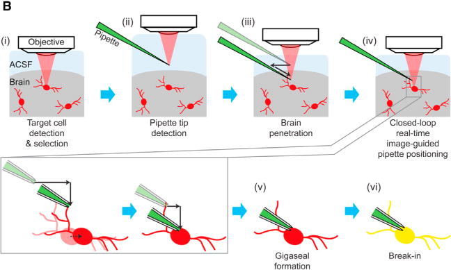
In our original Figure 1B, the appearance of Figures 1Bii and 1Biii incorrectly made it look like the pipette descended into the brain in the downward direction (implying that it would cause brain damage), and furthermore in the original Figure 1Biv the black arrows incorrectly implied that the pipette descends into the brain further downward. In reality, the pipette enters the brain at a diagonal angle, along the axis of the pipette, and furthermore as the pipette approaches the cell, it does so in staggered downward and lateral steps that simulate diagonal movement. We have now corrected Figure 1B to reflect the real configuration of the system. In particular, the pipette enters into the brain (Figure 1Biii) at a diagonal, and toward the end of the process, the pipette moves in staggered downward and lateral steps to approach the cell (we have removed the downward black arrows from Figure 1Biv, so that the lower left of Figure 1B provides the full explanation of the final stages of pipette movement). The figure has now been corrected online. The authors apologize for any confusion this error may have caused.
Figure 1B.

Imagepatching: Closed-Loop Real-Time Image-Guided Patch Clamping In Vivo (original)
Figure 1B.
Imagepatching: Closed-Loop Real-Time Image-Guided Patch Clamping In Vivo (corrected)