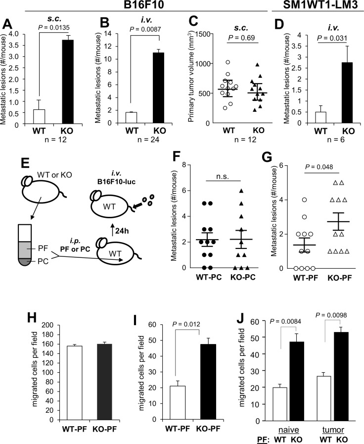
Figure 1. SSeCKS-null (KO) mice exhibit increased potential for peritoneal metastasis.
(A) The percent of WT vs. KO mice exhibiting macroscopic peritoneal metastases after s.c. injection of B16F10 cells. (B) The average number of macrometastases/mouse after i.v. injection of B16F10-luc. (C) Tumor volumes induced by B16F10-luc injected s.c. in WT vs. KO mice. (D) The average number of macrometastases/mouse after i.v. injection of SM1WT1-LM3-luc. (E) Schematic of adoptive transfer of either peritoneal fluid (PF) or peritoneal cells (PC) from naïve WT or KO mice to WT hosts (via i.p. injection), followed the next day by tail vein i.v. injection of B16F10-luc cells. (F) Number of metastatic lesions/mouse following the adoptive transfer of WT- or KO-PC. (G) Number of metastatic lesions/mouse following the adoptive transfer of WT- or KO-PF. (H) Migration of B16F10-luc in wound-healing assays containing 20% PF from WT- or KO-mice. The number of cells migrating into 6 microscopic fields were quantified and averaged. (I) Chemotaxis assays (through Boyden chambers) by B16F10-luc cells towards media (no serum) containing 20% PF from WT- or KO-mice (fluid flushed and pooled from 3 mice each, normalized for total protein content). (J) Similar chemotaxis assay as in panel H except using PF from naïve or tumored (s.c.) WT- or KO-mice.

