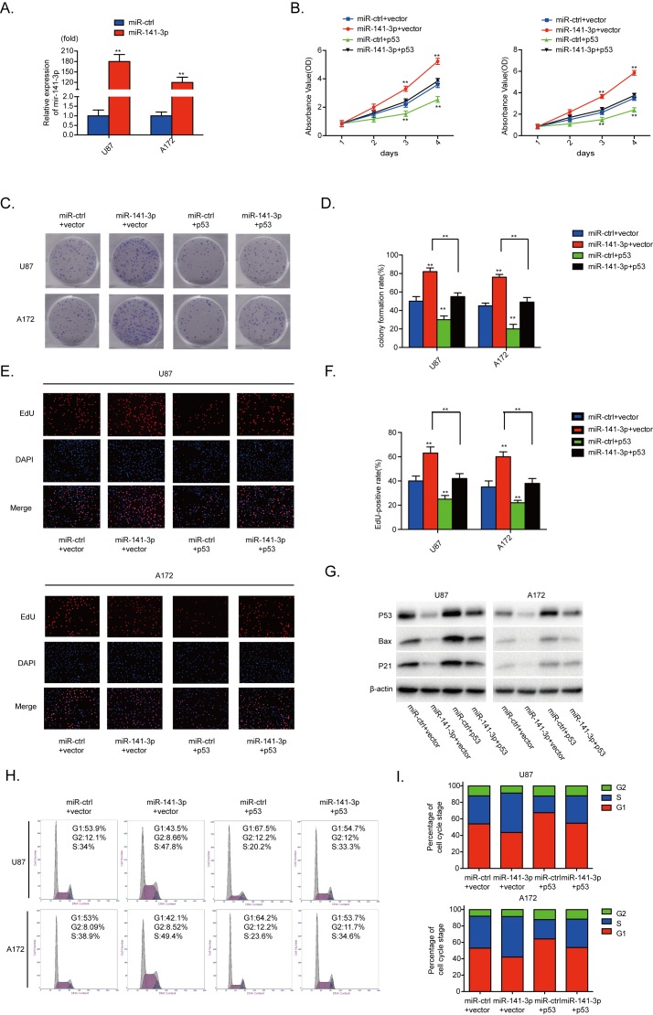
Figure 5. p53 reintroduction reverses the promotional effect of miR-141-3p.
(A) Relative expression of miR-141-3p in U87 and A172 cells was analyzed by qRT-PCR after transfection. (B) Cell proliferation was detected by CCK8 assays after U87 and A172 cells transfected with miR-ctrl or miR-141-3p with or without p53 expression plasmid. (C and D) Colony formation assays in U87 and A172 transfected with miR-ctrl or miR-141-3p with or without p53 expression plasmid. Scale bar: 500 μm. (E and F) Respective merged images of U87 and A172 cells in EDU transfected with miR-ctrl or miR-141-3p with or without p53 expression plasmid after 48 h. Representative images are shown (original magnification, 200×). (G) Downstream proteins of p53 in U87 and A172 cells transfected with miR-ctrl or miR-141-3p with or without p53 expression plasmid were examined by immunoblotting. (H and I) The cell cycle of U87 and A172 cells transfected with miR-ctrl or miR-141-3p with or without p53 expression plasmid was detected after 48 h by flow cytometry. All experiments were performed three times using triplicate samples. Average values are indicated with error bars in the histogram. Results are presented as the mean ± S.D. *P<0.05, **P<0.01, and ***P<0.001.

