Genetic variations in ion channels influence human hair color
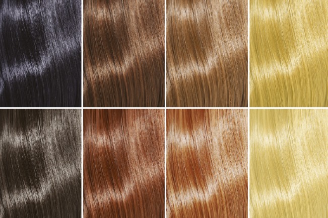
Genetic variations in TPC ion channels influence hair color. Image courtesy of iStockphoto/Sasha_Litt.
Levels of the human hair color pigment eumelanin, produced by cells found in the skin’s basal epidermis and hair follicles, depend on the cells’ internal pH. Previous studies have found that genetic variations in a human cation-transporting channel called two-pore channel (TPC) 2 can alter the pH of pigment-producing cells, causing hair color to shift from brown to blond. Yu-Kai Chao et al. (pp. E8595–E8602) used electrophysiological methods and molecular dynamics simulations to uncover how the genetic variations affect channel function. Compared with the wild-type channel, the M484L variant, in which a methionine residue is replaced by leucine, exhibited not only increased basal activity and response to the channel’s lipid ligand PI(3,5)P2, but also larger current amplitudes stemming from a dilated pore. Analysis of DNA samples from more than 100 donors with blond, brown, or black hair revealed that 7.2% of samples from blond-haired donors were homozygous for the M484L variation, whereas only 2.9% of samples from donors with brown, dark brown, or black hair were homozygous for the same variation. Patch-clamp experiments on skin cells from donors revealed heightened PI(3,5)P2-driven TPC2 activity in M484L cells, suggesting that the variation ramps up channel activity, ratchets down eumelanin synthesis, and renders hair blond. Because TPCs are implicated in blood vessel formation, cholesterol transport and metabolism, and cancer cell migration, genetic variations in TPCs might have broad impacts on human health, according to the authors. — P.N.
How dinosaurs developed bird beaks
Numerous theropod dinosaur lineages, including birds, lost their teeth during evolution. However, the developmental mechanisms linking the evolutionary loss of teeth and beak formation remain unresolved. Shuo Wang et al. (pp. 10930–10935) examined jaw fossils from one of the smallest known dinosaurs from the group Caenagnathidae and the early Cretaceous bird Sapeornis. Computer tomography and synchrotron scans indicated the presence of vestigial bony tooth sockets and other jaw morphologies similar to another beaked theropod, Limusaurus. Previous research indicates that Limusaurus transitioned from toothed juveniles to toothless beaked adults, suggesting that a reduction of dentition during maturation may explain the similar dental morphologies of the three theropods. Character correlation analyses indicate that terrestrial egg laying, the presence of keratinous egg teeth, and the acquisition of toothless beaks are correlated in tetrapods, potentially explaining the propensity for beak evolution in Archosauromorpha, which includes birds, crocodiles, and pterosaurs. The authors suggest that a protein called bone morphogenetic protein 4 may have simultaneously mediated embryonic tooth loss and formation of beak keratin. According to the authors, the evolutionary progression of postnatal and embryonic dental reduction may have driven the repeated evolution of beaks in nonavian theropods and birds as a response to diet specialization. — L.C.
Treating postdiet body weight rebound

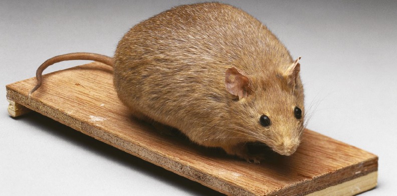
Postdieting hormonal changes to appetite often lead to weight regain. Image courtesy of Wellcome Trust, Wellcome Images.
Obesity contributes to many chronic conditions that decrease life quality and longevity, and its global prevalence is increasing. Caloric restriction can lower body weight rapidly in the short term, but weight tends to subsequently rebound as a result of compensatory metabolic changes, such as increased appetite. Vicky Ping Chen et al. (pp. 10960–10965) explored a strategy for preventing postdieting weight gain in mice by targeting the appetite-stimulating hormone ghrelin. Mice were fed a high-fat diet from 8–20 weeks of age before being switched to a diet with 40% lower calorie content than their previous diet, leading to substantial weight loss. Upon beginning the calorie-restricted (CR) diet, mice were injected with an adeno-associated virus expressing either the enzyme butyrylcholinesterase (BChE), which inactivates ghrelin, or a control enzyme, luciferase. After 3 weeks on the CR diet, mice were switched to an unrestricted low-fat diet. Following the end of the CR diet, mice treated with the BChE virus expressed lifelong high plasma levels of BChE and lower levels of acyl-ghrelin, the active form of ghrelin, than the control-treated mice. BChE-treated mice also consumed fewer calories, regained less weight, and had better glucose tolerance than control-treated mice. According to the authors, the results suggest the potential of BChE gene therapy for obesity treatment. — B.D.
How fingers interact with surfaces

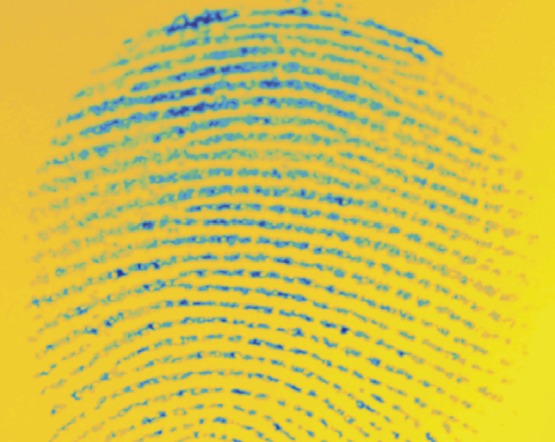
Small junctions between fingerprint ridges and smooth surface that grow and interconnect with increasing contact time.
The outer layer of human finger pads is stiff and rough at a small scale, but deforms under pressure to form stable contacts with surfaces. Brygida Dzidek et al. (pp. 10864–10869) used high-resolution imaging to monitor contact formation between two participants’ fingers and glass or rubber surfaces. For a hard glass surface, the area of the finger in contact with the surface was initially small and increased gradually over a period of tens of seconds. The coefficient of friction between the finger and the surface increased on a similar time scale. For a soft rubber surface, both the contact area and the coefficient of friction reached their maximum values within 2 seconds. Contact formation with a hard surface requires hydration of the keratin in the outer skin layer by sweat, which increases skin plasticity. Therefore, the slow contact formation observed with the glass surface reflects the rate of sweat secretion from the pores, whereas contact formation with a soft surface is not limited by hydration. According to the authors, differences in the rate of contact formation might help people to distinguish different types of materials by touch and may have implications for designing tactile displays. — B.D.
Microglial response in Alzheimer’s disease
Alzheimer’s disease (AD) is associated with dysfunction of microglia, which are macrophage-like cells of the central nervous system. Researchers recently identified a subtype of disease-associated microglia (DAM) that may modulate AD pathogenesis, but the underlying mechanisms are still unclear. Dimitry Ofengeim et al. (pp. E8788–E8797) investigated the role of RIPK1—a kinase that is highly expressed in the microglial cells of AD brains—in AD pathogenesis using an amyloid precursor protein/presenilin 1 transgenic mouse model of AD. The authors found that pharmacological or genetic inhibition of RIPK1 led to reduced amyloid burden and lower levels of inflammatory cytokines. Inhibiting RIPK1 also reduced behavioral deficits and improved memory function in the transgenic mice. The authors found that the kinase activity of RIPK1 plays a role in upregulating microglial expression of Cst7, which encodes a cathepsin inhibitor called cystatin F. RIPK1-dependent induction of Cst7 leads to impairments in the lysosomal pathway. RIPK1 inhibition restores normal levels of Cst7 and appears to inhibit the DAM response and promote microglial degradation of amyloid- in vitro. The results indicate a mechanism by which RIPK1 might mediate the induction of a DAM state that reduces microglial phagocytic activity and promotes an inflammatory response, and the authors suggest that RIPK1 could serve as a therapeutic target for AD. — S.R.


