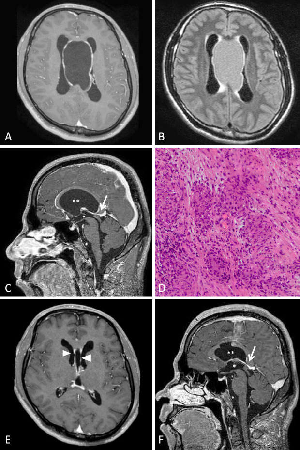
A 39-year-old apparently healthy man was hospitalized due to headache that had persisted for ten days. He had no neurological deficits except for small-step and/or magnet gait. T1-weighted contrast-enhanced magnetic resonance imaging (T1-WI) revealed a massive lobular cyst with an irregularly enhanced, thickened wall (Picture A). The cyst contained fluid that was isointense to cerebrospinal fluid (CSF) on T2-WI and slightly more intense than CSF on fluid-attenuated inversion recovery (FLAIR) images (Picture B), suggesting that it was rich in protein or cellular components.
Picture.
Sagittal contrast-enhanced T1-WI (Picture C) showed that the huge cyst (double asterisk) compressed the internal cerebral vein (arrow) and the third ventricle (asterisk), resulting in hydrocephalus, which presented as enlargement of the lateral ventricles (Picture A, B).
Hematoxylin and eosin staining of pathological specimens of the cyst wall, which had been collected after craniotomy, revealed non-caseating epithelioid granuloma (Picture D); these findings indicated a diagnosis of sarcoidosis with extra-neural involvement. Residual enhanced wall thickening was still present after surgery (Picture E, arrowheads); however, the patient's gait disturbance disappeared along with the resolution of hydrocephalus and the shrinking of the huge cyst (Picture F).
The authors state that they have no Conflict of Interest (COI).


