
Figure 8.
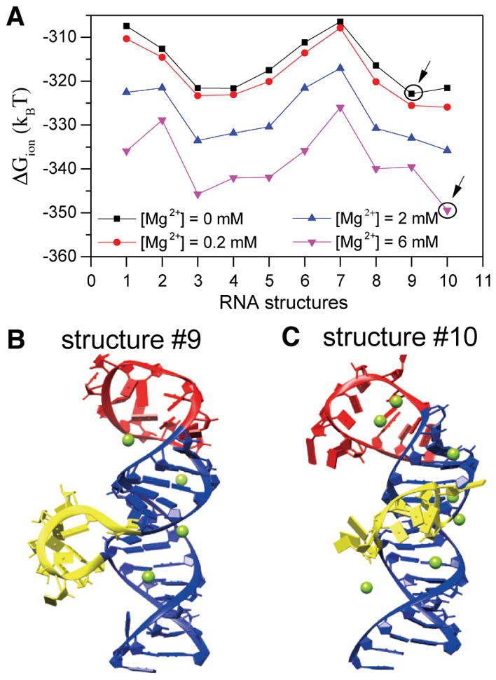
Structure of 5BSL3.2 is sensitive to Mg2+ concentration. (A) Ion electrostatic free energies for the different 3D structures of 5BSL3.2 at different Mg2+ concentrations. The structures that have the lowest free energy are labeled with circles at 0 and 6 mM Mg2+. (B and C) Structures 9 and 10, respectively, with the predicted bound Mg2+ ions at 2 mM Mg2+. Loop residues U9280–G9291 are colored red, bulge residues A9298–G9304 yellow, and residues in the main helix blue.