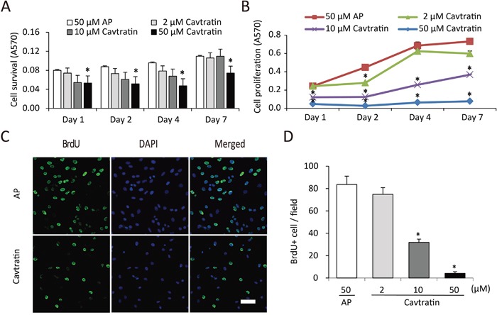
Figure 1. Cavtratin inhibits the survival and proliferation of endothelial cells.

(A) Cavtratin decreased HUVEC survival in an MTT assay with 0.5% of FBS in the culture medium. Cells were treated with different concentrations (2 μM, 10 μM and 50 μM) of cavtratin or with 50 μM AP as a control. Absorbance at 570 nm (A570) indicated the cell survival. (B) HUVEC proliferation was arrested under treatment with cavtratin in an MTT assay with 10% of FBS in the culture medium. Cells were treated with 2 μM, 10 μM or 50 μM cavtratin, or with 50 μM AP as a control. A570 represented the cell proliferation. (C, D) Treatment with cavtratin significantly decreased BrdU incorporation into HUVECs. Cells were treated with 50 μM cavtratin or AP for 4 days. BrdU was also added to the medium to label the proliferating cells, which were later visualized by anti-BrdU staining. Images were captured under the same confocal setting. All BrdU+ cells were counted, including those with weak fluorescence. All data represents the mean ± SEM. *, P < 0.05 versus the AP group under the same conditions. Scale bar, 50 μm.
