Abstract
Tunable interparticle interactions in colloidal suspensions are of great interest because of their fundamental and practical significance. In this paper we present a new experimental setup for self-assembly of colloidal particles in two-dimensional systems, where the interactions are controlled by external rotating electric fields. The maximal magnitude of the field in a suspension is 25 V/mm, the field homogeneity is better than 1% over the horizontal distance of 250 μm, and the rotation frequency is in the range of 40 Hz to 30 kHz. Based on numerical electrostatic calculations for the developed setup with eight planar electrodes, we found optimal experimental conditions and performed demonstration experiments with a suspension of 2.12 μm silica particles in water. Thanks to its technological flexibility, the setup is well suited for particle-resolved studies of fundamental generic phenomena occurring in classical liquids and solids, and therefore it should be of interest for a broad community of soft matter, photonics, and material science.
Introduction
Self-assembly of colloidal particles with sizes of to is a very common phenomenon in nature. Hierarchical (multiscale) order occurring through self-assembly is governed by local interactions between particles, which can be used in prospective materials, devices, and technologies. The assembled materials have emergent properties, making them appealing for photonics, electronics, and sensing technologies1.
Self-assembly of particles can be driven by different mechanisms, such as their interaction energy, entropy, and external fields2. The process strongly depends both on specific properties of individual particles and their surrounding, and can be due to gravitational field3,4, confinement at structured substrates5,6 and interfaces7–10, chemical heterogeneities in solvents2,11, electrophoresis12–15, dielectrophoresis16–18, depletion forces19, optical fields20,21, magnetic fields22–29, and alternating anisotropic and rotating electric fields6,30–40. Self-assembly occurs relatively easy at nano- and micro-scales2. However, various technological applications need controllable self-assembly at scales over mm. The use of alternating electric fields provides a number of advantages for this purpose. For instance, electric fields can be created and controlled accurately at large spatial scales by a system of compact electrodes. Contrary to magnetically-driven self-assembly, where specific magnetic properties of colloids and solvent are required, the only necessary condition for electrically-induced interparticle interactions is a contrast between complex dielectric permittivities of the particles and solvent.
The mechanism of the field-induced attraction between particles can be generally understood as follows: External field polarizes colloidal particles (electric field also induces currents of cations and anions in the solvent, thus polarizing the surrounding ion clouds), as illustrated in Fig. 1. The energy of dipole-dipole interaction between two identical particles located at the relative distance is , where is the induced dipole moment of a particle, is the angle between and , is the vacuum permittivity, and is the dielectric permittivity of the solvent. If the field rotates in the particle plane with a sufficiently high frequency (larger than the inverse relevant diffusion timescale of a particle), one can average over and obtain the effective interaction energy. This leads to isotropic tunable attraction31, . Due to the long-range nature of dipolar interactions, the particle self-assembly can be realized over a large area.
Figure 1.
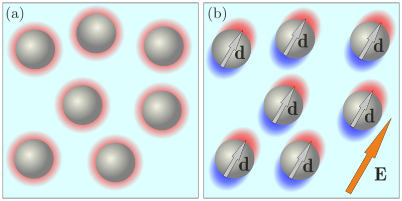
Schematic illustration of interparticle interactions induced by an external electric field. (a) In the absence of the field, each particle (typically charged negatively) is surrounded by a spherical cloud of screening ions (predominantly positive cations, shown in red). (b) External field polarizes each particle and also the surrounding ion cloud (as indicated by the red-blue shading displaced along the field direction).
Apart from their practical applications, colloidal suspensions are known to be used as model systems for particle-resolved studies of molecular systems41–46. Colloidal suspensions with tunable attractive interactions are particularly well suited for investigating generic phenomena in liquids and solids, e.g., for studying the role of attraction in melting and crystallization, binodal decay, dynamics of dislocations, solid-solid phase transitions, nucleation, etc.41,45. However, an external rotating field controlling the long-range attraction should be very homogeneous across the suspension, to ensure quasi-identical interactions in the whole system.
Here, we present a new experimental setup for tunable self-assembly of particles in horizontal two-dimensional (2D) colloidal suspensions. The interparticle attraction is driven by a horizontally rotating electric field, with the maximal magnitude in a suspension of 25 V/mm, homogeneity better than 1% over the horizontal distance of 250 μm, and the rotation frequency in the range of 40 Hz to 30 kHz. Using numerical calculations, we carefully investigate the spatial distribution of the field generated by eight planar electrodes (located at the same height). The distribution changes qualitatively with the vertical distance from the electrode plane, revealing a remarkable horizontal homogeneity at a certain height and thus providing optimal experimental conditions. To demonstrate this regime, we perform experiments on self-assembly of 2.12 μm silica particles in water. Our simulations and experiments show that the proposed setup has excellent characteristics for prospective studies–in particular, a high homogeneity and magnitude of the field, as well as tunability of the frequency and form of the rotating field. The setup has been primarily developed for controlled particle-resolved studies of large 2D colloidal systems. However, based on the achieved characteristics, we suggest that it can also be utilized for applied research in soft matter, photonics, material science, chemical physics, microfluidics, and biological systems.
Results
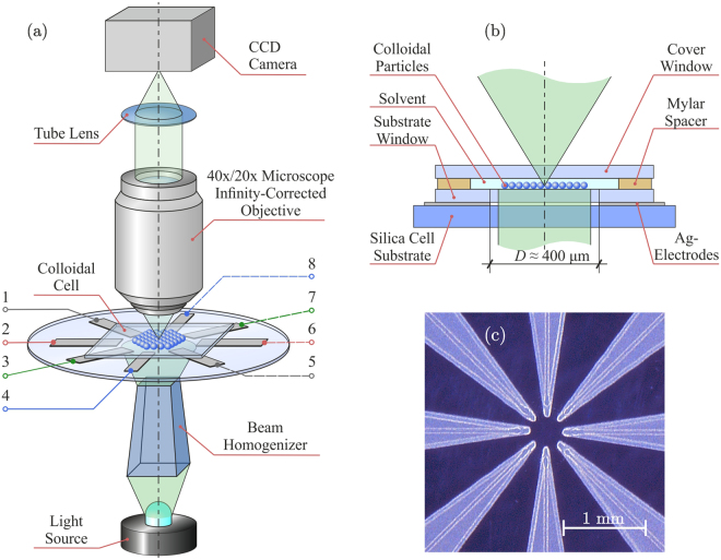
Figure 2 presents the developed experimental setup for tunable self-assembly of colloidal particles in external rotating electric fields. The setup includes an optical module and colloidal cell shown in the figure, as well as a module of electrical signal generation described in Methods. Through the optically transparent cell, the colloidal suspension is illuminated using light source and beam homogenizer. The video-imaging is provided by CCD Camera (8051C-GE, Thorlabs) using a (RMS40X-PF, Olympus) or (RMS20X-PF, Olympus) microscope infinity-corrected objective and a tube lens. A translation stage enables accurate positioning of the cell both in the vertical and lateral directions.
Figure 2.
Schematic representation of the experimental setup for tunable self-assembly of two-dimensional colloidal suspensions in rotating electric fields: Panel (a) shows an overall scheme of the setup with details presented in (b), panel (c) is a photo of eight-electrodes cell (8EC). Potentials at electrodes 1–8 in panel (a) are controlled by a module of electric signal generation (see Methods for technical details).
The colloidal cell is sketched in Fig. 2(b). The work space with a colloidal suspension is bounded by substrate and cover windows (with sizes of ), which are separated by -μm-Mylar polymer spacer placed at the periphery of the windows. A silica substrate, at which the -μm-thick silver electrodes are deposited by aerosol sputtering method, is attached to the substrate window from below. The electrodes have a form of arrows with angle of 15° and radius of curvature of 25 μm in the vertex, converging radially to the center of the cell, as shown in Fig. 2(c). The distance between the vertexes of opposite electrodes is .
The vertical distance from the electrode plane to the plane of particle self-assembly can be easily controlled by varying the thickness of the substrate window. Four pairs of independent signals with frequencies and magnitudes of and , respectively, can be applied to the electrodes using the module of electric signal generation (see Methods). With the presented experimental setup one can flexibly tune the magnitude, homogeneity, rotation frequency, and hodograph of the electric field in a suspension.
The spatial distribution of the electric field generated in the cell plays the crucial role in self-assembly of particles. The main advantage of the proposed eight-electrode cell (8EC) is the ability to produce homogeneous electric fields. To study the field distribution, we performed simulations using boundary element method (see Methods for details). Since the charge distributions at biased neighboring electrodes are mutually affected, the electrostatic boundary problem has to be solved for a given electrode configuration. As a basic example, we considered a circular distribution of potentials applied to the electrodes,
| 1 |
where is the voltage amplitude, is the electrode number (see Fig. 2(a)), and is a time-dependent phase shift. For uniformly rotating fields , where is the rotation frequency.
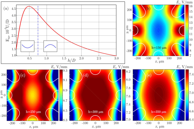
Figure 3 shows the calculated distribution of electric field in 8EC. At a certain height, presented in Fig. 3(d), we observe a remarkable homogeneity of the field over a large horizontal area. To check the predictions, we carried out a demonstration experiment with suspensions of silica particles in water (see details in Methods). A typical cluster observed in the experiment is illustrated in Fig. 4.
Figure 3.
Numerical simulation of the electric field in 8EC: Panel (a) shows the magnitude of the tangential electric field in the cell center, , versus the normalized thickness of substrate window, . The vertical dashed line at marks the critical (optimum) height where the field configuration in the center changes its curvature. Panels (b)–(e) demonstrate the horizontal distributions of for different . The white solid lines indicate contours of the electrodes from the top view. Calculations are performed for and , the voltage distribution at the electrodes is given by Eq. (1) for . The optimal field configuration is obtained for (): The field homogeneity is better than 1% over the horizontal distance of , as one can see from panel (d).
Figure 4.
A demonstration experiment with a self-assembly of colloidal particles in external rotating electric fields: Panels (a)–(d) depict about 450 silica particles of diameter, undergoing a transition from a (dilute) fluid state to a crystalline state upon a gradual increase of the field ( kV, respectively). In the shown example the field rotated with the frequency of .
Discussion
Figure 3 demonstrates results of the calculations of the horizontal electric field, representing our typical experimental conditions. We found that the vertical component of electric field is much smaller than the horizontal one, because of two reasons: (i) the electric field is horizontal in the center due to symmetry, and (ii) a large dielectric contrast between the substrate window and aqua-based solvent leads to a significant reduction of the vertical field component in the solvent. For instance, the absolute value of the vertical-to-horizontal field ratio is less than for substrate window thickness .
Figure 3(a) shows the dependence of the tangential (in-plane) magnitude of electric field in the center of self-assembly plane on thickness of the substrate window. One can see that the used planar electrodes yield a non-monotonous dependence, reaching a maximum at . Figures 3(b)–(e) present distributions of the tangential electric field in the plane of self-assembly at , corresponding, respectively, to , in Fig. 3(a). At small , the tangential field has a local minimum in the center, as shown in Fig. 3(b), while for thick substrate windows, Fig. 3(e), the field attains a maximum in the center. Thus, varying provides a simple way for optimization of experimental conditions.
The most homogeneous distribution of the electric field, presented in Fig. 3(d), is obtained at . Near this value the surface of the constant field magnitude changes its curvature, as shown schematically in Fig. 3(a). We found that in this case changes in the center by less than 1% over the horizontal distance of (for ).
Although the maximum field is reached at , the cell configuration with provides the most promising conditions for experimental studies: the largest area of a homogeneous electric field in the plane of self-assembly combined with a reasonably high magnitude of the field. Indeed, according to Fig. 3(a), the maximum field corresponds to V/mm for and kV. For the optimal configuration shown in Fig. 3(d), the magnitude of the field is only slightly smaller, , corresponding to V/mm.
During the rotation, the field varies periodically between two configurations corresponding to and in Eq. (1). Calculations performed for reveal practically the same field configuration as in Fig. 3, just rotated by .
In our demonstration experiment illustrated in Fig. 4, we observed in total about 10 very isotropic clusters, as follows from the analysis of the angular distributions of distances between the neighbor particles. Results of self-assembly at different values of are presented in Figs. 4(a)–(d) for a typical equilibrium cluster (the system was equilibrated over 3–5 min after each voltage change). Here, we provide snapshots of the center of a self-assembly plane, with the field of view of . Figures 4(a)–(d) demonstrate that, by applying in-plane rotating electric field of increasing magnitude, particles can be assembled in a complex two-dimensional liquid cluster which crystallizes when is sufficiently large. The spatial distribution of electric field in the experiment corresponds to that shown in Fig. 3(d). The crystal melts after the field is turned off, then the whole process of self-assembly can be repeated. Note that the field-controlled reversibility distinguishes this mechanism of self-assembly from other mechanisms, typically driven by chemical reactions.
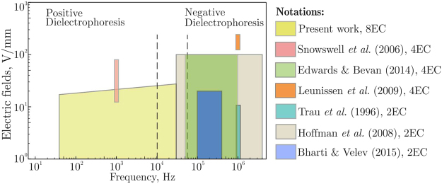
Let us compare the main characteristics of our experimental setup with those reported in other papers. In particular, we consider the magnitude of electric field, rotation frequency, and field homogeneity achieved in the plane of self-assembly. Figure 5 presents the operation regimes of the setups in terms of the field magnitude and frequency. We have chosen these characteristics since the sign of dielectrophoresis may change with the frequency17,18, while the field strength determines the magnitude of induced interparticle interactions. The maximal voltage generated by amplifiers used in our setup slightly increases with the frequency (see Methods for details), as indicated in Fig. 5. Using available data of refs.12,15,30,31,34,40,47, the electric field in solvent for other experimental setups is estimated as , where is a voltage amplitude at electrodes and is the distance between them. In Table 1 we summarize relevant parameters, such as the cell size, characteristic size of area with a homogeneous electric field, range of frequencies, and maximum magnitude of the field in solvent.
Figure 5.
Comparison of 8EC with other setups in terms of the field frequency and magnitude.
Table 1.
Main parameters of the present setup and setups used in previous studies.
| Paper | Cell size, | Frequencies, Hz | Maximal field, | ||
|---|---|---|---|---|---|
| Present work | 8 | ||||
| Snowswell et al. (2006)30,31 | 4 | ||||
| Edwards & Bevan (2014)34 | 4 | ||||
| Leunissen et al. (2009)40 | 4 | — | |||
| Trau et al. (1996)15 | 2 | — | |||
| Hoffman et al. (2008)12 | 2 | — | |||
| Bharti & Velev (2015)47 | 2 | — |
* is the number of electrodes in the setup.
** is the characteristic size of area within the cell, where the field homogeneity is better than 1%.
Frequencies in Fig. 5 are divided into the low- and high-frequency regimes, corresponding to the essentially positive and negative dielectrophoresis emerging at and 17,18, respectively. One can see that in the low-frequency regime our setup enables particle manipulation via a positive dielectrophoresis (where particles move toward the field maximum). In the frequency range between kHz and kHz the dielectrophoresis is practically negligible, providing the best conditions for self-assembly due to the field-induced interactions. For instance, the demonstration experiment in Fig. 4, corresponding to the optimal thickness , was performed at frequency .
According to our simulations, the number of electrodes and their geometry strongly affect the field configuration: For four-electrode cells, the horizontal field varies by less than 1% is in the central area of size (compared to in 8EC with otherwise the same conditions). Hence, the proposed setup provides much more homogeneous electric fields and therefore enables self-assembly over much larger area than in setups reported in refs.6,30,31,34,43. Furthermore, 8EC is more flexible technologically, since it can be restricted to operation in regimes with four and two electrodes. Therefore, in Fig. 5 and in Table 1 we also present the reference data for 2EC, although the assembled structures were essentially anisotropic in these cases. Note that Leunissen et al.40 used four-electrode capillary cell, but the particles were assembled in the vertical plane.
From Fig. 5 one can see that the magnitude of electric field in the developed setup is smaller than that in refs.30,34. This is because we used insulated electrodes, while in the other setups the electrodes were in direct contact with solvent. We point out that insulated electrodes prevent electrolysis, discharge in solvent30, and can be used with biological systems. Generation of strong fields is particularly important for self-assembly of small (nano) particles and particles with a small dielectric contrast (with respect to solvent), since the strength of electrically-induced interactions drops rapidly with decrease of these parameters2. However, our setup provides good conditions for self-assembly of even sub-micrometer particles in crystalline clusters: For instance, clusters in a suspension of - particles can be crystallized in the rotating field of 30,31, which is well below the maximum value of for our setup (in the optimal configuration shown in Fig. 3(d)). For - particles, crystallization occurs at . In addition, 8EC allows us to generate rotating fields with a complex (angularly-modulated) hodograph, to induce anisotropic interparticle interactions. This provides additional opportunities for tunable self-assembly of structures with complicated geometries.
The cluster shown in Fig. 4 contains more than 450 particles, which is almost twice as much as reported in refs.12,15,31,34,40,47. The colloidal crystals can be used, e.g., to grow photonic crystals, as structured substrates for sensors and light conversion, as matrices for optical composites, and for spectroscopy using optical field localization48–51. The same principle of field-induced self-assembly can be applied to bubbles stabilized by ions in aqua solutions of electrolytes52–55, or generated by intensive electric fields56,57. This mechanism may be utilized for novel technologies of deep degassing of aqua solutions58. However, such studies are beyond the scope of the present paper and should be considered in future works.
We hope that the presented experimental setup can be employed for fundamental and applied research in different fields, where tunable interactions and self-assembly in complex media are important. Apart from technological applications of 2D colloidal clusters, the setup is well suited for fundamental particle-resolved studies of collective phenomena in many-body systems. The ability to form large homogeneous systems of particles is crucial for experiments aiming to better understand the role of interparticle attraction in elementary processes governing nucleation, melting, crystallization, and dislocation dynamics.
Methods
Module of electric signal generation
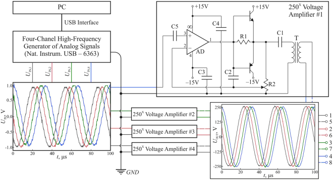
The principal scheme of the module of electric signal generation is shown in Fig. 6. The module consists of a personal computer (PC) with a specially developed software, digital-to-analog converter (DAC), and 4-channel electrical voltage amplifier. The software allows us to generate predetermined profiles of the voltage signals at the electrodes. In addition, the frequency and magnitude of DAC signals can be varied in the ranges and , respectively. Magnitude of the signal for each channel is amplified by a voltage amplifier. Each channel of amplifier consists of operational amplifier AD826 (Analog Devices, USA), current buffer based on complementary transistors BD139 (npn-type, ST Microelectronics, Switzerland), BD140 (pnp-type, ST Microelectronics, Switzerland), and setup transformer. Operational amplifier, working in non-inverting mode, and current buffer have the joint negative feedback, which essentially linearizes the system. Crossover distortion is prevented due to the feedback, which encloses transistors and operational amplifier. Resistor R1 (50 Om) is supplied for the best result of getting rid of the distortion. The amplification coefficient can be tuned by the potentiometer R2 (). The ceramic capacitor C2 () prevents a high-frequency self-excitation of the operational amplifier. The voltage amplification occurs in transformer T (Iron-core plate-type power transformer TP-114–1, produced by Scientific and industrial company “Complex”; input/output voltage 220 V/6 V, nominal load current 2.1 A), which does not pass the high-frequency harmonics, because at 30 kHz (our main operating frequency) the gain reaches its maximum and then drops at higher frequencies, as shown in Fig. 5. The capacitor C1 plays a role of a separating capacitor, which blocks an undesirable constant component of the signal and prevents an overload of the transistors. The capacitor C5 is also a separating one, in the amplifier input. The values of C1 and C5 (in our case, ) were chosen from the condition that their reactive resistance at low frequencies does not affect the propagation of alternating signal. The ceramic capacitors C3 and C4 play a role of source filters and have a capacity of . The maximal voltage amplification of 250x is reached at frequency , providing the maximum voltage difference of 2.5 kV at opposite electrodes. The input and output signals have the same form (when superimposed on the oscilloscope screen and scaled linearly to magnitude), the total harmonic distortion is less than 3%.
Figure 6.
The principal scheme of electric signal generation.
Electrostatic fields in a colloidal cell
The complex dielectric permittivity of solvent can be written as59 , where is the vacuum permittivity and is a real part of the dielectric permittivity of water. The conductivity of deionized water (used in our experiment) is . At (experimental operation frequency), . Therefore, our solvent can be considered as an insulator, and the electric fields in the cell can be calculated in the quasistatic limit.
Figure 7(a) shows a 3D model of the cell used for the calculations. Since the planar electrodes are very thin () in comparison to the substrate window thickness and diameter , one can solve the electrostatic problem assuming that the electrodes are located at .
Figure 7.
Example of electrostatic calculations: Panel (a) depicts a 3D model of 8EC, potential at electrode is given by Eq. (1). Panel (b) shows the field distribution in the vertical plane (for and ), through points A and B indicated in panel (a). Planar electrodes are marked at by the thick red solid lines, the interface between solvent and substrate window is the black solid line at . The blue arrows represent the electric field, with the blue solid lines being the force lines.
The electrostatic potential is given by the solution of the Laplace equation
| 2 |
with the boundary condition for the potential in the plane of electrodes and at large distances:
| 3 |
| 4 |
The standard boundary conditions at the interface between dielectric areas 1 and 2 are59
| 5 |
where are the dielectric permittivities of the contacting media, is the electric field, and indices and correspond to the normal and tangential field components, respectively. In our calculations, the dielectric permittivities (aqua solvent) and (glass window) were used60.
Solution of Eqs. (2)–(5) can be obtained by using boundary element method61. According to Eq. (2), potential is a harmonic function and, therefore, satisfies relation62
| 6 |
Here, the integration is performed over boundary (plane ), is a surface element, is the external normal vector to the surface, is the derivative along ; we use for the potential at boundary and for the calculation in the volume.
To find the electric field near the surface of electrodes, the plane is triangulated to small boundary elements. The potential at the boundary is related to the normal component of the electric field in the substrate window, . Assuming that the magnitude of the normal field within a given boundary element is constant, we can apply Eq. (6) to each element. This yields
| 7 |
where are indices of the boundary elements, is unit matrix, is the center of boundary element , is a surface of boundary element , is inverse matrix to , and the summation is over repeated indices. Note that the horizontal size of a system should be much larger than thickness and diameter , to exclude the horizontal boundary effects.
Using Eq. (7) at boundary , potential in the volume of the substrate window is readily obtained from Eq. (6) with . At , Eq. (6) with boundary conditions (5) yield the potential and electric field at the solvent interface. Then one can apply Eq. (6) again, to calculate in the solvent.
Figure 7(b) presents a typical distribution of the electric force lines in the area of interest. The results correspond to . One can see that the electric field is indeed planar in the central area.
Details of demonstration experiment
All glass surfaces were treated with ethanol and deionized water, and then dried at temperature of °C during 30 minutes. Afterwards, the optical interfaces were treated to form a hydrophobic coating. The hydrophobization was performed with deposition of 3% solution of polymethylsiloxane oil (PMS-200) in benzol, followed by annealing at 250 °C during 120 minutes.
The colloidal suspension, used for the demonstration experiment shown in Fig. 4, contained silica particles with diameter of (, Microparticles GmbH, Germany), dispersed in deionized water with resistance of , produced after the purification of distilled water in ion-exchanging resins. The particles were dispersed in the solvent by ultrasonic mixing.
Acknowledgements
Study is supported by Russian Science Foundation (RSF), Grant No. 17-19-01691. Electrostatic calculations were performed using software developed in the framework of RSF Grant No. 14-29-00277. Authors are grateful to Acad. V. Brazhkin and Dr. O. Tsiok for valuable discussions.
Author Contributions
K.I.Z., A.V.I., and S.O.Y. conceived the research and wrote the manuscript; E.V.Y., K.I.Z., and S.O.Y. developed the concept of the experimental setup and analysed the results; K.A.K., N.P.K., and S.O.Y. conducted electrostatic simulations; K.I.K., D.A.S., and V.L.T. developed the electrical module for setup; A.K.Z., V.N.K., and E.V.Y. made the cell and performed experiment with self-assembly. All authors reviewed the manuscript.
Competing Interests
The authors declare that they have no competing interests.
Footnotes
Publisher's note: Springer Nature remains neutral with regard to jurisdictional claims in published maps and institutional affiliations.
References
- 1.Fernandez-Nieves, A. & Puertas, A. M. Fluids, colloids, and soft materials: an introduction to soft matter physics (Wiley, 2016).
- 2.Grzelczak M, Vermant J, Furst EM, Liz-Marzán LM. Directed self-assembly of nanoparticles. ACS Nano. 2010;4:3591–3605. doi: 10.1021/nn100869j. [DOI] [PubMed] [Google Scholar]
- 3.Davis KE, Russel WB, Glantschnig WJ. Disorder-to-order transition in settling suspensions of colloidal silica: X-ray measurements. Science. 1989;245:507–510. doi: 10.1126/science.245.4917.507. [DOI] [PubMed] [Google Scholar]
- 4.Lee W, Chan A, Bevan MA, Lewis JA, Braun PV. Nanoparticle-mediated epitaxial assembly of colloidal crystals on patterned substrates. Langmuir. 2004;20:5262–5270. doi: 10.1021/la035694e. [DOI] [PubMed] [Google Scholar]
- 5.Fernandes GE, Beltran-Villegas DJ, Bevan MA. Spatially controlled reversible colloidal self-assembly. The Journal of Chemical Physics. 2009;131:134705. doi: 10.1063/1.3243686. [DOI] [PubMed] [Google Scholar]
- 6.Edwards TD, Yang Y, Everett WN, Bevan MA. Reconfigurable multi-scale colloidal assembly on excluded volume patterns. Scientific Reports. 2015;5:13612. doi: 10.1038/srep13612. [DOI] [PMC free article] [PubMed] [Google Scholar]
- 7.Terao T, Nakayama T. Crystallization in quasi-two-dimensional colloidal systems at an air-water interface. Phys. Rev. E. 1999;60:7157–7162. doi: 10.1103/PhysRevE.60.7157. [DOI] [PubMed] [Google Scholar]
- 8.Bonales LJ, et al. Freezing transition and interaction potential in monolayers of microparticles at fluid interfaces. Langmuir. 2011;27:3391–3400. doi: 10.1021/la104917e. [DOI] [PubMed] [Google Scholar]
- 9.Garbin V, Jenkins I, Sinno T, Crocker JC, Stebe KJ. Interactions and stress relaxation in monolayers of soft nanoparticles at fluid-fluid interfaces. Phys. Rev. Lett. 2015;114:108301. doi: 10.1103/PhysRevLett.114.108301. [DOI] [PubMed] [Google Scholar]
- 10.Poulichet V, Garbin V. Ultrafast desorption of colloidal particles from fluid interfaces. Proceedings of the National Academy of Sciences. 2015;112:5932–5937. doi: 10.1073/pnas.1504776112. [DOI] [PMC free article] [PubMed] [Google Scholar]
- 11.Bianchi E, Likos CN, Kahl G. Tunable assembly of heterogeneously charged colloids. Nano Letters. 2014;14:3412–3418. doi: 10.1021/nl500934v. [DOI] [PMC free article] [PubMed] [Google Scholar]
- 12.Hoffman PD, Sarangapani PS, Zhu Y. Dielectrophoresis and ac-induced assembly in binary colloidal suspensions. Langmuir. 2008;24:12164–12171. doi: 10.1021/la8013392. [DOI] [PubMed] [Google Scholar]
- 13.Prieve DC, Sides PJ, Wirth CL. 2-d assembly of colloidal particles on a planar electrode. Current Opinion in Colloid & Interface Science. 2010;15:160–174. doi: 10.1016/j.cocis.2010.01.005. [DOI] [Google Scholar]
- 14.Hayward RC, Saville DA, Aksay IA. Electrophoretic assembly of colloidal crystals with optically tunable micropatterns. Nature. 2000;404:56–59. doi: 10.1038/35003530. [DOI] [PubMed] [Google Scholar]
- 15.Trau M, Saville DA, Aksay IA. Field-induced layering of colloidal crystals. Science. 1996;272:706–709. doi: 10.1126/science.272.5262.706. [DOI] [PubMed] [Google Scholar]
- 16.Gangwal S, Cayre OJ, Velev OD. Dielectrophoretic assembly of metallodielectric janus particles in ac electric fields. Langmuir. 2008;24:13312–13320. doi: 10.1021/la8015222. [DOI] [PubMed] [Google Scholar]
- 17.Tsukahara S, Sakamoto T, Watarai H. Positive dielectrophoretic mobilities of single microparticles enhanced by the dynamic diffusion cloud of ions. Langmuir. 2000;16:3866–3872. doi: 10.1021/la980441k. [DOI] [Google Scholar]
- 18.Gascoyne PRC, Vykoukal J. Particle separation by dielectrophoresis. Electrophoresis. 2002;23:1973–1983. doi: 10.1002/1522-2683(200207)23:13<1973::AID-ELPS1973>3.0.CO;2-1. [DOI] [PMC free article] [PubMed] [Google Scholar]
- 19.Fernandes GE, Beltran-Villegas DJ, Bevan MA. Interfacial colloidal crystallization via tunable hydrogel depletants. Langmuir. 2008;24:10776–10785. doi: 10.1021/la802025d. [DOI] [PubMed] [Google Scholar]
- 20.Han X, Luo H, Xiao G, Jones PH. Optically bound colloidal lattices in evanescent optical fields. Opt. Lett. 2016;41:4935–4938. doi: 10.1364/OL.41.004935. [DOI] [PubMed] [Google Scholar]
- 21.Korda PT, Grier DG. Annealing thin colloidal crystals with optical gradient forces. The Journal of Chemical Physics. 2001;114:7570–7573. doi: 10.1063/1.1359741. [DOI] [Google Scholar]
- 22.Zahn K, Méndez-Alcaraz JM, Maret G. Hydrodynamic interactions may enhance the self-diffusion of colloidal particles. Phys. Rev. Lett. 1997;79:175–178. doi: 10.1103/PhysRevLett.79.175. [DOI] [Google Scholar]
- 23.Zahn K, Lenke R, Maret G. Two-stage melting of paramagnetic colloidal crystals in two dimensions. Phys. Rev. Lett. 1999;82:2721–2724. doi: 10.1103/PhysRevLett.82.2721. [DOI] [Google Scholar]
- 24.Osterman N, et al. Field-induced self-assembly of suspended colloidal membranes. Phys. Rev. Lett. 2009;103:228301. doi: 10.1103/PhysRevLett.103.228301. [DOI] [PubMed] [Google Scholar]
- 25.Alert R, Casademunt J, Tierno P. Landscape-inversion phase transition in dipolar colloids: Tuning the structure and dynamics of 2d crystals. Phys. Rev. Lett. 2014;113:198301. doi: 10.1103/PhysRevLett.113.198301. [DOI] [PubMed] [Google Scholar]
- 26.Du D, Li D, Thakur M, Biswal SL. Generating an in situ tunable interaction potential for probing 2-d colloidal phase behavior. Soft Matter. 2013;9:6867–6875. doi: 10.1039/c3sm27620a. [DOI] [Google Scholar]
- 27.Carstensen H, Kapaklis V, Wolff M. Phase formation in colloidal systems with tunable interaction. Phys. Rev. E. 2015;92:012303. doi: 10.1103/PhysRevE.92.012303. [DOI] [PubMed] [Google Scholar]
- 28.Helseth LE. Self-assembly of colloidal pyramids in magnetic fields. Langmuir. 2005;21:7276–7279. doi: 10.1021/la051140v. [DOI] [PubMed] [Google Scholar]
- 29.Bharti B, Kogler F, Hall CK, Klapp SHL, Velev OD. Multidirectional colloidal assembly in concurrent electric and magnetic fields. Soft Matter. 2016;12:7747–7758. doi: 10.1039/C6SM01475E. [DOI] [PubMed] [Google Scholar]
- 30.Snoswell DRE, et al. Dynamic control of lattice spacing within colloidal crystals. New Journal of Physics. 2006;8:267. doi: 10.1088/1367-2630/8/11/267. [DOI] [Google Scholar]
- 31.Elsner N, Royall CP, Vincent B, Snoswell DRE. Simple models for two-dimensional tunable colloidal crystals in rotating ac electric fields. The Journal of Chemical Physics. 2009;130:154901. doi: 10.1063/1.3115641. [DOI] [PubMed] [Google Scholar]
- 32.Lumsdon SO, Kaler EW, Velev OD. Two-dimensional crystallization of microspheres by a coplanar ac electric field. Langmuir. 2004;20:2108–2116. doi: 10.1021/la035812y. [DOI] [PubMed] [Google Scholar]
- 33.Li N, Newman HD, Valera M, Saika-Voivod I, Yethiraj A. Colloids with a tunable dipolar interaction: equations of state and order parameters via confocal microscopy. Soft Matter. 2010;6:876–880. doi: 10.1039/B909953K. [DOI] [Google Scholar]
- 34.Edwards TD, Bevan MA. Controlling colloidal particles with electric fields. Langmuir. 2014;30:10793–10803. doi: 10.1021/la500178b. [DOI] [PubMed] [Google Scholar]
- 35.Juàrez JJ, Bevan MA. Feedback controlled colloidal self-assembly. Advanced Functional Materials. 2012;22:3833–3839. doi: 10.1002/adfm.201200400. [DOI] [Google Scholar]
- 36.Juàrez JJ, Cui J-Q, Liu BG, Bevan MA. kt-scale colloidal interactions in high frequency inhomogeneous ac electric fields. i. single particles. Langmuir. 2011;27:9211–9218. doi: 10.1021/la201478y. [DOI] [PubMed] [Google Scholar]
- 37.Juàrez JJ, Liu BG, Cui J-Q, Bevan MA. kt-scale colloidal interactions in high-frequency inhomogeneous ac electric fields. ii. concentrated ensembles. Langmuir. 2011;27:9219–9226. doi: 10.1021/la2014804. [DOI] [PubMed] [Google Scholar]
- 38.Juarez JJ, Feicht SE, Bevan MA. Electric field mediated assembly of three dimensional equilibrium colloidal crystals. Soft Matter. 2012;8:94–103. doi: 10.1039/C1SM06414B. [DOI] [Google Scholar]
- 39.Gong T, Wu DT, Marr DWM. Electric field-reversible three-dimensional colloidal crystals. Langmuir. 2003;19:5967–5970. doi: 10.1021/la0340697. [DOI] [Google Scholar]
- 40.Leunissen ME, Vutukuri HR, van Blaaderen A. Directing colloidal self-assembly with biaxial electric fields. Advanced Materials. 2009;21:3116–3120. doi: 10.1002/adma.200900640. [DOI] [Google Scholar]
- 41.Ivlev, A., Löwen, H., Morfill, G. & Royall, C. P. Complex plasmas and Colloidal dispersions: particle-resolved studies of classical liquids and solids (Series in soft condensed matter) (Singapore: Word Scientific, 2012).
- 42.Janai, E. et al. Dipolar colloids in apolar media direct microscopy of two-dimensional suspensions. Scientific Reports6 (2016). [DOI] [PMC free article] [PubMed]
- 43.Edwards TD, Yang Y, Beltran-Villegas DJ, Bevan MA. Colloidal crystal grain boundary formation and motion. Scientific Reports. 2014;4:6132. doi: 10.1038/srep06132. [DOI] [PMC free article] [PubMed] [Google Scholar]
- 44.Wang Z, Wang F, Peng Y, Han Y, Axel A. Direct observation of liquid nucleus growth in homogeneous melting of colloidal crystals. Nature Communications. 2015;6:6942. doi: 10.1038/ncomms7942. [DOI] [PMC free article] [PubMed] [Google Scholar]
- 45.Li B, Zhou D, Han Y. Assembly and phase transitions of colloidal crystals. Nature Reviews Materials. 2016;1:15011. doi: 10.1038/natrevmats.2015.11. [DOI] [Google Scholar]
- 46.Peng Y, et al. Diffusive and martensitic nucleation kinetics in solid-solid transitions of colloidal crystals. Nature Communications. 2017;8:14978. doi: 10.1038/ncomms14978. [DOI] [PMC free article] [PubMed] [Google Scholar]
- 47.Bharti B, Velev OD. Multidirectional, multicomponent electric field driven assembly of complex colloidal chains. Zeitschrift für Physikalische Chemie. 2015;229:1075–1088. doi: 10.1515/zpch-2014-0543. [DOI] [Google Scholar]
- 48.Zaytsev KI, Yurchenko SO. Enhancement of second harmonic generation in NaNO2-infiltrated opal photonic crystal using structural light focusing. Applied Physics Letters. 2014;105:051902. doi: 10.1063/1.4892363. [DOI] [Google Scholar]
- 49.Zaytsev KI, Katyba GM, Yakovlev EV, Gorelik VS, Yurchenko SO. Band-gap nonlinear optical generation: The structure of internal optical field and the structural light focusing. Journal of Applied Physics. 2014;115:213505. doi: 10.1063/1.4880299. [DOI] [Google Scholar]
- 50.Yurchenko SO, et al. Enhanced third-harmonic generation in photonic crystals at band-gap pumping. Journal of Physics D: Applied Physics. 2017;50:055105. doi: 10.1088/1361-6463/aa53d4. [DOI] [Google Scholar]
- 51.Tang X, et al. Optimal feedback controlled assembly of perfect crystals. ACS Nano. 2016;10:6791–6798. doi: 10.1021/acsnano.6b02400. [DOI] [PubMed] [Google Scholar]
- 52.Bunkin NF, et al. Formation and dynamics of ion-stabilized gas nanobubble phase in the bulk of aqueous nacl solutions. The Journal of Physical Chemistry B. 2016;120:1291–1303. doi: 10.1021/acs.jpcb.5b11103. [DOI] [PubMed] [Google Scholar]
- 53.Bunkin NF, Yurchenko SO, Suyazov NV, Shkirin AV. Structure of the nanobubble clusters of dissolved air in liquid media. Journal of Biological Physics. 2012;38:121–152. doi: 10.1007/s10867-011-9242-8. [DOI] [PMC free article] [PubMed] [Google Scholar]
- 54.Alheshibri M, Qian J, Jehannin M, Craig VSJ. A history of nanobubbles. Langmuir. 2016;32:11086–11100. doi: 10.1021/acs.langmuir.6b02489. [DOI] [PubMed] [Google Scholar]
- 55.Yurchenko SO, et al. Ion-specific and thermal effects in the stabilization of the gas nanobubble phase in bulk aqueous electrolyte solutions. Langmuir. 2016;32:11245–11255. doi: 10.1021/acs.langmuir.6b01644. [DOI] [PubMed] [Google Scholar]
- 56.Postnikov AV, Uvarov IV, Prokaznikov AV, Svetovoy VB. Observation of spontaneous combustion of hydrogen and oxygen in microbubbles. Applied Physics Letters. 2016;108:121604. doi: 10.1063/1.4944780. [DOI] [Google Scholar]
- 57.Postnikov AV, Uvarov IV, Lokhanin MV, Svetovoy VB. Observation of spontaneous combustion of hydrogen and oxygen in microbubbles. Scientific Reports. 2016;6:39381. doi: 10.1038/srep39381. [DOI] [PMC free article] [PubMed] [Google Scholar]
- 58.Bunkin NF, Ninham BW, Babenko VA, Suyazov NV, Sychev AA. Role of dissolved gas in optical breakdown of water: Differences between effects due to helium and other gases. The Journal of Physical Chemistry B. 2010;114:7743–7752. doi: 10.1021/jp101657f. [DOI] [PubMed] [Google Scholar]
- 59.Landau, L. & Lifshitz, E. Electrodynamics of Continuous Media (Vol. 8). A Course of Theoretical Physics (Pergamon Press, 1960).
- 60.Kasap, S. & Capper, P. Springer Handbook of Electronic and Photonic Materials (Springer, 2007).
- 61.Brebbia, C., Telles, J. & Wrobel, L. Boundary Element Techniques (Berlin, Neidelberg, New-York, Tokyo: Springer-Verlag, 1984).
- 62.Tikhonov, A. & Samarskii, A. Equations of Mathematical Physics. Dover Books on Physics (Dover Publications, 2013).


