

Abstract
To describe the patient population, etiology, and complications associated with thigh compartment syndrome (TCS). TCS is a rare (0.3% of trauma patients) condition of elevated pressure within a constrained space that may cause necrosis of all tissues within the compartment resulting in severe local (infection, amputation) and systemic complications (renal insufficiency, even death). Retrospective cohort This study examines the course of treatment of nine consecutive patients with thigh compartment syndrome sustained during an eight-year period at our Level 1 trauma centre, admitting more than 2,000 trauma patients yearly. Patients developing TCS were young (average 34.8 years) and likely to have a vascular injury on presentation (55.5%). A tense and edematous thigh was the most consistent clinical exam finding prompting the compartment release (77.8%). Average time from admission to the operating room was 19.8 ± 6 hours and 3/9 (33%) were noted to have ischemic muscle changes upon compartment releases. Complications ranging from infection to amputation developed in 4/9 (44.4%) patients.
TCS is associated with high energy trauma and it is difficult to diagnose in non-cooperative-obtunded and polytrauma patients. Vascular injuries are a common underlying cause and require prompt recognition and team work including surgical intensive care, interventional radiology, vascular and orthopaedic surgery in order to avoid severe medical and legal consequences.
Keywords: thigh injuries, compartment syndrome, amputation, renal insufficiency
INTRODUCTION
Compartment syndróme of the thigh is a serious condition resulting from increased pressures and muscle damage within any of the three thigh fascial compartments. The most common aetiologies include blunt trauma, with or without fracture, vascular injuries with ischemia reperfusion, or frank bleeding into the myofascial spaces (1,2,3). While the mechanism of compartment syndrome has been well described in the literature, the outcomes of those affected by thigh compartment syndrome have not. A review of the English literature reveals only two series, aside from isolated case reports, which document the outcomes of this condition. Schwartz et al. reported on their results of 17 patients and Mithöfer et al. on 28 patients with thigh compartment syndrome (4,5). In some patients this syndrome leads to significant morbidity and mortality with others experiencing complete recovery. The disparity in outcomes may result from different mechanisms of injury, severity of soft tissue trauma, fracture, and/or the timing of treatment. Once thigh compartment syndrome is identified, immediate and complete compartment releases are required to prevent further ischemic insult to the tissues. This may not be the case when the diagnosis is delayed more than twelve hours, as the complication rate increases precipitously. There are many reasons the diagnosis or intervention may be delayed including prolonged extrication, transfer time to definitive treatment facility, or other emergent medical or surgical life threatening injuries. The obtunded and/or intubated patient, if the treating physician is not vigilant, is the most likely to experience a delay in diagnosis and subsequently the clinical outcome for this group is poor. Sheridan et al. showed that when the fasciotomy is performed more than twelve hours after diagnosis, complication rate increased from 4.5% to 54% with 1 in 5 patients requiring amputation (6). To further elucidate the timing of optimal intervention and better understand the impact of injury mechanism on outcomes, we present our cohort of nine patients with thigh compartment syndrome.
MATERIALS AND METHODS
We performed a retrospective review of trauma registry at Thomason Hospital, the only Level 1 Trauma Centre in El Paso, Texas. Approximately 2,000 severely injured patients are admitted to our centre yearly, and we looked specifically for those patients diagnosed with thigh compartment diagnosis during the period of September 1999 to March of 2007. The Institutional Review Board approved the study protocol. Nine patients with thigh compartment syndrome were identified. The data collected included time and mechanism of injury, time to surgical decompression, associated injuries, vital signs and GCS (Glasgow Coma Score) on presentation, compartment pressure measurements, muscle appearance at the time of surgery, subsequent surgical interventions, hospital length of stay, and ultimate outcome following definitive closure to include infections, nerve damage, chronic pain, and amputation. In the awake and alert patient, the diagnosis of compartment syndrome was made most often using clinical criteria to include pain out of proportion to injury, pain with passive stretch (can be complicated by the presence of fractures), palpation of compartment tension (anterior-quadriceps, posterior-hamstrings and medial-adductors compartments-muscles), and hypoes-thesia or changes in motor function in the distribution of the nerves traversing the compartments in question (femoral, sciatic and obturator nerves) (7). The presence or absence of distal pulses was noted but not used as a sole criterion for compartment release as several patients had vascular injuries below the level of Hunters canal. The changes in distal pulses may result from a late compartment syndrome or an acute vascular injury. For non-cooperative, obtunded and polytrauma patients in-tubated prior to examination, both compartment pressure measurements and clinical exam finding of palpably tense compartments were used in the decision making process. Absolute compartment pressures greater than thirty millimetres of mercury were considered diagnostic, especially in the critically patient where blood pressure fluctuations may alter tissue perfusion pressures acutely (8). In those patients not at risk for development of hemorrhagic shock, a delta pressure (Δp) of less than 30 mm Hg was used as an indication for fasciotomies (9). The compartments of the thigh were released through a single, long, lateral incision to access the anterior compartment directly and posterior compartment through the lateral intermuscular septum (10). After these compartments were released, a repeat evaluation of the medial (adductor) compartment was performed. If pressures remained elevated, the medial compartment was released through a separate incision. Assessment of muscle viability was made at the time of surgical decompression using the bovie for electrical stimulation in conjunction with contractility when grasping muscle tissue with forceps. If muscle twitch was not noted following stimulation with electro-cautery, then appropriate debridement was carried out until bleeding and contractile muscle was encountered. Most incisions following fasciotomy were left open and treated with either non-adhesive dressings or a vacuum assisted device. Split-thickness skin grafting or delayed primary closure was performed after subsidence of swelling, usually in timeframe of five to seven days. If necrotic muscle was encountered, a thorough debridement was carried out, a drain placed, and primary closure easily performed (without tension) to prevent contamination or infection.
RESULTS
The average patient age was 34.8 years. The primary mechanism for development of thigh compartment syndrome was motor vehicle collision in 4/9 (44.4%), blunt injuries to the thigh including those with pelvic fractures 3/9 (33%), isolated gunshot wound to the thigh 1/9 (11.1%), and intramuscular injection of drugs 1/9 (11.1%). A vascular injury which may have contributed to development of compartment syndrome was present in 5/9 (55.6%). This includes patients with injury to the femoral arteries (Figures 1-5), external iliac vessels and retro-peritoneal vessels that may have contributed to abdominal compartment syndrome and thus decreased venous return from the lower extremity. Tense compartments were noted in 7/9 patients (77.8%), diminished pulses to the lower extremities in 3/9 (33%), pain with passive stretch in 3/9 (33%), and paresthesias in 2/9 (22.2%), of patients. Compartment pressures were measured in 8/9 patients (88.9%) with the average compartment pressure measurement being 55 ± 27 mmHg. In general, the anterior compartment had the highest pressures. Average time from admission to compartment releases in the operating room was 19.8 ± 6 hours. Nonviable muscle was noted in 3/9 patients (33.3%) and follow-up information was available on all nine patients. Of these, 1 patient required an above knee amputation for complications relating to infection, closed femur fracture, open fracture of the ipsilateral tibia, and compartment syndrome of the leg. Three minor complications were also reported to include hematoma formation, superficial infection, and local tissue necrosis treated with limited debridement. Ipsilateral femur fractures were noted in 4/9 patients (44.4%), three closed and one open. Three fractures were treated with intramedullary fixation at time of thigh compartment released and one with external fixation. There were no fatalities in this series. Wound closure data was available on 8/9 thighs. Primary closure of the fasciotomy sites was performed in 2/9 (22.2%) thighs, delayed primary closure in 1/9 (11.1%), split thickness skin grafting in 3/9 (33%), full thickness skin grafting in 1/9 (11.1%), above knee amputation with split thickness skin grafting in 1/9 (11.1%).
FIGURE 1.
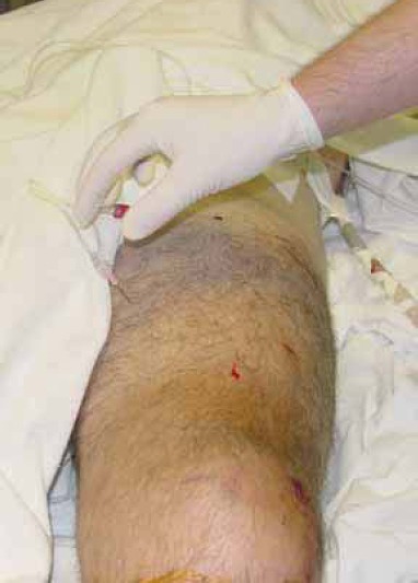
Measuring thigh compartment pressures with arterial line in polytrauma patient with femur fracture and arterial injury
FIGURE 2.

(A) Angiography showing injury to the deep femoral artery with contrast leak and after coil embolization (B)
FIGURE 3.

Negatíve wound pressure therapy as preparation for split thickness grafting.
FIGURE 4.
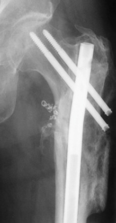
Healed fracture fixed with cephalomedullary nail.
FIGURE 5.

Good clinical result, back to bicycling.
DISCUSSION
Thigh compartment syndrome remains a rare clinical entity with only two complete series reported in the English literature comprised of 45 patients (4,5). The variability in patient outcomes following treatment of TCS is not only a function of injury mechanism but also the timely and accurate diagnosis of reversible muscular ischemia and immediate surgical intervention. In a canine model, Matava et al. showed that eight hours of increased intracompartmental pressures to within 20 mm Hg of the diastolic blood pressure (Δp) was the critical threshold for ischemic muscle necrosis (11). In an earlier study, Heppenstall et al. also showed that ischemic changes may be present in a four to six hour time frame when the Δp approached 20 mm Hg. They also suggest that periods of hypotension may result in muscle damage at even lower compartment pressures (12). These studies highlight the need for increased emphasis to be placed on compartment pressure monitoring or serial examinations in those patients who are at risk for developing TCS as the window for successful treatment may be very narrow. Whereas fracture is the leading cause of compartment syndrome in the leg, thigh compartment syndrome is more commonly associated with blunt trauma or vascular injury (4,5,13). Hope et al. reported on 151 cases of acute compartment syndromes, including both upper and lower extremities, noting that only 40% of those developing compartment syndromes of thigh could be attributed to a fracture (14). This contrasts sharply with the 77.8% of his patients whose compartment syndromes of the leg were attributed to tibia fractures. In addition, compartment syndrome of the leg was diagnosed in 59.6% of this cohort while thigh compartment syndrome was present in only 6.6%. This makes selection of those who are at risk for developing TCS more difficult as the sentinel event may be more obscure than a displaced femoral shaft fracture and the frequency at which this is seen is considerably lower than that of the leg. Case reports of thigh compartment syndrome highlight the diverse mechanisms of injury to include exercise induced, quadriceps tendon rupture (1), drug popping, crush injury, thigh contusion, aggressive resuscitation in the trauma setting, positional ischemia, aneurysm, following joint replacement, deep venous thrombosis, vascular injury, and of course fracture (2, 5,10, 27, 29, 31, 33). In our study, the vast majority of TCS resulted from blunt trauma to the pelvis or lower extremities in 6/9 (66.7%) thighs. Of these patients, 4/9 (44.4%) had fractures of the ipsilateral femur. Vascular injury was the most common mechanism in our cohort. Those were present in the ipsilateral extremity in 5/9 (55.6%) cases, making this diagnosis likely in the face of a developing TCS, though only 3/9 (33%) involved the superficial femoral, iliac, or deep femoral vessels. This rate of vascular injuries is higher than that previously published by Mithöfer et al. 4/28 patients (14%) and Schwartz et al. with 4/17 patients (23.5%) (4.5). The most consistent objective exam finding leading to diagnosis of TCS was a tense and oedematous thigh noted in 7/9 (77.8%) compartment syndromes. Pain and paresthesias to the effected extremity have also been well supported in the literature as an indication for impending compartment syndrome. This was documented in only 6/9 (66.7%) of thighs lending to the high energy mechanisms and multiple systems involved in these patients. These findings will only be useful in the awake and alert patient. Two of 9 (22.2%) patients were intubated either on scene or upon arrival to our trauma centre due decreased GCS. The difficulty in managing patients who are at risk for developing TCS is early recognition, especially in the polytrauma patient who is intubated and sedated. While TCS may be obvious on initial exam in the trauma bay, it also may develop insidiously over the next 24-48 hours as seen in two patients in our series. It is critical to identify at what time intracompartmental pressures have reached the critical, tissue “suffocating” level. Our average arrival to the operating room for compartment releases was 19.8 ± 6 hours from the time of admission to our facility (clearly documented). This wide range resulted from two patients who developed thigh compartment syndrome after 30 and 45 hours respectively. They were closely monitored during their hospital course and were both noted to have viable compartments at time of decompression, resulting in excellent outcomes. At surgical decompression 3/9 (33%) of our patients had nonviable or dusky muscle noted and one of those patients required an amputation. The other two patients with dusky musculature did not develop a wound infection. The overall complication rate was high (44.4%): 1 hematoma requiring second procedure and evacuation, 1 deep wound infection, and 1 amputation. Of patients who experienced complications, 75% sustained a vascular injury. Mithöfer et al. reported similar duration (measured from time of injury) of 11.1 ± 3 hours for all patients and 14.5 ± 5.8 hours in those patients presenting without fractures, though they reported a much lower complication rate of 18% (5). Schwartz et al. did not report the time from injury to fasciotomy but the time of diagnosis averaging 4 hours. They reported a much higher wound complication rate of 66% though there were no reports of amputations (4). Wound closure data was available in 8/9 of our patients. Definitive treatment of the fasciotomy sites often (50% of patients) required skin grafting for closure which is comparable to the Schwartz et al. cohort (41%) and significantly higher than the Mithöfer cohort (12%). Delayed or primary closure of the incision sites was possible in 3/8 (37.5%) thighs. We had no fatalities in this series, though previous series have shown mortality rates to be between 11%-47% depending on the study (4,5).
CONCLUSION
The key to successful treatment of compartment syndrome in any location depends on a prompt diagnosis of pending muscle and nerve ischemia and expedient compartment releases. While these large surgical wounds will often require secondary procedures for definitive closure, the benefits of early release surely outweigh the risks, evidenced by the poor results in those who had necrotic muscle at the time of compartment release. One out of three such patients in our study required an amputation. A careful clinical exam at the time of admission and diligence with serial examinations of the extremity at risk may identify the majority of TCS in the awake and alert patient, but other objective measures need to be employed in the obtunded or multiply injured patient. The side port needle used with the Stryker system remains a mainstay in the measurement of compartment pressures at our institution. Though it provides only a single data point regarding a condition that is both continuous and dynamic, its accuracy and simplicity often help to confirm compartment syndrome in patients with a confusing exam or in those who are unresponsive. Continuous monitoring of extremities at risk using an arterial line manometer as described by Matsen et al. should be considered when a more comprehensive set of data is required (8). Newer technologies include infrared imaging of the extremities in the trauma setting, using temperature differences between the proximal and distal skin surfaces in order to make the diagnosis (31). This technology is promising though requires additional equipment in the emergency room setting, software, and personnel for data interpretation.
Regardless of the technology employed, strong consideration for early compartment releases should be given to those patients sustaining high energy injury mechanisms to the thigh, with or without fractures. The treating physician needs to be well versed in detecting the signs and symptoms of TCS and strongly consider continuous intracompartmental monitoring in the uncooperative or sedated patients. For the cooperative patient, a clinical diagnosis is sufficient to move forward with urgent compartment releases. For patients in whom the diagnosis has been delayed for more than twelve hours (or even 6 hours in the face of high intracompartmental pressures), strong consideration should be given to avoid exposing these damaged tissues to the environment due to increased infection risk (32). In this subset of patients, aggressive management of medical issues to prevent renal damage may better serve these patients, allowing for preservation of limb, life, and late reconstructions (33).
REFERENCES
- 1.Kuri J.A, 2nd, Difelice GS. Acute compartment syndrome of the thigh following rupture of the quadriceps tendon. A case report. J. Bone Joint Surg Am. 2006;88(2):418–420. doi: 10.2106/JBJS.D.03048. [DOI] [PubMed] [Google Scholar]
- 2.Reis N.D, Better O.S. Mechanical muscle-crush injury and acute muscle-crush compartment syndrome: with special reference to earthquake casualties. J. Bone Joint Surg. Br. 2005;87(4):450–453. doi: 10.1302/0301-620X.87B4.15334. [DOI] [PubMed] [Google Scholar]
- 3.Mallik K, Diduch D.R. Acute noncontact compartment syndrome. J. Orthop. Trauma. 2000;14(7):509–510. doi: 10.1097/00005131-200009000-00009. [DOI] [PubMed] [Google Scholar]
- 4.Schwartz J.T, Jr, Brumback R.J, Lakatos R, Poka A, Bathon G.H, Burgess A.R. Acute compartment syndrome of the thigh. A spectrum of injury. J. Bone Joint Surg. Am. 1989;71(3):392–400. [PubMed] [Google Scholar]
- 5.Mithöfer K, Lhowe D.W, Vrahas M.S, Altman D.T, Altman G.T. Clinical spectrum of acute compartment syndrome of the thigh and its relation to associated injuries. Clin. Orthop. Relat Res. 2004;425:223–229. doi: 10.1097/00003086-200408000-00032. [DOI] [PubMed] [Google Scholar]
- 6.Sheridan G.W, Matsen F.A., 3rd Fasciotomy in the treatment of the acute compartment syndrome. J. Bone Joint Surg. Am. 1976;58(1):112–115. [PubMed] [Google Scholar]
- 7.Mubarak S.J, Owen C.A, Hargens A.R, Garetto L.P, Akeson WH. Acute compartmentsyndromes: diagnosis and treatment with the aid of the wick catheter. J. Bone Joint Surg. Am. 1978;60(8):1091–1095. [PubMed] [Google Scholar]
- 8.Matsen F.A, 3rd, Winquist RA, Krugmire RB., Jr Diagnosis and management of compartmental syndromes. J. Bone Joint Surg. Am. 1980;62(2):286–291. [PubMed] [Google Scholar]
- 9.McQueen M.M, Court-Brown C.M. Compartment monitoring in tibial fractures. The pressure threshold for decompression. J. Bone Joint. Surg Br. 1996;78(1):99–104. [PubMed] [Google Scholar]
- 10.Tarlow S.D, Achterman C.A, Hayhurst J, Ovadia D.N. Acute compartment syndrome in the thigh complicating fracture of the femur. A report of three cases. J. Bone Joint Surg. Am. 1986;68(9):1439–1443. [PubMed] [Google Scholar]
- 11.Matava M.J, Whitesides T.E, Jr, Seiler J.G, 3rd, Hewan-Lowe K, Hutton W.C. Determination of the compartment pressure threshold of muscle ischemia in a canine model. J. Trauma. 1994;37(1):50–58. doi: 10.1097/00005373-199407000-00010. [DOI] [PubMed] [Google Scholar]
- 12.Heppenstall R.B, Sapega A.A, Izant T, Fallon R, Shenton D, Park Y.S, Chance B. Compartment syndrome: a quantitative study of high-energy phosphorus compounds using 31P-magnetic resonance spectroscopy. J Trauma. 1989;29(8):1113–1139. [PubMed] [Google Scholar]
- 13.McQueen M.M, Gaston P, Court-Brown C.M. Acute compartment syndrome. Who is at risk? J. Bone Joint Surg Br. 2000;82(2):200–203. [PubMed] [Google Scholar]
- 14.Hope M.J, McQueen M.M. Acute compartment syndrome in the absence of fracture. J. Orthop. Trauma. 2004;18(4):220–224. doi: 10.1097/00005131-200404000-00005. [DOI] [PubMed] [Google Scholar]
- 15.Lynch J.R, Jenkins M.V, Smith D.G, Bellabarba C. Bilateral exercise-induced compartment syndrome of the thigh and leg associated with massive heterotopic ossification. A case report. J. Bone Joint Surg. Am. 2006;88(10):2265–2269. doi: 10.2106/JBJS.E.01441. [DOI] [PubMed] [Google Scholar]
- 16.Nau T, Menth-Chiari W.A, Seitz H, Vécsei V. Acute compartment syndrome of the thigh associated with exercise. Am. J. Sports Med. 2000;28(1):120–122. doi: 10.1177/03635465000280010601. [DOI] [PubMed] [Google Scholar]
- 17.Kahan J.S, McClellan R.T, Burton D.S. Acute bilateral compartment syndrome of the thigh induced by exercise. A case report. J. Bone Joint. Surg. Am. 1994;76(7):1068–1071. doi: 10.2106/00004623-199407000-00017. [DOI] [PubMed] [Google Scholar]
- 18.Robinson D, On E, Halperin N. Anterior compartment syndrome of the thigh in athletes-indications for conservative treatment. J. Trauma. 1992;32(2):183–186. doi: 10.1097/00005373-199202000-00012. [DOI] [PubMed] [Google Scholar]
- 19.Swan M.C, Lam D, Giele H.P. Intravascular ecstasy: an unusual cause of thigh compartment syndrome. J. Trauma. 2006;60(5):1129–1131. doi: 10.1097/01.ta.0000217247.90726.bd. [DOI] [PubMed] [Google Scholar]
- 20.Mithöfer K, Lhowe D.W, Altman G.T. Delayed presentation of acute compartment syndrome after contusion of the thigh. J. Orthop. Trauma. 2002;16(6):436–438. doi: 10.1097/00005131-200207000-00014. [DOI] [PubMed] [Google Scholar]
- 21.Rööser B, Bengtson S, Hägglund G. Acute compartment syndrome from anterior thigh muscle contusion: a report of eight cases. J. Orthop. Trauma. 1991;5(1):57–59. doi: 10.1097/00005131-199103000-00010. [DOI] [PubMed] [Google Scholar]
- 22.Viegas S.F, Rimoldi R, Scarborough M, Ballantyne G.M. Acute compartment syndrome in the thigh. A case report and a review of the literature. Clin. Orthop. Relat. Res. 1988;234:232–234. [PubMed] [Google Scholar]
- 23.Rööser B. Quadriceps contusion with compartment syndrome. Evacuation of hematoma in 2 cases. Acta Orthop Scand. 1987;58(2):170–172. doi: 10.3109/17453678709146462. [DOI] [PubMed] [Google Scholar]
- 24.Tremblay L.N, Feliciano D.V, Rozycki G.S. Secondary extremity compartment syndrome. J. Trauma. 2002;53(5):833–837. doi: 10.1097/00005373-200211000-00005. [DOI] [PubMed] [Google Scholar]
- 25.McLaren A.C, Ferguson J.H, Miniaci A. Crush syndrome associated with use of the fracture-table. A case report. J. Bone Joint Surg. Am. 1987;69(9):1447–1449. [PubMed] [Google Scholar]
- 26.Karkos C.D, Hughes R, Prasad V, D’Souza S.P. Thigh compartment syndrome as a result of a false aneurysm of the profunda femoris artery complicating fixation of an intertrochanteric fracture. J. Trauma. 1999;47(2):393–395. doi: 10.1097/00005373-199908000-00033. [DOI] [PubMed] [Google Scholar]
- 27.Nadeem R.D, Clift B.A, Martindale J.P, Hadden W.A, Ritchie I.K. Acute compartment syndrome of the thigh after joint replacement with anticoagulation. J. Bone Joint Surg. Br. 1998;80(5):866–868. doi: 10.1302/0301-620x.80b5.8324. [DOI] [PubMed] [Google Scholar]
- 28.Rahm M, Probe R. Extensive deep venous thrombosis resulting in compartment syndrome of the thigh and leg. A case report. J. Bone Joint Surg Am. 1994;76(12):1854–1857. doi: 10.2106/00004623-199412000-00013. [DOI] [PubMed] [Google Scholar]
- 29.Blasier R.B, Pape J.M. Simulation of compartment syndrome by rupture of the deep femoral artery from blunt trauma. Clin. Orthop. Relat. Res. 1991;266:214–217. [PubMed] [Google Scholar]
- 30.Boody A.R, Wongworawat M.D. Accuracy in the measurement of compartment pressures: a comparison of three commonly used devices. J. Bone Joint Surg. Am. 2005;87(11):2415–22. doi: 10.2106/JBJS.D.02826. [DOI] [PubMed] [Google Scholar]
- 31.Katz L.M, Nauriyal V, Nagaraj S, Finch A, Pearlstein K, Szymanowski A, Sproule C, Rich P.B, Guenther B.D, Pearlstein R.D. Infrared imaging of trauma patients for detection of acute compartment syndrome of the leg. Crit. Care Med. 2008;36(6):1756–1761. doi: 10.1097/CCM.0b013e318174d800. [DOI] [PubMed] [Google Scholar]
- 32.Finkelstein J.A, Hunter G.A, Hu R.W. Lower limb compartment syndrome: course after delayed fasciotomy. J. Trauma. 1996;40(3):342–344. doi: 10.1097/00005373-199603000-00002. [DOI] [PubMed] [Google Scholar]
- 33.Manoli A, 2nd, Smith D.G, Hansen S.T., Jr Scarred muscle excision for the treatment of established ischemic contracture of the lower extremity. Clin Orthop. Relat Res. 1993;292:309–314. [PubMed] [Google Scholar]