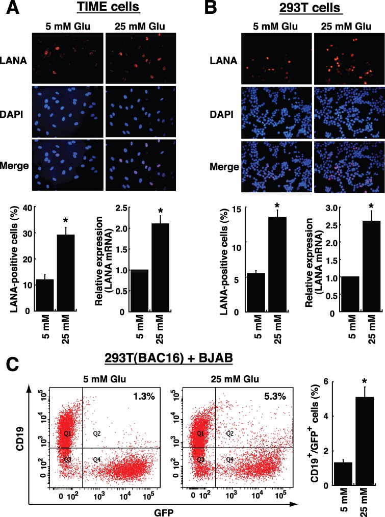
Figure 6. High glucose increases cell susceptibility to KSHV infection.
TIME cells (A) or 293T cells (B) were cultured under normal (5 mM) or high glucose (25 mM) conditions for 2 days, and then infected with KSHV for 2 hours. After removing unbound viruses, cells were cultured for another 24 hours in normal or high glucose. Representative images of LANA immunofluorescence staining are shown in the upper panel. The calculated percentages of LANA-positive cells (n= 5) and the relative mRNA levels of LANA (n= 3) in infected TIME cells and 293T cells are showed in bottom panels. (C) BJAB cells were cocultured with 293T(BAC16) cell in a 3:1 ratio, and the cocultures were exposed to normal glucose or high glucose for 4 days. The percentages of BJAB cells infected with BAC16-KSHV (CD19 and GFP double-positive cells) were examined by flow cytometry (n= 3). All data are represented as mean ± SEM. Symbol * represents significant difference vs. the normal glucose treatment (P < 0.05).

