

Abstract
Panfolliculoma is a benign neoplasm of follicular differentiation, and its morphological characteristics are similar to those of trichoblastoma, but it shows greater follicular differentiation. A 55-year-old female patient, visited for a dermatological consultation, presented comedo-like lesions on the eyelids, which were accompanied by moderate pruritus that spread gradually. On examination, the patient had localized dermatosis on the face, which symmetrically affected both upper eyelids and to a lesser extent the lower eyelids. The lesions consisted of papulonodular neoformations, comedones, and redundant skin, and a biopsy was performed. The histopathological report mainly described the presence of atrophic epidermis and dermis that was occupied entirely by a neoplasm of epithelial strain, comprising cystic structures that were surrounded by infundibular epithelium of an internal radicular sheath. Panfolliculoma is a rare benign neoplasm of follicular differentiation, and its morphological characteristics are similar to those of trichoblastoma, the pathologist must perform the clinicopathological correlation to establish the correct diagnosis.
Key words: Follicular differentiation, panfolliculoma, rare benign neoplasm
INTRODUCTION
Panfolliculoma is a benign neoplasm of follicular differentiation that was described initially by Ackerman et al. in 1993. Its morphological characteristics are similar to those of trichoblastoma, but it shows greater follicular differentiation. Clinically, it presents as a solitary tumor in the head or trunk[1] and is composed primarily of germinal follicular cells and matrix cells, differentiating toward the outer root sheath of the hair follicle at the level of the isthmus.[2] Panfolliculoma is a rare follicular neoplasia; thus, the objective of this paper is to report the first such case in Mexico.
CASE REPORT
A 55-year-old female patient, originally from Apango Guerrero, Mexico, visited for a dermatological consultation, presented comedo-like lesions on the eyelids, which were accompanied by moderate pruritus that spread gradually.
On examination, the patient had localized dermatosis on the face, which symmetrically affected both upper eyelids and to a lesser extent the lower eyelids. The lesions consisted of papulonodular neoformations, comedones, and redundant skin [Figure 1a].
Figure 1.
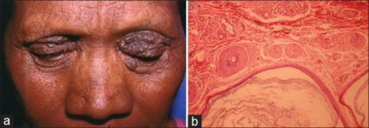
(a) Papulonodular neoformations, comedones, and redundant skin affecting both eyelids. (b) H and E, presence of cystic structures surrounded by infundibular epithelium and monomorphic cells with a basaloid aspect that resembled germinative follicles
At the consultation, she reported having observed the lesions for 5 years, and a 20-year-old female relative (daughter) had similar characteristics in the same location. Thus, a biopsy was performed, and the patient was transferred to the plastic surgery service for surgical correction.
The histopathological report described the presence of atrophic epidermis and dermis that was occupied entirely by a neoplasm of epithelial strain, comprising cystic structures that were surrounded by infundibular epithelium of an internal radicular sheath. We also identified solid constituents that were composed of small monomorphic cells with a basaloid aspect [Figure 1b] that resembled germinative follicles, surrounded by a dense stroma, prompting a diagnosis of cystic panfolliculoma. Consequently, the patient was monitored postoperatively, without no clinical lesions appeared.
DISCUSSION
Follicular neoplasms can be categorized using several methods. A common classification system is based on the part of the follicle in which the differentiation occurs; the location of the tumor in the epidermis, dermis, or both can be useful in making a differential histological diagnosis. Panfolliculoma presents as differentiation in the upper and lower portions of the hair follicle and lies primarily in the dermis.[3]
A 2016 study by Neill et al. noted that there are likely fewer than forty cases of panfolliculoma in the literature – lesions that usually present as a neoformation or a slow increase in growth, resembling other lesions, such as basal cell carcinoma, infundibular cyst, and trichoepithelioma.[4] Idriss et al. mentioned that some of the reported tumors occurred in patients aged older than 50 years, located in the extremities and trunk, with clinical diagnoses of keratosis and wart.[5]
Our patient presented with neoformations with a papular and nodular aspect and a long evolution, corresponding to cystic panfolliculoma.[4] Certain benign neoplasms of the hair follicle usually develop in the head-and-neck region in adult subjects.[6]
We do not have enough information about any gender-related predisposition to this condition. Huang and Wu presented two cases of panfolliculoma: a 41-year-old woman with neoplasia in the skin over several months of evolution and a 51-year-old male who had had an asymptomatic tumor in the left eyebrow for several years.[7] Our patient was a 55-year-old woman whose 20-year-old daughter presented with similar lesions in the same location, which we should corroborate to determine whether this case is also a neoplasia.
With regard to histopathological characteristics, Shan and Guoclassified 19 cases into three subtypes: nodular, superficial, and cystic, with a variant that presented as sebaceous differentiation. This group demonstrated the presence of differentiation in all parts of the hair follicle (infundibulum, isthmus, stem, and bulb/papilla). The lesions were well circumscribed and lacked cellular atypia; within the cellular components, they identified granules of trichohyalin, pale/clear cells, and various corneocytes.[8]
Terushkin et al. reported a case of panfolliculoma with an endophytic and solid pattern, which, at lower magnification, resembled a hair follicle that formed keratin, similar to the hair shaft.[9] We identified an epithelial neoplasm in the dermis and cystic structures that were surrounded by infundibular epithelium and monomorphic cells that resembled germinative follicles. The definitive diagnosis was panfolliculoma as per the Shan's classification and corresponding to the cystic variety.
CONCLUSION
Panfolliculoma is a rare benign neoplasm of follicular differentiation that was described initially by Ackerman et al. in 1993. Its morphological characteristics are similar to those of trichoblastoma, but it shows greater follicular differentiation; the pathologist must perform the clinicopathological correlation to establish the correct diagnosis.
Declaration of patient consent
The authors certify that they have obtained all appropriate patient consent forms. In the form the patient(s) has/have given his/her/their consent for his/her/their images and other clinical information to be reported in the journal. The patients understand that their names and initials will not be published and due efforts will be made to conceal their identity, but anonymity cannot be guaranteed.
Financial support and sponsorship
Nil.
Conflicts of interest
There are no conflicts of interest.
REFERENCES
- 1.Marini M, Starck F, Duhm G, Baldrich MA, Magariños G. Panfolliculoma a case report. Arch Argent Dermatol. 2006;56:71–3. [Google Scholar]
- 2.González-Guerra E, Requena L, Kutzner H. Immunohistochemical study of calretinin in normal hair follicles and tumors with follicular differentiation. Actas Dermosifiliogr. 2008;99:456–63. [PubMed] [Google Scholar]
- 3.Harris A, Faulkner-Jones B, Zimarowski MJ. Epidermal panfolliculoma: A report of 2 cases. Am J Dermatopathol. 2011;33:7–10. doi: 10.1097/dad.0b013e3181d94d3e. [DOI] [PubMed] [Google Scholar]
- 4.Neill B, Bingham C, Braudis K, Zurowski S. A rare cutaneous adnexal neoplasm: Cystic panfolliculoma. J Cutan Pathol. 2016;43:1183–5. doi: 10.1111/cup.12807. [DOI] [PubMed] [Google Scholar]
- 5.Idriss MH, Khalil A, Long W, Elston DM. Epidermal panfolliculoma: An adnexal proliferation with advanced follicular differentiation confined to the epidermis. J Cutan Pathol. 2013;40:409–12. doi: 10.1111/cup.12088. [DOI] [PubMed] [Google Scholar]
- 6.Tellechea O, Cardoso JC, Reis JP, Ramos L, Gameiro AR, Coutinho I, et al. Benign follicular tumors. An Bras Dermatol. 2015;90:780–96. doi: 10.1590/abd1806-4841.20154114. [DOI] [PMC free article] [PubMed] [Google Scholar]
- 7.Huang CY, Wu YH. Panfolliculoma: Report of two cases. Dermatol Sín. 2010;28:73–6. [Google Scholar]
- 8.Shan SJ, Guo Y. Panfolliculoma and histopathologic variants: A study of 19 cases. Am J Dermatopathol. 2014;36:965–71. doi: 10.1097/DAD.0000000000000108. [DOI] [PubMed] [Google Scholar]
- 9.Terushkin V, Meehan S, Shahabi L, Brinster N. Panfolliculoma with an endophytic architecture resembling a hair follicle: A report of three cases. J Cutan Pathol. 2016;43:1074–6. doi: 10.1111/cup.12799. [DOI] [PubMed] [Google Scholar]