

Abstract
It has been reported that intake of ω-3 polyunsaturated fatty acids (PUFAs) reduces the risk of coronary heart disease. It also influences bile composition, decreasing biliary cholesterol saturation in the bile of patients with gallstones. In addition to bile composition disturbances, gallbladder hypomotility must be a cofactor in the pathogenesis of cholelithiasis, as it leads to the prolonged nucleation phase. Our current knowledge about gallbladder motility has been enhanced by the study of a population of newly described interstitial (stromal) cells—telocytes (TCs). The purpose of this study was to determine whether TC loss, reported by our team recently, might be related to bile lithogenicity, expressed as cholesterol saturation index or the difference in biliary PUFA profiles in patients who suffer from cholecystolithiasis and those not affected by this disease. We determined biliary lipid composition including the fatty acid composition of the phospholipid species in bile. Thus, we investigated whether differences in biliary fatty acid profiles (ω-3 PUFA and ω-6 PUFA) in gallbladder bile may influence its lithogenicity and the quantity of TCs within the gallbladder wall. We conclude that the altered PUFA concentrations in the gallbladder bile, with elevation of ω-6 PUFA, constitute important factors influencing TC density in the gallbladder wall, being one of the possible pathophysiological components for the gallstone disease development. This study established that altered bile composition in patients with cholelithiasis may influence TC quantity within the gallbladder muscle, and we concluded that reduction in TC number may be a consequence of the supersaturated bile toxicity, while some other bile components (ω-3 PUFA, glycocholic, and taurocholic acids) may exert protective effects on TC and thus possibly influence the mechanisms regulating gallbladder and extrahepatic bile duct motility. Thus, ω-3 PUFA may represent a possible option to prevent formation of cholesterol gallstones.
Keywords: Telocytes (TCs), Interstitial Cajal-like cells, Gallstones, Cholesterol saturation index (CSI), Bile lithogenicity, Polyunsaturated fatty acids (PUFAs), Biliary fatty acids
Introduction
Gallstone disease constitutes a significant health problem in developed societies, affecting 10% to 15% of the adult population1. The prevalence of cholesterol gallstones has increased in recent years, especially in the Western world2. Cholelithiasis (gallstone formation) results from a combination of several factors, including supersaturation of bile with cholesterol, accelerated nucleation of cholesterol monohydrate in bile, biliary stasis, and delayed gallbladder emptying due to impaired gallbladder motility. Cholesterol supersaturation can result from an excessive concentration of cholesterol in bile, a deficiency of substances that keep cholesterol in solution (i.e., bile salts and phospholipids), or a combination of these factors3. Western diet, characterized by a high total calorie content, high level of cholesterol, saturated fatty acids, refined carbohydrates, proteins, and low fiber, has been proven to be lithogenic4,5. It has been reported that intake of ω-3 polyunsaturated fatty acids (PUFAs) reduces the risk of coronary heart disease and decreases biliary cholesterol saturation in the bile of gallstone patients6. ω-3 PUFA prevents gallstone formation in mice being on a lithogenic diet by increasing the levels of bile phospholipids and suppressing bile mucin formation7. PUFAs are a specific family of fatty acids. The most bioactive ω-3 PUFAs are C20:5ω3 eicosapentaenoic acid (EPA) and C22:6ω3 docosahexaenoic acid (DHA), which are found predominantly in oily fish such as salmon, mackerel, and sardines8,9. ω-6 PUFA promotes the production of inflammatory cyto kines that, in turn, can stimulate multiple pathways such as cell proliferation, apoptosis, and angiogenesis, which favor tumor growth. The ω-6 PUFA, arachidonic acid, is found in the diet or is formed from the conversion of the ω-6 PUFA linoleic acid. Linoleic and α-linolenic acids are the fatty acids designated as “essential” since they are not synthesized by mammalian cells and must be provided in the diet. Increased dietary ω-6 polyunsaturated triglycerides accelerate cholesterol gallstone formation in the prairie dog10. In addition to bile composition disturbances, gallbladder hypomotility must be a cofactor in the pathogenesis of cholelithiasis, as it leads to the prolonged nucleation phase (i.e., time for cholesterol micro-crystals to precipitate from lithogenic bile)11,13. Recently, our knowledge about gallbladder motility has been enhanced by the study of a population of newly described cells, the so-called telocytes (TCs). TCs are a novel interstitial (stromal) cell type described in many tissues and organs (www.telocytes.com). TCs are characterized by a small cell body (9–15 μm) and a variable number (one to five) of extremely long and thin telopodes (Tps), with alternating regions of podomers (∼80 nm) and podoms (250–300 nm)14. Tps are interconnected by homo- and heterocellular junctions and form three-dimensional networks. TCs are suggested to be involved in signaling processes15,16. TCs were already discovered in the wall of the human gallbladder3,17. Previously, we reported a significant decrease in c-Kit+ TC density in the gallbladder wall in patients suffering from cholelithiasis18. The purpose of this study was to determine whether TC loss was related to bile lithogenicity, which was expressed as a lithogenic index [cholesterol saturation index (CSI)] or to the difference in biliary PUFA profiles in patients who suffer from cholecystolithiasis and those who are not affected by this disease. We determined biliary lipid composition including the fatty acid composition of the phospholipid species in bile. Thus, we investigated whether differences in biliary fatty acid profiles (ω-3 PUFA and ω-6 PUFA) in gallbladder bile may influence its lithogenicity and the quantity of TCs within the gallbladder wall.
Materials and Methods
Subjects
Twenty-five consecutive patients with symptomatic gallstone disease were scheduled for elective surgery (laparoscopic cholecystectomy) and selected for the study group (5 males, mean age: 54.2 ± 13.7 years; 20 females, mean age: 55.9 ± 16.9 years). Gallstones were visualized in the gallbladder on ultrasound examination before the operation. Patients presented with mild, recurrent episodes of biliary colic. None of these patients had associated choledocholithiasis or acute cholecystitis. The control group consisted of 15 consecutive patients (8 males, mean age: 62.0 ± 7.6 years; 7 females, mean age: 61.0 ± 9.8 years) who were electively treated for pancreatic head tumors and had no pre- or intraoperative signs of cholelithiasis and jaundice. Pancreaticoduodenectomy was performed according to the standard Whipple procedure or the pylorus-sparing Traverso–Longmire technique in patients with resectable lesions. For patients with nonresectable lesions, bypass (gastroenterostomy) was carried out for palliative purposes. Gallbladders that were not affected by primary tumors and did not contain any concrements were removed. Serum bilirubin levels were measured preoperatively and were normal in both groups. All patients were surgically treated in the First Department of General, Oncological and Gastrointestinal Surgery at the Jagiellonian University Medical College from 2010 to 2011.
Ethical Approval
The study was conducted in accordance with the moral, ethical, regulatory, and scientific principles governing clinical research. Every patient gave informed consent to the cholecystectomy procedure and to the clinical study. All surgical samples were retrieved with the approval of the Jagiellonian University Bioethics Committee using procedures that conformed to the Declaration of Helsinki guidelines (Protocol No. KBET/30/B/2010).
Tissue Processing
Tissue samples from fresh cholecystectomy specimens were collected and rinsed thoroughly with phosphate-buffered saline (PBS; 0.01 M, pH 7.4; Sigma-Aldrich, St. Louis, MO, USA), fixed in 4% phosphate-buffered paraformaldehyde (PFA; Sigma-Aldrich), routinely processed, and embedded in paraffin (Sigma-Aldrich). Serial sections were cut and mounted on poly-L-lysine-coated glass slides (Sigma-Aldrich) and stained with hematoxylin and eosin (H&E; Sigma-Aldrich) for routine histopathology.
Immunohistochemistry
Indirect double immunofluorescence after heat-induced epitope retrieval was used to allow the simultaneous visualization of two antigens. A preincubation step was performed with 5% normal goat serum (Sigma-Aldrich) and 0.5% Triton X-100 (Sigma-Aldrich) for 20 min to reduce nonspecific binding and to increase penetration of the antibodies. For the simultaneous visualization of two antigens, an indirect double immunofluorescence procedure was used. The sections were incubated for 17 h at room temperature in humidified chambers with a mixture of either a rabbit polyclonal anti-c-Kit antibody (anti-CD117; A4502; diluted 1:150; Dako, Glostrup, Denmark) and a mouse monoclonal anti-mast cell tryptase antibody (M7052; 1:800; Dako) or the anti-CD117 antibody and a mouse monoclonal anti-CD34 antibody (NCL-ENDO; 1:50; Novocastra, Newcastle, UK). The sections were rinsed in PBS and incubated for 1 h at room temperature (RT) with a mixture of a Cy3-conjugated goat anti-rabbit antibody (111-165-144; 1:600; Jackson ImmunoResearch, West Grove, PA, USA) and a biotinylated goat anti-mouse antibody (115-065-146; 1:600; Jackson ImmunoResearch). The primary and secondary antibodies were diluted in the same solution used in the preincubation step. After washing in PBS, the slides were incubated with (4,6-dichlorotriazinyl)aminofluorescein (DTAF)-conjugated streptavidin (016-010-084; 1:500 in PBS; Jackson ImmunoResearch) for 1 h. After a final rinse in PBS, the nuclei were counterstained with 4′,6-diamidino-2-phenylindole (DAPI) (D9542; 1:30,000; Sigma-Aldrich) for 30 s. The sections were mounted in Vectashield medium (H-1000; Vector Laboratories, Burlingame, CA, USA) to minimize fluorophore photobleaching.
Microscopic Examination and Quantification of TCs
Slides were examined using an Olympus BX50 epifluorescence microscope (Olympus, Tokyo, Japan) equipped with an Olympus DP71 digital CCD camera. The use of mast cell tryptase staining enabled c-Kit+ mast cells to be distinguished from TCs. The distribution of TCs in the gallbladder corpus was quantitatively assessed. TCs were considered to be c-Kit+ and tryptase negative concurrently. These cells were counted in 10 consecutive highpower fields (400×). The data are expressed as the mean number of cells per 1 field of view (FOV) of gallbladder muscularis propria. The thickness of the muscularis propria in the same region of the gallbladder was also measured using an image analysis software (Multiscan v.18; Computer Scanning Systems II, Warsaw, Poland).
Biliary Lipid Composition
During surgery, bile samples were aspirated under sterile conditions by puncture of the gallbladder after ligation of the cystic duct. Cholesterol, phospholipid, and bile acid concentrations as well as percentage of individual fatty acids (FAs) of phospholipid (PL) fraction in bile samples were determined.
Bile samples were extracted on SEP-PACK-NH2 columns (500 mg; Waters, Milford, MA, USA). Each column was first activated using 6 ml of n-hexane (Sigma-Aldrich), then 0.1 ml of centrifuged bile sample was applied to the column, and the flow-through was discarded. The samples were eluted in three 1-ml volumes of a chloroform–isopropanol (Sigma-Aldrich) mixture (3:1, v/v), followed by three 1-ml volumes of methanol (Sigma-Aldrich). The eluted fractions from each column were collected and dried at 50°C under nitrogen. Dry residues were reconstituted in 0.5 ml of isopropanol and mixed vigorously. Cholesterol (Randox Laboratories Ltd., Crumlin, UK) and phospholipid (Wako Chemicals, Neuss, Germany) concentrations were measured by enzymatic methods. The intra- and interassay coefficients of variations were 3% and 4.8% for cholesterol and 5% and 6% for phospholipids, respectively. All determinations were performed with a Cobas-Bio analyzer (Roche, Atlanta, GA, USA). Individual bile acids were measured by reverse-phase high-performance liquid chromatography (HPLC) with an isocratic solvent system (Waters). Prior to chromatographic separation, bile acids were extracted from bile samples using SEP-PACK C18 columns (Waters). All columns were activated using 5 ml of methanol and 5 ml of water, after which 0.1 ml of bile mixed with phosphate buffer (0.07 mmol/L, pH 7.0; Sigma-Aldrich) and 0.1 ml of internal standard (2′,3′,4′-trihydroxyacetophenone; Sigma-Aldrich) were applied. Then the columns were washed with 10 ml of water, 3 ml of 10% acetonitrile (Sigma-Aldrich), and another 10 ml of water. Bile acids were eluted in 3 ml of methanol. The eluates were dried at 37°C under nitrogen, and dry residues were redissolved in 1 ml of an acetonitrile– water mixture (1:1, v/v). Each sample was filtered using a Millex GN filter (13 mm; Sigma-Aldrich) and separated chromatographically using an XTERRA RPC-18 column (18.5 μm × 3.9 mm × 150 mm; Waters) with detection at 200 nm. The mobile phase (flow rate: 2 ml/min) contained 10% acetonitrile in a mixture of methanol and 0.1 M monobasic potassium phosphate (60:40, v/v, pH 4.50; Sigma-Aldrich). Before use, the solvent was filtered through a 0.45-μm filter (type HV; Millipore, Bedford, MA, USA). An elution profile of conjugated bile acid standards (Sigma-Aldrich) was obtained by injecting 20 μl of a standard bile acid mixture in methanol that contained glycocholic acid (GCA; 1.640 mmol/L; Sigma-Aldrich), taurocholic acid (TCA; 1.488 mmol/L; Sigma-Aldrich), glycochenodeoxycholic acid (1.696 mmol/L; Sigma-Aldrich), glycodeoxycholic acid (1.696 mmol/L; Sigma-Aldrich), taurochenodeoxycholic acid (1.532 mmol/L; Sigma-Aldrich), and taurodeoxycholic acid (1.532 mmol/L; Sigma-Aldrich).
CSI was calculated by dividing the cholesterol concentration by the maximum cholesterol solubility according to Carey and Small19 and corrected for the total lipid content of each individual bile20. Bile samples with a CSI equal to 1 or more were considered supersaturated.
Total FA, total saturated fatty acids (SFAs), total monounsaturated fatty acids (MUFAs), n-6 PUFA, and n-3 PUFA concentrations, as well as the ratio of n-6 PUFA to n-3 PUFA in bile, were calculated. The analytical procedure for FA of phospholipid fraction in bile consisted of a few separate steps: extraction of bile total lipids by the method of Folch et al.21; separation of lipid fraction on SEP-PACK-NH2 columns22; methylation23,24; and separation of the FA from phospholipid fraction by gas chromatography (GC) (6890N Network GC Systems; Agilent Technologies, Santa Clara, CA, USA) equipped with HP-88 capillary column (100 m, 0.250 mm, 0.20 μm). 1,2-Dipentadecanoil-sn-glicero-3-phosphocholine (Sigma-Aldrich, Munich, Germany) was used as an internal standard. Levels of n-7 (palmitoleic, C16:1), n-9 (oleic, C18:1), n-3 (alfa-linolenic, C18:3; eicosapentanoic, C20:5; and docosahexaenoic, C22:6), n-6 (linoleic, C18:2; eicosadienoic C20:2; and arachidonic, C20:4), and saturated (lauric, C12; myristic, C14; palmitic, C16; and stearic, C18) acids of the phospholipid fraction were measured. The results of individual FAs were expressed as the percentage of total FA.
Data Analysis and Statistical Evaluation
The data were expressed as the mean ± standard deviation (SD). The results were analyzed using a one-way analysis of variance (ANOVA), followed by a post hoc least significant difference (LSD) test. Pearson's correlation test was used to examine the relationship between continuous variables. Values of p < 0.05 were considered to indicate statistical significance. All statistical analyses were performed using STATISTICA 9.0 software (StatSoft, Tulsa, OK, USA).
Results
Histopathological Findings
The histopathological evaluations showed chronic cholecystitis of varying intensities in both groups of patients. The inflammation was assessed and designated as mild, moderate, or severe (Fig. 1B). In the study group, severe inflammation was predominant. In contrast, only mild to intermediate inflammation was observed in the control group (Fig. 1A). In the study group, the thickness of the gallbladder muscularis propria was significantly increased compared with that of the control group (4.35 ± 0.6 mm vs. 3.2 ± 0.3 mm; p < 0.01). In immu nostained slides, the c-Kit+/mast cell tryptase− cells were considered to be TCs. We found them predominantly located in the corpus, but these cells were also observed in the gallbladder fundus and neck. TCs had a centrally located nucleus and were mostly fusiform in shape with small branches, which were rarely visible in some sections; however, sparse, round tryptase/c-Kit+ cells were also present. Numerous TCs were detected, mostly in the muscularis propria, and some TCs were observed in the connective tissue separating the smooth muscle bundles (Fig. 2A).
Figure 1.

Cross section of the gallbladder wall from the control group (A) and the cholelithiatic group (B) showing signs of severe chronic inflammation. Note: Intense infiltration with inflammatory cells and thickened muscular layer. Hematoxylin and eosin (H&E) staining. l.p., lamina propria; m.p., muscularis propria.
Figure 2.

Cross section of the gallbladder wall from the control group (A) and the cholelithiatic group (B) presenting CD117+ and tryptase− telocytes (TCs) (arrows) and mast cells (CD117+ and tryptase+; arrowheads). Immunofluorescence, CD117 [cyanine 3 (Cy3)], tryptase [fluorescein isothiocyanate (FITC)], nuclei counterstained with 4′,6-diamidino-2-phenylindole (DAPI).
The number of TCs in the wall of the gallbladder corpus was significantly lower in the study group than in the control group (2.77 ± 1.21 vs. 6.72 ± 1.82 cell/FOV in the muscularis propria; p < 0.001) (Fig. 2B). CD34+ cells were visualized in the gallbladder; however, most of them appeared to be mainly of vascular origin. However, rare CD34+ cells were encountered in the muscularis propria. On the basis of the CD34 and CD117 staining, these sparse CD34+ cells were c-Kit− concurrently, and no prolongations were observed. Unfortunately, reliable quantitative analysis was not possible due to the limited number of cells.
Bile Composition Analysis
No significant difference for concentrations of cholesterol, bile salts, and phospholipids was obtained between bile samples with and without cholesterol crystals. How ever, the CSI was significantly higher (1.45 ± 0.21 vs. 0.81 ± 0.12; p < 0.01) in gallbladder bile with crystals than in bile without crystals. We observed significantly lower mean concentrations of GCA (35.66 ± 16.66 vs. 43.44 ± 32.40; p < 0.02) and TCA (9.00 ± 7.66 vs. 15.18 ± 11.09; p < 0.05) in the bile from cholelithiatic gallbladder, compared to the control group, respectively.
The study revealed significant disturbances of the FA content in the phospholipid fraction of gallbladder bile. The specific FAs assessed in this work are listed in the Materials and Methods section. SFA concentrations in the phospholipid fraction of the bile in gallstone patients were slightly decreased compared with control (47.7 ± 3.0% vs. 50.7 ± 7.5%; p = 0.07, respectively). The MUFA concentrations in the phospholipid fraction of the bile in gallstone patients remained unchanged between both groups (13.7 ± 2.5% for cholelithiasis group vs. 15.2 ± 54.1% for controls; p = 0.17). Surprisingly, however, the PUFA concentrations in the phospholipid fraction of the bile in gallstone patients were significantly higher in patients suffering from cholelithiasis compared with the control subjects (38.6±4.7% vs. 34.0±6.8%; p = 0.0046) (Fig. 3A and B). The ω-3 PUFA levels did not differ between both groups (3.68±1.1% for gallstone patients, 3.79±1.6% for controls; p = 0.81), whereas the ω-6 PUFA concentrations were significantly elevated in gallstone patients (35.8 ± 4.3% for gallstone patients, 30.2±2.6% for controls; p = 0.0015) (Fig. 4A). Such increase originated mainly from the significant elevation of the C18:2 (n-6) (linoleic acid) serum concentrations (28.8±4.8% vs. 22.8±4.9%, respectively, for cho lelithiasis and controls; p = 0.0005). In addition, the ω-6/Ω-3 PUFA ratio was significantly higher in gallstone patients compared to controls (11.0±3.1 vs. 8.41±2.3; p = 0.0121). The study also revealed the moderate correlation of TC counts in the gallbladder muscularis propria with the total PUFA concentrations (r = −0.543; p < 0.05), ω-6 PUFA levels (r = −0.415; p < 0.05), and ω-6/ω-3 PUFA ratio (r = −0.559; p < 0.05). The detailed data from the FA concentration assessment in the phospholipid fraction of the bile in gallstone patients are presented in Table 1.
Figure 3.
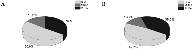
Concentrations of saturated fatty acids (SFAs), monounsaturated fatty acids (MUFAs), and polyunsaturated fatty acids (PUFAs) in gallbladder bile from control subjects (A) (n = 15) and from patients with cholelithiasis (B) (n = 25). The results of individual fatty acids (FAs) expressed as the percentage of total FAs. The data are expressed as the mean ± standard deviation (SD).
Figure 4.

The ω-6 polyunsaturated fatty acid (PUFA) concentrations (A) and the number of telocytes (TCs) (B) in control subjects (n = 15) and in patients with cholelithiasis (study group, n = 25). The results of ω-6 PUFA expressed as the percentage of total fatty acids (FAs). The data are expressed as the mean ± standard deviation (SD).
Table 1.
The Polyunsaturated Fatty Acid (PUFA), Monounsaturated Fatty Acid (MUFA), and Saturated Fatty Acid (SFA) Concentrations in the Phospholipid Fraction of the Bile in Cholelithiasis Patients (n = 25) and Control Subjects (n = 15)
| The Polyunsaturated Fatty Acids (%) | ||||||||
| Group: | C18:2 (n-6) | C20+CC18:3 (n-6) | C18:3 (n-3) | C20:2 (n-6) | C20:4 (n-6) | C20:5 (n-3) | C22:6 (n-3) | Total PUFA |
| Controls | 22.88 ± 4.93 | 0.33 ± 0.20 | 0.38 ± 0.19 | 0.12 ± 0.03 | 6.92 ± 3.26 | 1.24 ± 0.95 | 2.16 ± 0.75 | 34.04 ± 6.83 |
| Cholelithiasis | 28.85 ± 4.84 | 0.34 ± 0.21 | 0.59 ± 0.20 | 0.18 ± 0.09 | 6.46 ± 1.25 | 1.15 ± 0.58 | 1.94 ± 0.67 | 38.53 ± 4.70 |
| Monounsaturated Fatty Acids (%) | ||||||||
| Group: | C16:1 | C18:1 | Total MUFA | |||||
| Controls | 2.36 ± 1.09 | 12.84 ± 3.17 | 15.21 ± 4.11 | |||||
| Cholelithiasis | 2.09 ± 0.96 | 11.66 ± 2.21 | 13.75 ± 2.54 | |||||
| Saturated Fatty Acids (%) | ||||||||
| Group: | C12 | C14 | C16 | C18 | Total SFA | |||
| Controls | 0.03 ± 0.02 | 0.43 ± 0.18 | 44.22 ± 6.79 | 6.04 ± 1.383 | 50.74 ± 7.46 | |||
| Cholelithiasis | 0.03 ± 0.02 | 0.50 ± 0.18 | 40.74 ± 1.46 | 6.43 ± 2.17 | 47.7 ± 3.00 | |||
Results of individual fatty acids (FAs) are expressed as the percentage (%) of total FAs in the phospholipid fraction of the bile. Data are expressed as the mean ± standard deviation (SD).
Discussion
In observational studies, higher intake of saturated fat or trans FAs was associated with an increased incidence of gallstones26,28. In contrast, higher intake of polyunsaturated or monounsaturated FAs was associated with decreased risk29. The apparent protective effect of PUFAs is consistent with experimental observations, in which hamsters fed an essential FA-deficient diet had a high incidence of cholesterol gallstones and lithogenic bile (diets low in essential FAs are, in general, also low in PUFAs)30,31. Additionally, in patients with gallstones, supplementation with 11.3 g per day of fish oil (which is high in PUFAs) decreased the cholesterol saturation of bile by 25%32. While both ω-3 and ω-6 PUFAs may be protective or noxious, further research is needed to determine the optimal amounts and ratios of these FAs. Hence this study was undertaken to evaluate the biliary lipid composition and its relation to TC density in gallbladder wall (Fig. 4B). Our results revealed that the biliary ω-6 PUFA concentrations are strongly related to the number of TCs within the human gallbladder. Significant loss of TCs in the gallbladder wall in patients with gallstones was associated with an increased CSI of bile and high biliary ω-6 PUFA concentrations. We assume that, in humans, ω-6 PUFAs could increase CSI and prolong nucleation time contributing to TC loss and gallstone formation. The recent dietary shift toward the consumption of n-6 at the expense of n-3 PUFAs is thought to be a primary cause of many diseases related to the Western diet33. The body converts linoleic acid to arachidonic acid and derives EPA from α-linolenic acid. Ideally, the effects of these FAs and their eicosanoid derivatives are tailored to the specific biological needs of the body. The balance between ω-3 and ω-6 PUFAs is essential for metabolism and maintenance of the functions of both classes. Derangement of biliary ω-6/ω-3 PUFA ratio may impact the histological pattern of cholelithiatic gallbladder through modulation of the amount of biliary lipids. Moreover, derangement of the ω-6/ω-3 PUFA ratio could influence the synthesis of various eicosanoids. In the presence of large amounts of dietary ω-6 PUFAs, the eicosanoids derived from arachidonic acid are synthesized in larger quantities than those from EPA. The arachidonic acid-derived eicosanoids are biologically active even in small quantities. In high concentrations, they contribute to the formation of thrombi and the development of inflammatory disorders34. The influence of PUFAs and their eicosanoid products on organ microcirculation and ischemia/reperfusion injury has been demonstrated in many studies, and it could also be related to gallbladder microcirculation and TC injury.
Another important mechanism underlying TC loss concerns the chronic inflammatory processes involving the gallbladder wall. Portincasa et al. described impaired gallbladder motility caused by mild inflammation35. Indeed, we observed inflammatory infiltration, predominantly localized in the lamina propria, in the gallbladders of patients with gallstones, which was associated with a significant increase in the mast cell count. However, as we reported previously, TC loss does not correlate with inflammatory grade or mast cell count36.
We realize that additional stainings need to be considered in better understanding the role of TCs in the human gallbladder. For example, we observed CD34+ cells in the gallbladder wall, including muscularis propria; however, they appeared to be predominantly of vascular origin. These CD34+ cells were c-Kit−, and no prolongations were observed. Unfortunately, we were unable to present reliable quantitative analysis due to the limited number of cells. Coexpression of newly described markers, like CD34 and vimentin or platelet-derived growth factor receptor a (PDGFRa), is of great interest recently37. A growing number of publications on TCs along with discovery of new markers for their identification attracted an increasing interest among researchers regarding TC function and their potential applications in the treatment of various diseases including cholelithiasis. This knowledge will certainly help to explain the pathogenesis of this disease.
ω-3 PUFAs can inhibit cholesterol nucleation and cholesterol monohydrate crystal precipitation without affecting mixed micelles; they do this by stabilizing the phospholipid vesicle, which is another solubilizer of cholesterol. ω-3 PUFAs inhibit cholesterol monohydrate crystal precipitation and gallstone formation in prairie dogs by increasing the stability of biliary phospholipid vesicles through decreasing ionized calcium and protein in the bile of the gallbladder without changing either the concentration of biliary lipids or the CSI25. ω-3 PUFAs might alter vesicular stability and cholesterol-carrying capacity by modifying the composition of phospholipids in bile, thereby suppressing cholesterol precipitation. Derangement of ω-6/ω-3 PUFA ratio could negatively influence this protective effect.
Our results showed that there were no significant differences between two groups of patients in the mean concentrations of total bile acids, phospholipids, or cholesterol in vesicular bile, except for the lower mean concentrations of GCAs and TCAs in the cholelithiatic group. The lower amounts of these compounds were associated with the increased lithogenicity index (CSI). Moreover, in patients with gallstones, a significant positive correlation between the mean number of TCs and the concentrations of GCAs and TCAs was found. We conclude that GCAs and TCAs are somehow protective to TCs. However, it is not clear whether this is an artifact of the statistical analysis or whether TCs are indeed preserved by these acids, and the exact mechanism of this possible protective effect should be examined further, including in an experimental model. We acknowledge that possible mechanisms underlying the destructive influence of bile on TCs remain speculative rather than empiric, as reports on the role of TCs in gallstone pathophysiology are sparse. A study by Xu and Shaffer38 reported that gallbladder hypomotility was impaired by the increased bile cholesterol level. A study by Lavoie et al.39 showed that excess cholesterol in the smooth muscle of the gallbladder attenuates the ability of the muscle to contract as a result of changes in signal transduction and ion channel activity, decoupled membrane receptor–ligand interactions, and disturbances in contractile protein activity. Moreover, in a subsequent study on guinea pigs fed a lithogenic diet, Lavoie et al.40 reported cholesterol accumulation in gallbladder smooth muscles in the plasma membrane, especially membrane caveolae, leading to a decrease in membrane fluidity and a subsequent change in rhythmic electric activity.
We conclude that the disrupted PUFA concentration in the gallbladder bile, with elevation of ω-6 PUFA, is an important factor influencing TC density in the gallbladder wall and is one of the possible pathophysiological components for gallstone disease development. This study established that altered bile composition in patients with cholelithiasis may influence TC quantity within the gallbladder muscle. We conclude that a reduction in TC number may be a consequence of the toxicity of the supersaturated bile, while some other bile components (ω-3 PUFAs, GCAs, and TCAs) may exert protective effects on TCs and thus possibly influence the mechanisms regulating gallbladder and extrahepatic bile duct motility. We are aware that the present study only indicates a potential relationship between altered bile acid composition and reduced TC count in cholelithiasis using Pearson's correlation analysis. The direct evidence needs to be further obtained with gain of loss-of-function assays. However, ω-3 PUFAs may represent a new option for cholesterol gallstone disease management.
Acknowledgment
This study was financed by the Jagiellonian University grant K/ZDS/005450.3 The authors declare no conflicts of interest.
References
- 1.Stinton L.M., Shaffer E.A.. Epidemiology of gallbladder disease: Cholelithiasis and cancer. Gut Liver 2012; 6(2): 172–87. [DOI] [PMC free article] [PubMed] [Google Scholar]
- 2.Van Erpecum K.J.. Pathogenesis of cholesterol and pigment gallstones: An update. Clin Res Hepatol Gastroenterol. 2011; 35: 281–7. [DOI] [PubMed] [Google Scholar]
- 3.Matyja A., Gil K., Pasternak A., Sztefko K., Gajda M., Tomaszewski K.A., Matyja M., Walocha J.A., Kulig J., Thor P.. Telocytes: New insight into the pathogenesis of gallstone disease. J Cell Mol Med. 2013; 17(6): 734–42. [DOI] [PMC free article] [PubMed] [Google Scholar]
- 4.Diehl A.K.. Epidemiology and natural history of gallstone disease. Gastroenterol Clin North Am. 1991; 20: 1–19. [PubMed] [Google Scholar]
- 5.Mendez-Sanchez N., Zamora-Valdes D., Chavez-Tapia N.C., Uribe M.. Role of diet in cholesterol gallstone formation. Clin Chim Acta 2007; 376: 1–8. [DOI] [PubMed] [Google Scholar]
- 6.Méndez-Sánchez N., González V., Aguayo P., Sánchez J.M., Tanimoto M.A., Elizondo J., Uribe M.. Fish oil (n-3) polyunsaturated fatty acids beneficially affect biliary cholesterol nucleation time in obese women losing weight. J Nutr. 2001; 131(9): 2300–3. [DOI] [PubMed] [Google Scholar]
- 7.Kim J.K., Cho S.M., Kang S.H., Kim E., Yi H., Yun E.S., Lee D.G., Cho H.J., Paik Y.H., Choi Y.K., Haam S.J., Shin H.C., Lee D.K.. N-3 polyunsaturated fatty acid attenuates cholesterol gallstones by suppressing mucin production with a high cholesterol diet in mice. J Gastroenterol Hepatol. 2012; 27(11): 1745–51. [DOI] [PubMed] [Google Scholar]
- 8.Hull M.A.. Omega-3 polyunsaturated fatty acids. Best Pract Res Clin Gastroenterol. 2011; 25: 547–54. [DOI] [PubMed] [Google Scholar]
- 9.Lee D.K., Jang S.I.. Can fish oil dissolve gallstones? J Gastroenterol Hepatol. 2012; 27(11): 1649–51. [DOI] [PubMed] [Google Scholar]
- 10.LaMorte W.W., O'Leary D.P., Booker M.L., Scott T.E.. Increased dietary fat content accelerates cholesterol gallstone formation in the cholesterol-fed prairie dog. Hepatology 1993; 18(6): 1498–503. [PubMed] [Google Scholar]
- 11.Pomeranz I.S., Shaffer E.A.. Abnormal gallbladder emptying in a subgroup of patients with gallstones. Gastroenterology 1985; 88: 787–91. [DOI] [PubMed] [Google Scholar]
- 12.Sharma B.C., Agarwal D.K., Dhiman R.K., Baijal S.S., Choudhuri G., Saraswat V.A.. Bile lithogenicity and gallbladder emptying in patients with microlithiasis: Effect of bile acid therapy. Gastroenterology 1998; 115: 124–8. [DOI] [PubMed] [Google Scholar]
- 13.Portincasa P., Di Ciaula A., Vendemiale G., Palmieri V., Moschetta A., Vanberge-Henegouwen G.P., Palasciano G.. Gallbladder motility and cholesterol crystallization in bile from patients with pigment and cholesterol gallstones. Eur J Clin Invest. 2000; 30: 317–24. [DOI] [PubMed] [Google Scholar]
- 14.Cretoiu S.M., Popescu L.M.. Telocytes revisited. Biomol Concepts 2014; 5(5): 353–69. [DOI] [PubMed] [Google Scholar]
- 15.Gherghiceanu M., Popescu L.M.. Cardiac telocytes—Their junctions and functional implications. Cell Tissue Res. 2012; 348: 265–79. [DOI] [PMC free article] [PubMed] [Google Scholar]
- 16.Popescu L.M., Manole E., Serboiu C.S., Manole C.G., Suciu L.C., Gherghiceanu M., Popescu B.O.. Identification of telocytes in skeletal muscle interstitium: Implication for muscle regeneration. J Cell Mol Med. 2011; 15: 1379–92. [DOI] [PMC free article] [PubMed] [Google Scholar]
- 17.Hinescu M.E., Ardeleanu C., Gherghiceanu M., Popescu L.M.. Interstitial Cajal-like cells in human gallbladder. J Mol Histol. 2007; 38: 275–84. [DOI] [PubMed] [Google Scholar]
- 18.Pasternak A., Gil K., Matyja A., Gajda M., Sztefko K., Walocha J.A., Kulig J., Thor P.. Loss of gallbladder interstitial Cajal-like cells in patients with cholelithiasis. Neurogastroenterol Motil. 2013; 25: e17–24. [DOI] [PubMed] [Google Scholar]
- 19.Carey M.C., Small D.M.. The physical chemistry of cholesterol solubility in bile. J Clin Invest. 1978; 61: 998–1026. [DOI] [PMC free article] [PubMed] [Google Scholar]
- 20.Carey M.C.. Critical tables for calculating the cholesterol saturation of native bile. J Lipid Res. 1978; 19: 945–55. [PubMed] [Google Scholar]
- 21.Folch J., Lees M. Sloane Stanley GH. A simple method for the isolation and purification of total lipids from animal tissues. J Biol Chem. 1957; 226(1): 497–509. [PubMed] [Google Scholar]
- 22.Kaluzny M.A., Duncan L.A., Merritt M.V., Epps D.E.. Rapid separation of lipid classes in high yield and purity using bonded phase columns. J Lipid Res. 1985; 26(1): 135–40. [PubMed] [Google Scholar]
- 23.Morrison W.R., Smith L.M.. Preparation of fatty acid methyl esters and dimethylacetals from lipids with boron fluoride—Methanol. J Lipid Res. 1964; 5: 600–8. [PubMed] [Google Scholar]
- 24.Kang J.X., Wang J.. A simplified method for analysis of polyunsaturated fatty acids. BMC Biochem. 2005; 6: 5. [DOI] [PMC free article] [PubMed] [Google Scholar]
- 25.Magnuson T.H., Lillemoe K.D., High R.C., Pitt H.A.. Dietary fish oil inhibits cholesterol monohydrate crystal nucleation and gallstone formation in the prairie dog. Surgery 1995; 118: 517–23. [DOI] [PubMed] [Google Scholar]
- 26.Misciagna G., Centonze S., Leoci C., Guerra V., Cisternino A.M., Ceo R., Trevisan M.. Diet, physical activity, and gallstones—A population-based, case-control study in southern Italy. Am J Clin Nutr. 1999; 69: 120–6. [DOI] [PubMed] [Google Scholar]
- 27.Tsai C.J., Leitzmann M.F., Willett W.C., Giovannucci E.L.. Long-term intake of trans-fatty acids and risk of gallstone disease in men. Arch Intern Med. 2005; 165: 1011–5. [DOI] [PubMed] [Google Scholar]
- 28.Tsai C.J., Leitzmann M.F., Willett W.C., Giovannucci E.L.. Long-chain saturated fatty acids consumption and risk of gallstone disease among men. Ann Surg. 2008; 247: 95–103. [DOI] [PubMed] [Google Scholar]
- 29.Tsai C.J., Leitzmann M.F., Willett W.C., Giovannucci E.L.. The effect of long-term intake of cis unsaturated fats on the risk for gallstone disease in men: A prospective cohort study. Ann Intern Med. 2004; 141: 514–22. [DOI] [PubMed] [Google Scholar]
- 30.Rotstein O.D., Kay R.M., Wayman M., Strasberg S.M.. Prevention of cholesterol gallstones by lignin and lactulose in the hamster. Gastroenterology 1981; 81: 1098–103. [PubMed] [Google Scholar]
- 31.Robins S.J., Fasulo J.. Mechanism of lithogenic bile production: Studies in the hamster fed an essential fatty acid-deficient diet. Gastroenterology 1973; 65: 104–14. [PubMed] [Google Scholar]
- 32.Berr F., Holl J., Jungst D., Fischer S., Richter W.O., Seifferth B., Paumgartner G.. Dietary n-3 polyunsaturated fatty acids decrease biliary cholesterol saturation in gallstone disease. Hepatology 1992; 16: 960–7. [DOI] [PubMed] [Google Scholar]
- 33.El-Badry A.M., Graf R., Clavien P.A.. Omega 3–Omega 6: What is right for the liver? J Hepatol. 2007; 47(5): 718–25. [DOI] [PubMed] [Google Scholar]
- 34.Simopoulos A.P.. Essential fatty acids in health and chronic diseases. Forum Nutr. 2003; 56: 67–70. [PubMed] [Google Scholar]
- 35.Portincasa P., Di Ciaula A., Vendemiale G., Palmieri V., Moschetta A., Vanberge-Henegouwen G.P., Palasciano G.. Gallbladder motility and cholesterol crystallization in bile from patients with pigment and cholesterol gallstones. Eur J Clin Invest. 2000; 30: 317–24. [DOI] [PubMed] [Google Scholar]
- 36.Pasternak A., Gil K., Matyja A., Gajda M., Sztefko K., Walocha J.A., Kulig J., Thor P.. Loss of gallbladder interstitial Cajal-like cells in patients with cholelithiasis. Neurogastroenterol Motil. 2013; 25: e17–24. [DOI] [PubMed] [Google Scholar]
- 37.Aleksandrovych V., Walocha J.A., Gil K.. Telocytes in female reproductive system (human and animal). J Cell Mol Med. 2016; 20(6): 994–1000. [DOI] [PMC free article] [PubMed] [Google Scholar]
- 38.Xu Q.W., Shaffer E.A.. The potential site of impaired gallbladder contractility in an animal model of cholesterol gallstone disease. Gastroenterology 1996; 110: 251–7. [DOI] [PubMed] [Google Scholar]
- 39.Lavoie B., Nausch B., Zane E.A., Leonard M.R., Balemba O.B., Bartoo A.C., Wilcox R., Nelson M.T., Carey M.C., Mawe G.M.. Disruption of gallbladder smooth muscle function is an early feature in the development of cholesterol gallstone disease. Neurogastroenterol Motil. 2012; 24: e313–24. [DOI] [PMC free article] [PubMed] [Google Scholar]
- 40.Lavoie B., Balemba O.B., Nelson M.T., Ward S.M., Mawe G.M.. Morphological and physiological evidence for interstitial cell of Cajal-like cells in the guinea pig gallbladder. J Physiol. 2007; 579: 487–501. [DOI] [PMC free article] [PubMed] [Google Scholar]