Figure 3.
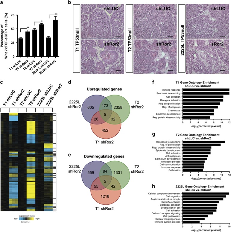
Depletion of Ror2 expression enhances canonical Wnt signaling across basal-like TP53-null models despite distinctions in both histopathology and gene expression outcomes. (a) Elevated Wnt/β-catenin signaling occurs in the absence of Ror2 in all three basal-like TP53-null models. Quantitation of fluorescence-activated cell sorting analysis of 7xTCF-eGFP positivity within shLUC and shRor2 tumors within T1, T2 and 2225L models (n=3 shLUC tumors and n=3 shRor2 tumors per basal-like model, *P<0.05, **P<0.001). (b) H&E staining of shLUC and shRor2 tumors showing squamous differentiation in T1 and T2 and disorganization in 2225L upon Ror2 loss. Scale 50 μm. (c) Supervised clustering and heat map showing gene expression changes (P<0.01 by t-test, fold change>1.4) across TP53-null basal-like models T1, T2 and 2225L, harboring shLUC vs shRor2 hairpins (derived from sorted transduced tdTomato-positive tumor cells). Within each model (T1, T2, 2225L), expression values were centered on the corresponding control. Bright yellow/blue represents minimum of two-fold change from the corresponding control. (d, e) Venn diagrams of (d) upregulated and (e) downregulated genes represented in shLUC and shRor2 groups within T1, T2 and 2225L, illustrating gene expression overlap between basal-like TP53-null models in response to Ror2 depletion. (f–h) Gene ontology analysis illustrating the enrichment of particular gene expression programs within (f) T1, (g) T2 and (h) 2225L.

