Abstract
The purpose of this research was to compare the accuracy of newly described P wave-related parameters (P wave area, Macruz index and mean electrical axis) with classical P wave-related parameters (voltage and duration of P wave) for the assessment of left atrial (LA) size in dogs with degenerative mitral valve disease. One hundred forty-six dogs (37 healthy control dogs and 109 dogs with degenerative mitral valve disease) were prospectively studied. Two-dimensional echocardiography examinations and a 6-lead ECG were performed prospectively in all dogs. Echocardiography parameters, including determination of the ratios LA diameter/aortic root diameter and LA area/aortic root area, were compared to P wave-related parameters: P wave area, Macruz index, mean electrical axis voltage and duration of P wave. The results showed that P wave-related parameters (classical and newly described) had low sensitivity (range=52.3 to 77%; median=60%) and low to moderate specificity (range=47.2 to 82.5%; median 56.3%) for the prediction of left atrial enlargement. The areas under the curve of P wave-related parameters were moderate to low due to poor sensitivity. In conclusion, newly P wave-related parameters do not increase the diagnostic capacity of ECG as a predictor of left atrial enlargement in dogs with degenerative mitral valve disease.
Keywords: dog, electrocardiography, left atrium, P wave
Left atrial enlargement (LAE) is a morphologic expression of the severity and chronicity of several heart diseases and have diagnostic, therapeutic, and prognostic importance in dogs [23]. The noninvasive gold standard test to assess left atrium (LA) size in dogs is the 2-dimensional echocardiography (2DE) image [2, 3, 5, 9, 11, 15, 35].
A common finding in dogs with degenerative mitral valve disease (DMVD) is LAE. Left atrial enlargement is strongly correlated to the development of left-sided congestive heart failure or death, as well as other serious complications (such as atrial fibrillation and other atrial tachyarrhythmias) [6, 23]. Therefore, the size of the LA is an important index of cardiac condition, and the assessment of LA size may be helpful in guiding dog management strategies, as it had been demonstrated lately [4].
There are different techniques used to assess LA dimensions, including thoracic radiography, 2DE and 3DE echocardiography, magnetic resonance imaging, computed tomography and angiography [9, 12, 13, 21, 30, 31]. However, 2DE is a reasonably accurate method to assess LA compared to the higher precision techniques of computed tomography and magnetic resonance imaging [24].
Electrocardiography (ECG) is a simple, non-invasive, cost-effective, accessible and reproducible test. It is accepted that P wave duration reflects the activation of the atrial muscle; that depends, primarily, upon the mass of tissue excited, and, when prolonged, P wave could represent a criterion for LAE in both human [18] and veterinary medicine [32].
In human medicine, the ECG patterns which suggest LA mass and chamber size abnormalities or reflect conduction delays within the atria are: (1) increased P wave amplitude, (2) double-peaked, notched P wave or increased interpeak duration [14], (3) increase in the product of the amplitude and the duration of the terminal negative component of the P wave in lead V1 (the P terminal force) [18], (4) left axis deviation of the terminal P wave, (5) increased P-wave area [36], (6) increase in the ratio of the P wave duration to the PR segment (Macruz Index [17]) and (7) increased P wave duration and PR interval ratio [18]. To the authors’ knowledge, these patterns have not been yet studied prospectively in dogs. In veterinary medicine, the ECG parameters related to LAE are: P wave duration and P wave electrical axis [6].
Several human studies have described the agreement between LAE detected by ECG and echocardiography [14, 17, 36] or magnetic resonance imaging [33], and there is some recently-published data on the diagnostic accuracy of ECG and thoracic radiography in the assessment of LAE in cats [27]. So far, the literature concerning the diagnostic accuracy of P wave-related parameter criteria, for both human and veterinary medicine, is limited. In human medicine, the comparison of various ECG abnormalities and echocardiographic criteria for LAE demonstrates limited sensitivity (SE) (8–78%) but high specificity (SP) (85–100%) for the ECG criteria [11, 19, 33]. Veterinary results reveal similar SE (12–60%) and SP (81–100%) values [6, 16, 27]. Studies in human medicine have demonstrated that P wave duration presents limited correlation with LA pressure and dimension [8].
The aim of this study was to compare the accuracy of newly described P wave-related parameters in dogs, such as P wave area, Macruz index and mean electrical axis, with classical P wave-related parameters, such as voltage or duration of P wave, for the assessment of left atrial size in dogs with DMVD. In addition, the study sought to check if the diagnostic accuracy of those P wave-related parameters varies with the degree of LAE.
MATERIALS AND METHODS
Study population
All dogs were prospectively studied between October 2005 and November 2009 at the Veterinary Teaching Hospital-Complutense University of Madrid (UCM), Spain.
All the procedures followed the ethical guidelines for animal studies and clinical care.
Healthy dogs: Thirty-seven healthy dogs without evidence of cardiovascular or systemic disease were classified as Group I. Subjects fulfilled the following criteria: (1) no history of heart disease or other current clinical abnormality; (2) normal physical examination; and (3) normal systolic blood pressure (non-invasive blood pressure monitor: Critikon Dinamap 1846SX NIBP) and echocardiogram including 2DE, M-mode, and Doppler variables within normal range. No dog in this group was receiving any medication.
Dogs with MVD: One hundred nine dogs with heart murmur confirmed as DMVD by echocardiographic evidence (increase thickness, nodularity and prolapse of mitral valve, in the absence of clinical signs of endocarditis) [23] were classified as Group II. All dogs were subjected to a complete thoracic radiographic study, including two perpendicular views (right lateral and dorsoventral views), a complete blood count and a basic metabolic panel including glucose, blood urea nitrogen and creatinine, sodium, potassium, chloride and alanine aminotransferase. None of this group was in congestive heart failure. Electrocardiographic and echocardiographic studies of each dog were made within 24 hr.
Echocardiography
Complete transthoracic 2D, M-mode and Doppler echocardiographic examinations were performed by two investigators (Soto-Bustos A. and Caro-Vadillo A.), following standard echocardiography guidelines [28, 30] using an Envisor CHD (Philips®). Left atrial diameter was measured with 2D echocardiography from a right parasternal short-axis view at the heart base, as previously described [25]. The aortic root diameter was measured at the level of the commissure between the non-coronary and right coronary aortic valve leaflet. The left atrial diameter was measured along a line extending from and parallel to the commissure between the non-coronary and left coronary aortic valve cusps to the distant margin of the left atrium [25]. Both measurements were performed on the first frame after aortic valve closure. The aortic area were determined by planimetry from the same short-axis view as describe previously. Left atrial area was measured with 2D echocardiography from a left apical four-chamber view by planimetry (Fig. 1). Left atrial diameter, LA area, aortic root diameter and area were measured using the ultrasound machine integrated software. All echocardiographic measurements were done in triplicate and the resulting mean of the 3 consecutive beats measurements was used. Two echocardiographic ratios were calculated: LA diameter/aortic root diameter (LAD/AoD) and LA area/aortic area (LAA/AoA). According to LAD/AoD results obtained by echocardiogram, this group was subdivided in two groups: B1 (26 dogs with LAD/AoD <1.55) and B2 (83 dogs with LAD/AoD ≥1.55), following ACVIM classification of dogs with DVMD [1].
Fig. 1.
Measurements of left aortic diameter and left atrial diameter from a right short axis view at the base (a), left aortic area from the same view (b) and left atrial area from a left apical four chamber view (c).
Electrocardiography
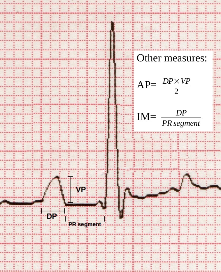
Electrocardiograms were recorded with a commercially available 3-channel ECG machine (Cardioline Mod AR1200view). During recordings, all dogs were fully conscious with no chemical restraint. The animals were placed in right lateral recumbency with the limbs held as nearly perpendicular to the body as possible. Standard 6-lead ECGs (leads I, II, III, aVR, aVL and aVF) were recorded for all dogs following standard electrocardiographic guidelines [6]. Recordings were made at a paper speed of 50 mm/sec and a vertical ECG calibration of 10 mm/mV. All ECG tracings were scanned at 300 ppi (pixels per inch) with a photo scanner (HP Scanjet G2710). All ECG measurements were done on scanned images amplified to 400% using an image manipulation program (GIMP 2.6.8 GNU Linux). All ECG measurements were done in triplicate on six consecutive P-QRS-T complexes from lead II using a screen caliper function included in the mentioned image software. Eight ECG variables were measured or calculated (Fig. 2) (with each corresponding range of accuracy appearing between brackets):
Fig. 2.

Measurements related to P wave performed in the ECG of all dogs included in the study. DP: duration of P wave (msec); DPR: duration of PR interval (msec); VP: voltage of P wave (mV); AP: area of P wave (mV·msec); IM: Macruz index.
-
1.
Voltage of the P wave (VP), expressed in mV [± 0.001 mV];
-
2.
Duration of the P wave (DP), expressed in milliseconds [± 0.1 msec];
-
3.
P wave area (AP) (expressed in mV·millisecond) as product of the voltage and half duration of the P wave [± 0.01mV·msec];
-
4.
Macruz Index (MI), which is the ratio of P wave duration and PR segment duration, both measured in msec, expressed without units [± 0.001];
-
5.
Mean electrical axis of the P wave (MEA) in the frontal plane, expressed in degrees [± 1°], calculated using P wave voltages from leads I & II as described by Singh et al. [29], following this formula:
 , where II and I
were the net voltage of P wave in those leads;
 -
6.
Heart rate (HR), expressed in beats per min (bpm) [± 1 bpm].
ECG measurements and calculations were performed blinded to echocardiographic assessment of LA size.
Statistical analyses
All statistical analyses were done with statistics software (IBM SPSS Statistics 22.0 & SAS 9.4). Data are expressed as median and range. In comparisons, a non-parametric Kruskal Wallis and Wilcoxon score (rank sums) test was used for continuous variables and a Chi-Square test was used for categorical variables. Studies of correlation were done between ECG, echocardiographic parameters and biometric variables using the Spearman correlation coefficient. A P<0.05 was considered statistically significant.
The number of true positives, true negatives, false positives and false negatives was calculated for the individual ECG criteria. Sensitivity, specificity, positive predictive value (PPV) and negative predictive value (NPV) were calculated.
The areas under the receiver (AUR) operating characteristic (ROC) curve for two LAE echocardiographic criteria (LAD/AoD and LAA/AoA) and the optimal cut-off value for each ECG parameter were calculated.
RESULTS
One hundred forty-six dogs were included in the study: 51 females (35%) and 95 males (65%). There were 46 mixed-breed dogs (30.5%), 21 poodles (14.4%), 15 Cocker Spaniels (10.3%) and 10 Yorkshire Terriers (6.8%), while the remaining 54 dogs (38%) belonged to 27 different breeds. The median and range of body weight (BW) were 9 kg (3.17–32 kg), with more dogs of low body weight in group B2. The median and range of age were 11 years (2–14.8 years), with more dogs older than 9 years in groups B1 and B2 (Table 1).
Table 1. Characteristics, electrocardiographic indexes and echocardiographic indexes of the dogs included in the study.
| Group I (N=37) | Group II (N=109) | |||||||||
|---|---|---|---|---|---|---|---|---|---|---|
| Group B1 (N=26) | Group B2 (N=83) | |||||||||
| Median | Min | Max | Median | Min | Max | Median | Min | Max | ||
| Characteristics | ||||||||||
| Age (years) | 7B1, B2 | 1 | 13 | 12A | 4.4 | 15.6 | 11A | 8 | 15 | |
| Weight (kg) | 14B, B2 | 1.7 | 45.5 | 8A | 4.1 | 27.2 | 8.5A | 3.5 | 22.5 | |
| Echocardiography | ||||||||||
| LAD/AoD | 1.363B2 | 0.99 | 1.55 | 1.39B2 | 1,022 | 1,541 | 2.337A, B1 | 1,593 | 4255 | |
| LAA/AoA | 2.273B2 | 0.97 | 3.8 | 2.364B2 | 1,952 | 3.92 | 5.607A, B1 | 2.31 | 17.84 | |
| Electrocardiography | ||||||||||
| DP (msec) | 43.4B2 | 27.7 | 54.4 | 43.3B2 | 34.0 | 55.7 | 48.3A, B1 | 28.8 | 76.0 | |
| VP (mV) | 0.238 | 0.106 | 0.369 | 0.23 | 0.106 | 0.406 | 0.26 | 0.059 | 0.622 | |
| AP (mV·msec) | 5.3B2 | 1.6 | 9.2 | 4.7B2 | 2.4 | 9.6 | 6.06 A, B1 | 0.9 | 18.3 | |
| MI | 0.942B2 | 0.356 | 1.85 | 1.002B2 | 0.453 | 1,977 | 1.168A, B1 | 0.51 | 6.24 | |
| MEA (°) | 53 | 10 | 83 | 36.5 | 1 | 122 | 54 | –13 | 136 | |
| HR (bpm) | 119B2 | 82 | 200 | 124.3 | 82 | 210 | 140.7A | 72 | 220 | |
Each row superscript represents the group from which there is statistically significant difference P<0.05. LAD/AoD: ratio left atrial dimension/aortic dimension; LAA/AoA: ratio left atrial area/aortic area; DP: duration of P wave; VP: voltage of P wave; AP: area of P wave; MI: Macruz index; MEA: mean axis of P wave; HR: Heart rate.
None of the dogs in group B1 were receiving treatment. None of the dogs in group B2 were receiving antiarrhythmic or diuretic drugs. Sixty-five percent of the dogs in group B2 (54/83) were in treatment either with an angiotensin converting enzyme inhibitor, or pimobendan, at normal established doses.
Thirty-seven dogs were considered healthy (Group I), whereas 109 dogs were affected by DMVD (Group II). Electrocardiographic and echocardiographic variables obtained from healthy dogs (Group I) are presented in Table 1.
The discrimination limits of the echocardiographic index LAD/AoD for LAE was >1.55 and the discrimination limits of the echocardiographic index LAA/AoA for LAE was >3.7 (Table 1).
Dogs with echocardiographic LAE (considering LAD/AoD) had significantly (P<0.05) increased DP, AP, MI and HR (Table 1).
In order to verify if there were differences in diagnostic accuracy according to severity in LAE, B2 dogs were divided in three groups: Mild LAE (1.55<LAD/AoD ≤2 or LAA/AoA≤5; N=27), Moderate LAE (2<LAD/AoD ≤2.5 or 5<LAA/AoA ≤7; N=25) and Severe LAE (LAD/AoD >2.5 or LAA/AoA >7; N=31). Electrocardiographic variables obtained from these three groups are presented in Table 2.
Table 2. Electrocardiographic indexes in B2 groups according to severity of LAE.
| Mild LAE (LAD/AoD >1.55 ≤ 2) (N=27) | Moderate LAE (LAD/AoD >2 ≤ 2.5) (N=25) | Severe LAE (LAD/AoD >2.5) (N=31) | |||||||
|---|---|---|---|---|---|---|---|---|---|
| Median | Min | Max | Median | Min | Max | Median | Min | Max | |
| DP (msec) | 45.8 | 28.9 | 56.9 | 48.1 | 30.1 | 55.4 | 50.8 | 37.3 | 76.1 |
| VP (mV) | 0.232severe | 0.06 | 0.58 | 0.20severe | 0.06 | 0.44 | 0.306mild, moderate | 0.094 | 0.622 |
| AP (mV·msec) | 5.1severe | 0.9 | 14.2 | 5.4severe | 1.3 | 11.9 | 7.5 mild, moderate | 1.8 | 18.3 |
| MI | 1,192 | 0.701 | 6.24 | 1,077 | 0.517 | 2.75 | 1.24 | 0.51 | 3.81 |
| MEA (°) | 49 | 7 | 136 | 56 | –13 | 121 | 56 | 1 | 90 |
| HR (bpm) | 128severe | 72 | 219 | 130severe | 83 | 219 | 164 mild, moderate | 96 | 209 |
| Mild LAE (LAA/AoA ≤5) (N=32) | Moderate LAE (LAA/AoA >5 ≤ 7) (N=22) | Moderate LAE (LAA/AoA >7) (N=29) | |||||||
| Median | Min | Max | Median | Min | Max | Median | Min | Max | |
| DP (msec) | 46.5 | 28.9 | 56.3 | 48.6 | 36.4 | 58.2 | 51 | 30.1 | 76.1 |
| VP (mV) | 0.23 | 0.06 | 0.42 | 0.25 | 0.06 | 0.58 | 0.3 | 0.087 | 0.62 |
| AP (mV·msec) | 5.1severe | 0.9 | 11.8 | 6 | 1.3 | 14.2 | 7mild | 1.8 | 18.3 |
| MI | 1,023 | 0.61 | 6.24 | 1.23 | 0.52 | 2.9 | 1.38 | 0.51 | 3.81 |
| MEA (°) | 45 | 7 | 136 | 56.5 | –13 | 83 | 58 | 1 | 121 |
| HR (bpm) | 126 | 71 | 219 | 149 | 83 | 219 | 158 | 96 | 204 |
Each row superscript represents the group from which there is statistically significant difference P<0.01. DP: duration of P wave; VP: voltage of P wave; AP: area of P wave; MI: Macruz index; MEA: mean axis of P wave; HR: Heart rate.
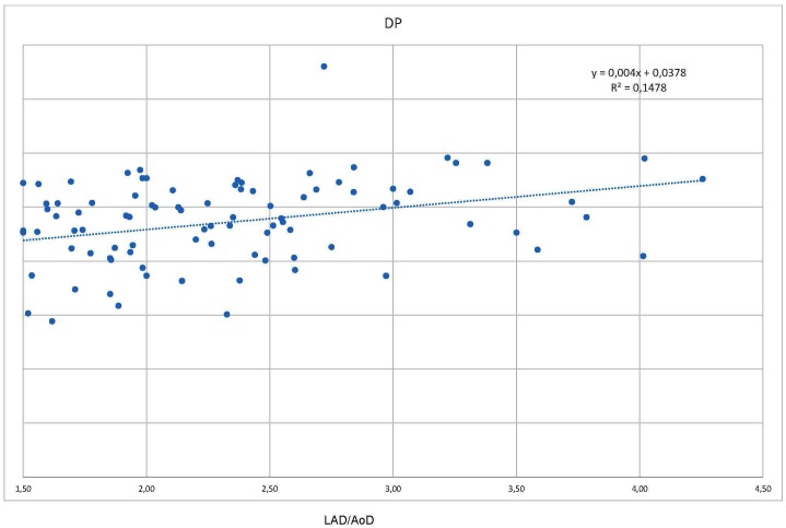
Results of correlation analyses are summarized in Table 3. Correlation analysis indicated evidence for a positive correlation, statistically significant, between echocardiographic ratios (LAD/AoD and LAA/AoA) and the ECG parameters DP, AP, MI and HR; hen considering all the dogs included in the study, although the Spearman coefficient is low (Fig. 3).
Table 3. Results of correlation analyses (Spearman correlation coefficient is expressed) between ECG, echocardiographic variables, age and weight for all dogs N=143.
| LAD/AoD | LAA/AoA | |
|---|---|---|
| Age (years) | 0.19 | 0.25 |
| Weight (kg) | –0.053 | –0.162 |
| DP (msec) | 0.394a) | 0.342a) |
| VP (mV) | 0.202 | 0.183 |
| AP (mV·msec) | 0.303a) | 0.274a) |
| MI | 0.218a) | 0.235a) |
| MEA (°) | 0.129 | 0.12 |
| HR (bpm) | 0.249a) | 0.262a) |
a) P<0.01. DP: duration of P wave; VP: voltage of P wave; AP: area of P wave; MI: Macruz index; MEA: mean axis of P wave; HR: Heart rate.
Fig. 3.
Scatter plot showing distribution of DP against LAD/AoD. DP: duration of P wave (msec), LAD/AoD: ratio left atrial dimension/aortic dimension.
There were no significant correlations between echocardiographic ratios and ECG parameters when considering the different groups of dogs according to severity of LAE.
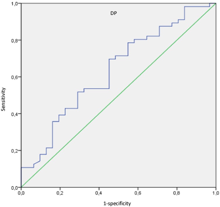
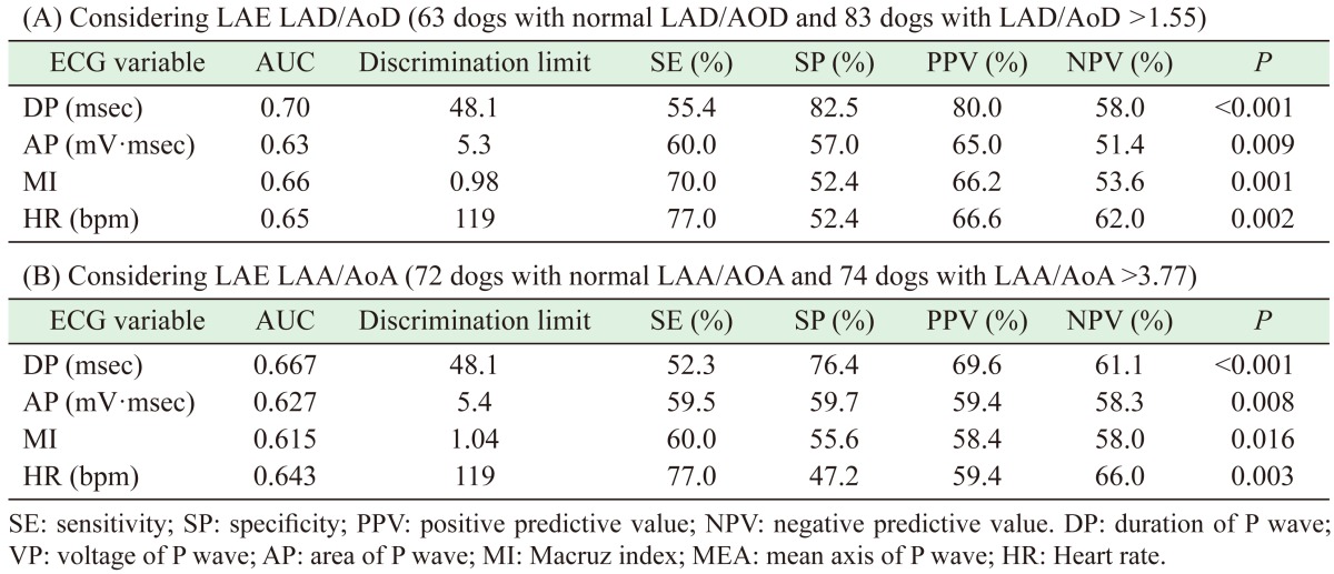
ECG parameters were analyzed using the area under the curve (AUC) in relationship to two previously defined normal echocardiographic LA indexes (LAD/AoD and LAA/AoA) to assess predictive power (Fig. 4). Variables characterizing diagnostic accuracy of different electrocardiographic indexes for the prediction of LAE, considering LAD/AoD and LAA/AoA, in both healthy dogs and dogs with DMVD are summarized in Table 4.
Fig. 4.
Receiver Operating Characteristics (ROC) curve for DP: P wave duration (msec).
Table 4. Diagnostic accuracy of different electrocardiographic indexes to predict LAE considering LAD/AoD (A) and LAA/AoA (B).
The ECG parameters DP, AP, MI and HR were found to be mildly sensitive (52.3–77%; median=60%) and mildly to moderately specific (47.2–82.5%; median=56.3%).
When considering only dogs included with severe LAE according to LAD/AoD (N=31), the sensitivity increased (61.3–87.1%; median=74.2%), but there was no increase in specificity (49.2–82.5%; median=65.85%).
DISCUSSION
To the authors’ knowledge, this is the first study investigating the diagnostic performance of P wave-related measurements, measured using an image manipulation software, to detect LAE in dogs with DMVD. In a previous report [26] the diagnostic accuracy of ECG, was analyzed measuring a single wave parameter (P wave duration) and using a metal caliper system. The prediction of LAE using the P wave area, Macruz index and mean electrical axis, has not been studied previously in veterinary medicine. The present investigation compared P wave-related measurements obtained from surface ECG with echocardiographic parameters (LAD/AoD and LAA/AoA) as the gold standard to estimate LA size. Furthermore, this is the first time that cut-off values of several P wave-related wave measurements have been assessed in normal dogs.
The on-screen measurements are consistent with other manual methods. A previous paper [7] also demonstrated that manual measurement of electrocardiographic patterns should preferably be performed with digital ECG recordings displayed on a high-resolution computer screen.
In the present study, the ECGs values of DP, VP and HR in the normal dogs (Group I) were found to coincide with the DP, VP, and HR reference values previously reported [6, 10, 32]. Newly P wave-related parameters (AP, MI, MEA) cannot be compared with other veterinary reports in dogs.
The value of DP was increased in B2 dogs compared to Group I dogs but not with respect to B1 dogs. This could indicate that, in spite of mitral regurgitation and LA volume overload in B1 dogs, there are no electrocardiographic consequences until LAE is detected.
The values of AP, MI and HR were increased in B2 dogs, there were differences only between B2 dogs and healthy dogs, not between B2 and B1 dogs or B1 and healthy dogs. These data could indicate that B1 dogs could be considered electrocardiographically normal with respect to AP, MI and HR.
In veterinary medicine, several authors [6, 32] have described a correlation between age and P wave duration. The results in the present study did not show that relationship. It is possible that the presence of remodeling and fibrosis in the aged-dog cardiac muscle does not alter any P wave-related electrocardiographic parameters [28].
There was a positive correlation between weight and DP. This correlation was reported in others studies [32]. The reason could be the relationship between weight and heart size in general and the relationship between weight and LA in particular. In this study, normal dogs of different weights, including large breed dogs were used. This fact could have affected the limits of DP for comparison. In spite of that, DP values of B1 dogs do not differ from those of normal dogs.
In the present study, an association between higher values in P wave-related parameters (DP, VP, AP and MI) and LA echocardiographic indexes (LAD/AoD and LAA/AoA) was observed, but the ability of each ECG parameter to predict LAE was poor because of the low SE level. Similarly, in humans, the monitoring of individual ECG P wave changes (especially DP, mean electrical axis and MI) does not reliably detect or predict the presence of LAE when compared to cardiac magnetic resonance imaging [33] or echocardiography [14].
The relationship between LA size and cardiac disease with the genesis of abnormal atrial electrical forces is complex and involves the interplay of several factors. These include (1) myocardial changes with intra-atrial conduction abnormalities such as ischemia, infarction and inflammation; (2) LA hypertension; (3) LA dilatation and stretch; (4) the extent of LA fibrosis; and (5) the chronicity of the disease. Although isolated intra-atrial conduction disturbances may be clinically observed as well as produced experimentally [32], the frequency of their occurrence in dogs is unknown, and therefore, for practical purposes, a prolonged P wave duration, especially the prolongation of the terminal portion of the P wave, should be indicative of LA involvement (most commonly dilatation). Drugs such as digitalis glycosides, other antiarrhythmics and vagal stimulation can all prolong intra-atrial conduction; however, their effects on P wave duration in people have not been found to be consistent [34]. No dog in this study was treated with antiarrhythmic drugs. In addition, independent factors such as the proximity of the LA to the recording electrodes, obesity, heart rate, autonomic tone, position of the heart in the chest and contact of the electrodes with the skin have been found to influence the amplitude of ECG deflections on the body surface [32]. The results of this study indicate that the electrocardiographic P wave alterations (in DP, AP and MI) are suggestive of LAE in dogs, although their absence does not rule out increased LA size. In particular, DP, AP, MI and HR were significantly correlated to high LAD/AoD ratio (Table 3), with low coefficients of correlation.
Results reported by Savarino et al. [26] show similar SE and SP percentages with a DP threshold >0.05 sec. Macruz index and HR were the most useful indexes that correctly identified 70% and 77% of patients respectively with LAE, considering LAD/AoD. However, the correlation between these ECG parameters and LAD/AoD was low (rS=0.218 and rS=0.249 (P<0.001)).
This study produced similar SP values to those of other studies [22, 27] and also yielded low SE data, as in other studies [11, 19, 27, 36]. O’Grady et al. [22] concluded that the SE of the ECG criterion for LAE (DP>40 msec) is rather low while the SP of this criterion is very high.
The low SE values obtained by the present study could be explained by the fact that it had a restricted number of patients with severe LAE, which tend to develop atrial fibrillation and there is no P wave-related parameters to measure, therefore these patients cannot be included in the study.
The ROC curve characteristics are concordant with previous studies in cats [20], dogs [26] and humans [34], in which the relatively poor diagnostic accuracy of ECG variables for LAE, due to low SE percentage, was reported.
This study presents certain limitations. Firstly, because of the observational nature of the investigation, true LA volume using 3D echocardiography, CT or MRI could not be assessed. Secondly, the presence of wandering pacemaker or possible intermittent inter- or intra-atrial conduction disorders can alter a P wave measurement [32] obtained from a single beat. In the present study, the decision to assess six consecutive beats was made in order to reduce the risk of over- or underestimating the length of the P wave-related waves. Thirdly, the LA appendage was not included in the echocardiographic measurements of LA size, yet it could represent a large chamber that contributes substantially to LAE, particularly in dogs with slightly high LAE affecting electrocardiographic measurements. Fourthly, the absence of hemodynamic and electrophysiological data in this study means we cannot rule out the presence of intra- or inter-atrial conduction disorders responsible for P wave changes. Precordial derivations to explore P terminal force in V1 had not been performed. Moreover, since this study was noninvasive, we were not able to measure atrial pressures, which may also affect the P wave [8]. Fifthly, we did not take into account the possibility of pulmonary hypertension in the patients, which could also influence the P wave (as pulmonary hypertension can influence the VP, AP and mean electrical axis).
In conclusion, using appropriate diagnostic criteria, the ECG results appear to be neither highly specific nor sensitive indicators of LAE in dogs with DMVD. A normal P wave on the ECG does not reliably exclude LAE in the individual dog. However, abnormal P wave duration does indicate LAE.
The ECG could be used as a first-phase, diagnostic tool for identifying in dogs with murmurs due to DMVD. The results indicate that the group of patients with high DP (>48.1 msec), AP (>5.3 mV·msec), MI (>0.98) and HR (>119 bpm) are more likely to have elevated LAD/AoD (>1.55), the predictive value of a positive test is high (Table 4).
The use of new P wave-related measurements (AP, mean electrical axis and MI) does not increase the diagnostic capacity of ECG, in spite of the more complex calculations needed to obtain them.
CONFLICT OF INTEREST
The authors declare that there is no conflict of interest.
Acknowledgments
The authors wish to thank the entire Complutense Veterinary Teaching Hospital staff for their support and assistance with this research. The authors also wish to thank the biostatistical collaboration of Mr. Ricardo García.
REFERENCES
- 1.Atkins C., Bonagura J., Ettinger S., Fox P., Gordon S., Haggstrom J., Hamlin R., Keene B., Luis-Fuentes V., Stepien R.2009. Guidelines for the diagnosis and treatment of canine chronic valvular heart disease. J. Vet. Intern. Med. 23: 1142–1150. doi: 10.1111/j.1939-1676.2009.0392.x [DOI] [PubMed] [Google Scholar]
- 2.Bélanger M. C.2007. Echocardiography. pp. 311–326. In: Textbook of Veterinary Internal Medicine, 6th ed. (Ettinger, S. J., Feldman, E. eds.) WB Saunders, Philadelphia. [Google Scholar]
- 3.Bonagura J. D., Luis-Fuentes V.2000. Echocardiography. pp. 834–874. In: Textbook of Veterinary Internal Medicine: Diseases of the Dog and Cat, 5th ed. (Ettinger, S. J., Feldman, E. eds.), WB Saunders, Philadelphia. [Google Scholar]
- 4.Boswood A., Häggström J., Gordon S. G., Wess G., Stepien R. L., Oyama M. A., Keene B. W., Bonagura J., MacDonald K. A., Patteson M., Smith S., Fox P. R., Sanderson K., Woolley R., Szatmári V., Menaut P., Church W. M., O’Sullivan M. L., Jaudon J. P., Kresken J. G., Rush J., Barrett K. A., Rosenthal S. L., Saunders A. B., Ljungvall I., Deinert M., Bomassi E., Estrada A. H., Fernandez Del Palacio M. J., Moise N. S., Abbott J. A., Fujii Y., Spier A., Luethy M. W., Santilli R. A., Uechi M., Tidholm A., Watson P.2016. Effect of Pimobendan in Dogs with Preclinical Myxomatous Mitral Valve Disease and Cardiomegaly: The EPIC Study-A Randomized Clinical Trial. J. Vet. Intern. Med. 30: 1765–1779. doi: 10.1111/jvim.14586 [DOI] [PMC free article] [PubMed] [Google Scholar]
- 5.Bouzas-Mosquera A., Broullón F. J., Álvarez-García N., Méndez E., Peteiro J., Gándara-Sambade T., Prada O., Mosquera V. X., Castro-Beiras A.2011. Left atrial size and risk for all-cause mortality and ischemic stroke. CMAJ 183: E657–E664. doi: 10.1503/cmaj.091688 [DOI] [PMC free article] [PubMed] [Google Scholar]
- 6.Côté E.2010. Electrocardiography and Cardiac Arrhythmias. pp. 1159–1187. In: Textbook of Veterinary Internal Medicine, 7th ed. (Ettinger, S. J. and Feldman, E. eds.) WB Saunders, Philadelphia. [Google Scholar]
- 7.Dilaveris P., Batchvarov V., Gialafos J., Malik M.1999. Comparison of different methods for manual P wave duration measurement in 12-lead electrocardiograms. Pacing Clin. Electrophysiol. 22: 1532–1538. doi: 10.1111/j.1540-8159.1999.tb00358.x [DOI] [PubMed] [Google Scholar]
- 8.Faggiano P., D’Aloia A., Zanelli E., Gualeni A., Musatti P., Giordano A.1997. Contribution of left atrial pressure and dimension to signal-averaged P-wave duration in patients with chronic congestive heart failure. Am. J. Cardiol. 79: 219–222. doi: 10.1016/S0002-9149(96)00720-5 [DOI] [PubMed] [Google Scholar]
- 9.Hansson K., Häggström J., Kvart C., Lord P.2002. Left atrial to aortic root indices using two-dimensional and M-mode echocardiography in cavalier King Charles spaniels with and without left atrial enlargement. Vet. Radiol. Ultrasound 43: 568–575. doi: 10.1111/j.1740-8261.2002.tb01051.x [DOI] [PubMed] [Google Scholar]
- 10.Hanton G., Rabemampianina Y.2006. The electrocardiogram of the Beagle dog: reference values and effect of sex, genetic strain, body position and heart rate. Lab. Anim. 40: 123–136. doi: 10.1258/002367706776319088 [DOI] [PubMed] [Google Scholar]
- 11.Hazen M. S., Marwick T. H., Underwood D. A.1991. Diagnostic accuracy of the resting electrocardiogram in detection and estimation of left atrial enlargement: an echocardiographic correlation in 551 patients. Am. Heart J. 122: 823–828. doi: 10.1016/0002-8703(91)90531-L [DOI] [PubMed] [Google Scholar]
- 12.Kühl J. T., Lønborg J., Fuchs A., Andersen M. J., Vejlstrup N., Kelbæk H., Engstrøm T., Møller J. E., Kofoed K. F.2012. Assessment of left atrial volume and function: a comparative study between echocardiography, magnetic resonance imaging and multi slice computed tomography. Int. J. Cardiovasc. Imaging 28: 1061–1071. doi: 10.1007/s10554-011-9930-2 [DOI] [PubMed] [Google Scholar]
- 13.LeBlanc N., Scollan K., Sisson D.2016. Quantitative evaluation of left atrial volume and function by one-dimensional, two-dimensional, and three-dimensional echocardiography in a population of normal dogs. J. Vet. Cardiol. 18: 336–349. doi: 10.1016/j.jvc.2016.06.005 [DOI] [PubMed] [Google Scholar]
- 14.Lee K. S., Appleton C. P., Lester S. J., Adam T. J., Hurst R. T., Moreno C. A., Altemose G. T.2007. Relation of electrocardiographic criteria for left atrial enlargement to two-dimensional echocardiographic left atrial volume measurements. Am. J. Cardiol. 99: 113–118. doi: 10.1016/j.amjcard.2006.07.073 [DOI] [PubMed] [Google Scholar]
- 15.Lester S. J., Ryan E. W., Schiller N. B., Foster E.1999. Best method in clinical practice and in research studies to determine left atrial size. Am. J. Cardiol. 84: 829–832. doi: 10.1016/S0002-9149(99)00446-4 [DOI] [PubMed] [Google Scholar]
- 16.Lombard C. W., Spencer C. P.1985. Correlation of radiographic, echocardiographic, and electrocardiographic signs of left heart enlargement in dogs with mitral regurgitation. Vet. Radiol. Ultrasound 26: 89–97. doi: 10.1111/j.1740-8261.1985.tb01389.x [DOI] [Google Scholar]
- 17.Macruz R., Perloff J. K., Case R. B.1958. A method for the electrocardiographic recognition of atrial enlargement. Circulation 17: 882–889. doi: 10.1161/01.CIR.17.5.882 [DOI] [PubMed] [Google Scholar]
- 18.Mirvis D. M., Goldberger A. L.2007. Electrocardiography. pp. 167–172. In: Braunwald’s Heart Disease: A Textbook of Cardiovascular Medicine. 8th ed., WB Saunders Co., Philadelphia. [Google Scholar]
- 19.Mishra A., Mishra C., Mohanty R. R., Behera M.2008. Study on the diagnostic accuracy of left atrial enlargement by resting electrocardiography and its echocardiographic correlation. Indian J. Physiol. Pharmacol. 52: 31–42. [PubMed] [Google Scholar]
- 20.Moïse N. S., Dietze A. E.1986. Echocardiographic, electrocardiographic, and radiographic detection of cardiomegaly in hyperthyroid cats. Am. J. Vet. Res. 47: 1487–1494. [PubMed] [Google Scholar]
- 21.Nakayama H., Nakayama T., Hamlin R. L.2001. Correlation of cardiac enlargement as assessed by vertebral heart size and echocardiographic and electrocardiographic findings in dogs with evolving cardiomegaly due to rapid ventricular pacing. J. Vet. Intern. Med. 15: 217–221. doi: 10.1111/j.1939-1676.2001.tb02314.x [DOI] [PubMed] [Google Scholar]
- 22.O’grady M., Difruscia R., Carley B., Hill B.1992. Electrocardiographic evaluation of chamber enlargement. Can. Vet. J. 33: 195–200. [PMC free article] [PubMed] [Google Scholar]
- 23.Olsen L. H., Häaggström J., Petersen H. D.2010. Acquired valvular heart disease. pp. 1299–1319. In: Textbook of Veterinary Internal Medicine, 7th ed. (Ettinger, S. J. and Feldman, E. eds.) WB Saunders, Philadelphia. [Google Scholar]
- 24.Poutanen T., Jokinen E., Sairanen H., Tikanoja T.2003. Left atrial and left ventricular function in healthy children and young adults assessed by three dimensional echocardiography. Heart 89: 544–549. doi: 10.1136/heart.89.5.544 [DOI] [PMC free article] [PubMed] [Google Scholar]
- 25.Rishniw M., Erb H. N.2000. Evaluation of four 2-dimensional echocardiographic methods of assessing left atrial size in dogs. J. Vet. Intern. Med. 14: 429–435. doi: 10.1111/j.1939-1676.2000.tb02252.x [DOI] [PubMed] [Google Scholar]
- 26.Savarino P., Borgarelli M., Tarducci A., Crosara S., Bello N. M., Margiocco M. L.2012. Diagnostic performance of P wave duration in the identification of left atrial enlargement in dogs. J. Small Anim. Pract. 53: 267–272. doi: 10.1111/j.1748-5827.2012.01200.x [DOI] [PubMed] [Google Scholar]
- 27.Schober K. E., Maerz I., Ludewig E., Stern J. A.2007. Diagnostic accuracy of electrocardiography and thoracic radiography in the assessment of left atrial size in cats: comparison with transthoracic 2-dimensional echocardiography. J. Vet. Intern. Med. 21: 709–718. doi: 10.1111/j.1939-1676.2007.tb03012.x [DOI] [PubMed] [Google Scholar]
- 28.Scott C. C., Leier C. V., Kilman J. W., Vasko J. S., Unverferth D. V.1983. The effect of left atrial histology and dimension on P wave morphology. J. Electrocardiol. 16: 363–366. doi: 10.1016/S0022-0736(83)80086-7 [DOI] [PubMed] [Google Scholar]
- 29.Singh P. N., Athar M. S.2003. Simplified calculation of mean QRS vector (mean electrical axis of heart) of electrocardiogram. Indian J. Physiol. Pharmacol. 47: 212–216. [PubMed] [Google Scholar]
- 30.Thomas W. P., Gaber C. E., Jacobs G. J., Kaplan P. M., Lombard C. W., Moise N. S., Moses B. L.1993. Recommendations for standards in transthoracic two-dimensional echocardiography in the dog and cat. Echocardiography Committee of the Specialty of Cardiology, American College of Veterinary Internal Medicine. J. Vet. Intern. Med. 7: 247–252. doi: 10.1111/j.1939-1676.1993.tb01015.x [DOI] [PubMed] [Google Scholar]
- 31.Tidholm A., Höglund K., Häggström J., Bodegård-Westling A., Ljungvall I.2013. Left atrial ejection fraction assessed by real-time 3-dimensional echocardiography in normal dogs and dogs with myxomatous mitral valve disease. J. Vet. Intern. Med. 27: 884–889. doi: 10.1111/jvim.12113 [DOI] [PubMed] [Google Scholar]
- 32.Tilley L. P.1992. Analysis of canine P-QRS-S deflections. pp. 59–99. In: Essentials of Canine and Feline Electrocardiography 3rd ed., Lea and Febiger, Philadelphia. [Google Scholar]
- 33.Tsao C. W., Josephson M. E., Hauser T. H., O’Halloran T. D., Agarwal A., Manning W. J., Yeon S. B.2008. Accuracy of electrocardiographic criteria for atrial enlargement: validation with cardiovascular magnetic resonance. J. Cardiovasc. Magn. Reson. 10: 7. doi: 10.1186/1532-429X-10-7 [DOI] [PMC free article] [PubMed] [Google Scholar]
- 34.Waggoner A. D., Adyanthaya A. V., Quinones M. A., Alexander J. K.1976. Left atrial enlargement. Echocardiographic assessment of electrocardiographic criteria. Circulation 54: 553–557. doi: 10.1161/01.CIR.54.4.553 [DOI] [PubMed] [Google Scholar]
- 35.Ware W. A.2009. Diagnostic tests for the cardiovascular system. pp. 12–52. In: Small Animal Internal Medicine. 4th ed. (Nelson, R. and Couto, G.), Mosby Elsevier, St. Louis. [Google Scholar]
- 36.Zeng C., Wei T., Zhao R., Wang C., Chen L., Wang L.2003. Electrocardiographic diagnosis of left atrial enlargement in patients with mitral stenosis: the value of the P-wave area. Acta Cardiol. 58: 139–141. doi: 10.2143/AC.58.2.2005266 [DOI] [PubMed] [Google Scholar]


