

Abstract
Histomonas meleagridis is a flagellate protozoan parasite living in the cecum of birds digestive system and is the causative agent of histomoniasis. In this study 110 poultry fresh stool samples were assessed in order to detect H. meleagridis, and egg or adult worm of Heterakis gallinarum in Lorestan province, Iran. The results showed that the prevalence of H. meleagridis was 31%. Also, 19.5% of infected poultry had a watery stool. The eggs or adult worms of H. gallinarum were not seen in any of the samples. The results showed the high prevalence of this parasite, and the factors such as temporary hosts, susceptible hosts, and cloacal transmission with the involvement of water containing the parasite are the effective risk factors in prevalence. It is better to considered H. meleagridis as waterborne parasites, so further epidemiological studies on other birds are suggested to determine the parasite pathogenic strains using molecular methods.
Keywords: Parasite, Transmission, Prevalence, Epidemiology, Cloaca
Introduction
Histomonas meleagridis is a flagellate protozoan parasite living in the cecum of birds’ (Callait-Cardinal et al. 2007; McDougald 2005). This parasite belongs to the order Trichomonadida with short life outside the host body and disintegrates within a few hours (Lotfi et al. 2012; McDougald 2005). This causative agent of histomoniasis (Blackhead disease) has been reported in many of the birds was only known in Europe, East Asia, and Australia, but now it is seen in all the countries with poultry breeding (Heelsbergen 1929; McDougald 2005; Patra et al. 2013). Although by PCR, the presence of parasite is proved in most tissues of infected birds, cecum and liver have shown to be the most important predisposing sites (Huber et al. 2006). Infection with this parasite has been reported in many birds, but infection in turkeys is the most critical case with occasionally mortality rate of 100%, with above two million dollars losses per year in some countries such as the United States, and then, the chickens with mortality rate of 10–20% (McDougald 1997, 2005). The main transmission is through the cecal worm (Heterakis gallinarum) eggs known as, considered as the parasite reservoir and protective (Aka et al. 2010; Lister 2010), proved by the presence of nematodes in birds infected with H. meleagridis, which is known as the potential carrier (Patra et al. 2013). The birds develop H. meleagridis infection by eating the H. gallinarum embryonated eggs containing H. meleagridis from farms or eating the earthworms infected with H. gallinarum containing the above parasite (Esquenet et al. 2003; Patra et al. 2013). However, since the parasite infection is reported without the presence of nematode in some fields (Chossat 2002), the various transmission ways are also discussed, directly through the mouth that is not so important, or the cloacal drinking or cloacal drop that are more important, since it is shown that the poultry absorb the watery or suspended materials through the cloaca and transfer them to the cecum through the Bursa of Fabricius (Huber et al. 2006; Patra et al. 2013; Sorvari and Sorvari 1977). Recently, a cyst structure of the parasite is found in the culture media, which is believed to be another transitional mechanism of the parasite, however, the form is so rare and transmission in this way has not also been proven (Zaragatzki et al. 2010).
This research aimed the epidemiological study of H. meleagridis in poultry for the first time in Khorramabad, Lorestan province, Iran, to be an introduction to further studies in pathogenicity to determine the pathogenic strains in that region.
Materials and methods
Study area
The city of Khorramabad with an area of 6447 km2 is the capital of Lorestan province that is the largest province in western Iran. Since the Zagros Mountains prevent the Northern winds to this region, the warm seasons are longer than the temperate and cold seasons including the biggest part of the year with temperature up to 42 °C in the hot days (Ali and Pirasteh 2004).
Sample collection
This descriptive–analytical study was conducted on poultry in Khorramabad city, Lorestan province, Iran, since April 2015 for a year. The sample size was estimated based on formula with 95% confidence interval in a number of 110 samples. Ten fresh stool samples were collected daily in the sterile disposable plastic containers and located at a temperature of 38 °C (temperature of the digestive system of birds).
Direct and concentration methods
Two slides were separately prepared from fresh stool samples, one in warm normal saline and another in lugol in order to assess for the presence of H. meleagridis trophozoite and H. gallinarum eggs or sometimes adult worms, separately. Formalin-ether concentration method was also done.
Staining technique
For suspicious cases, a thin spread was made from the sample and after fixing with methanol, it was stained with Giemsa for 20 min and then re-examined.
Culture method
Almost 1 g of stool samples was mixed in 10 ml of warm physiological saline, and after passing through the 400 µm sieve centrifuged in 3000g, the supernatant was discarded, and the sediment was incubated to 5 ml of RPMI 1640 medium supplemented with inactivated horse serum and an amount of starch without the use of antibiotics at 38 °C, and it was investigated using the microscopic for three consecutive days.
Results
The studied poultry included 80.5% hens, 12.5% chicks, and 7% roosters. The prevalence of H. meleagridis was 31% (Table 1). The lowest prevalence was reported in the direct method (20%) that compare to other diagnostic methods used, the difference was significant (p < 0.0.5).
Table 1.
Frequency of H. meleagridis infection in flocks of ten in the studied birds in Lorestan province
| Flocks | Frequency (%) | |||||||||||
|---|---|---|---|---|---|---|---|---|---|---|---|---|
| 1 | 2 | 3 | 4 | 5 | 6 | 7 | 8 | 9 | 10 | 11 | Total | |
| Infected cases | 1 (10) | 3 (30) | 2 (20) | 4 (40) | 6 (60) | 1 (10) | 5 (50) | 2 (20) | 3 (30) | 3 (30) | 4 (40) | 34 (31) |
| Noninfected cases | 9 (90) | 7 (70) | 8 (80) | 6 (60) | 4 (40) | 9 (90) | 5 (50) | 8 (80) | 7 (70) | 7 (70) | 6 (60) | 76 (69) |
The result showed 19.5% of the infected poultry with H. meleagridis had watery stools. The infected poultry infected had clinical signs of weight loss (8%), feather loss (8%), pasty stool (13%), and watery diarrhea (19.5%). H. meleagridis growth was lower in culture media containing antibiotics, which is probably caused by the reduction in bacterial synergistic effect for the growth of the parasite.
In some culture media, along with H. meleagridis, the flagellated parasites of Trichomonas spp. were also seen. In none of the samples, H. gallinarum eggs or other worms were not observed.
Discussion
The studied poultry was living in groups or small flocks to meet the daily necessities of households (eggs, meat, and chicks’ production). The storage conditions in cold seasons were the mud nests without fans at night, and sheltering with low mobility in a corner after breakfast of grains or flour at morning. In contrast, in the long warm seasons, they spent the nights in the open wall or wooden nests, and spent the days with activity in the meadow, or probably the surrounding wastewaters with neighbor birds or in the small water ponds, due to the overflowing of water extracted from wells or pipes in soil, sand, manure, or a mixture of them at the end of yard downhill for playing, clawing, pecking, eating liquids or foods, sitting and probably defecated (Fig. 1).
Fig. 1.
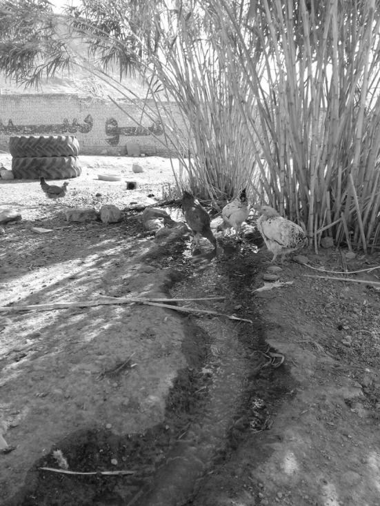
An example of location birds. A small flock of poultry
The results of this study showed that prevalence of H. meleagridis in the poultry was 31%, indicating a significant increase compared to the previous study, 11.9% in the poultry, and 6.5% in the birds (Badparva et al. 2015). In all the studied groups, there was also 10–60% of infection indicating a warning for the animals and economy of owners.
It is surprising that the epidemiological studies of this important pathogen parasite that can be considered as a basis for other studies have not been conducted in Iran, and been little in the world, while, the human parasites studies have been sufficiently studied (Badparva et al. 2014a; b), which can be due to the negligence of authorities towards the veterinary science and how to achieve clean and hygienic animal husbandry practices. Several methods have been reported for parasite transmission depending on risk factors associated such as the type of bird, habitat, geographic location, climatic conditions, nutrition, food consumption and even parasite strain. The high prevalence of parasite in the present study showed that the transmission facilities of this parasite are provided in the study area, but the type of transmission can be discussed. Although the cyst stage is reported, this type of transmission is not approved, so it cannot be invoked (Zaragatzki et al. 2010).
However, it has been shown that the oral administration of infected material was appropriate for introduction of histomoniasis (Tyzzer and Collier 1925), but the researchers have concluded that the birds cannot be infected by eating the new parasite-infected disposed material, which is probably due to the acidity of the gizzard and crop, because the infection is caused by eating the alkaline material, known as the modified infection (Horton-Smith and Long 1955; Lund 1956). Hence, it is accepted that the only way for transmission of H. meleagridis is through the cecal nematode eggs, H. gallinarum, to alive the parasite inside for several years as a resistant protection (Farr 1961; Hu et al. 2004). The eggs infected with parasite directly enter the avian digestive system or the birds are indirectly infected by eating the earthworms (Esquenet et al. 2003; Patra et al. 2013).
On one hand, the indices such as acidity of the gizzard, crop and parasite sensitivity, and on the other hand, the lack of eggs or H. gallinarum adult worms the oral transmission was ruled out, so the transmission should be direct since it is a sensitive parasite. At least three indices of infection source, transmission and host are required to transmit any disease agent. It was shown in this study that a percentage of each flock is the temporary host for other poultry, particularly, when there are clinical symptoms such as diarrhea or uncontrolled defecation reported. However, some studies introduced the ducks, an aquatic bird, as the host of this parasite (Takakuwa et al. 1998).
The cecum transmission was experimental and caused the infection (Hu et al. 2004). The sensitivity of H. meleagridis outside the body and lack of adult nematode or eggs indicating that the transmission should be performed quickly and directly, thus, there is no way except the water containing the parasite through the cloacal drinking. If accepted H. meleagridis such as Giardia lamblia, Cryptosporidium, and Entamoeba histolytica as the water-borne parasites, so it is influenced by the climatic environmental conditions such as heat (Slifko et al. 2000).
The high prevalence of parasites in the birds studied, indicating the susceptibility of the host for this parasite. If the source, host and transmission conditions are provided, the prevalence of infection is conducted easily, it might be an even epidemic, and the relative presence in all the studied area suggests the endemic state.
It is suggested first epidemiologic study conducted parasites in birds in order to determine the prevalence, transmission and the susceptible birds. Then the pathogenic strain is specified using the molecular methods, and the required prevention is achieved using the drugs with no side effects and necessary training.
Acknowledgements
The authors appreciate all people who helped in this research.
Authors’ contribution
Ebrahim Badparva designed the study. Ebrahim Badparva and Farnaz Kheirandish collaborated to the laboratory assays and the manuscript writing.
Compliance with ethical standards
Conflict of Interest
The authors declare that they have no conflict of interest.
References
- Aka J, Hauck R, Blankenstein P, Balczulat S, Hafez HM. Reoccurrence of histomonosis in turkey breeder farm. Berl Munch Tierarztl Wochenschr. 2010;124:2–7. [PubMed] [Google Scholar]
- Ali SA, Pirasteh S. Geological applications of landsat enhanced thematic mapper (ETM) data and geographic information system (GIS): mapping and structural interpretation in south-west Iran, Zagros structural belt. Int J Remote Sens. 2004;25:4715–4727. doi: 10.1080/01431160410001688295. [DOI] [Google Scholar]
- Badparva E, Kheirandish F, Ebrahimzade F. Prevalence of intestinal parasites in Lorestan province, west of Iran. Asian Pac J Trop Dis. 2014;4:S728–S732. doi: 10.1016/S2222-1808(14)60716-7. [DOI] [Google Scholar]
- Badparva E, Nayebzadeh H, Barkhordari MH, Ezatpour B. Epidemiological study of strongyloides stercoralis with a comparative diagnostic approach, in Lorestan, West of Iran. Arch Clin Infect Dis. 2014;9:e16815. doi: 10.5812/archcid.16815. [DOI] [Google Scholar]
- Badparva E, Ezatpour B, Azami M, Badparva M. First report of birds infection by intestinal parasites in Khorramabad, west Iran. J Parasit Dis. 2015;39:720–724. doi: 10.1007/s12639-014-0427-5. [DOI] [PMC free article] [PubMed] [Google Scholar]
- Callait-Cardinal M-P, Leroux S, Venereau E, Chauve C, Le Pottier G, Zenner L. Incidence of histomonosis in turkeys in France since the bans of dimetridazole and nifursol. Vet Rec. 2007;61:581–585. doi: 10.1136/vr.161.17.581. [DOI] [PubMed] [Google Scholar]
- Chossat L (2002) L’histomonose en production AOC “Dinde fermiere de Bresse” essai de prevention par phytotherapie. France These de Doctorat Veterinaire. Lyon 1
- Esquenet C, De Herdt P, De Bosschere H, Ronsmans S, Ducatelle R, Van Erum J. An outbreak of histomoniasis in free-range layer hens. Avian Pathol. 2003;32:305–308. doi: 10.1080/0307945031000097903. [DOI] [PubMed] [Google Scholar]
- Farr MM. Further observations on survival of the protozoan parasite, Histomonas meleagridis, and eggs of poultry nematodes in feces of infected birdes. Cornell Vet. 1961;51:3–13. [PubMed] [Google Scholar]
- Heelsbergen TV. Handbuch der Geflügelkrankheiten und der Geflügelzucht. Stuttgart: Ferdinand Enke; 1929. [Google Scholar]
- Horton-Smith C, Long P. The infection of chickens (Gallus gallus) with suspensions of the blackhead organism Histomonas meleagridis. Vet Res. 1955;67:79–90. [Google Scholar]
- Hu J, Fuller L, McDougald L. Infection of turkeys with Histomonas meleagridis by the cloacal drop method. Avian Dis. 2004;48:746–750. doi: 10.1637/7152. [DOI] [PubMed] [Google Scholar]
- Huber K, Reynaud M-C, Callait M, Zenner L. Histomonas meleagridis in turkeys: dissemination kinetics in host tissues after cloacal infection. Poult Sci. 2006;85:1008–1014. doi: 10.1093/ps/85.6.1008. [DOI] [PubMed] [Google Scholar]
- Lister S (2010) Histomoniasis in turkey breeders: a case report. Presented at 8th international symposium on turkey diseases, Berlin, Germany
- Lotfi A-R, Abdelwhab E, Hafez H. Persistence of Histomonas meleagridis in or on materials used in poultry houses. Avian Dis. 2012;56:224–226. doi: 10.1637/9519-090910-ResNote.1. [DOI] [PubMed] [Google Scholar]
- Lund EE. Oral transmission of Histomonas in turkeys. Poult Sci. 1956;35:900–904. doi: 10.3382/ps.0350900. [DOI] [Google Scholar]
- McDougald L. Other protozoan diseases of the intestinal tract. Dis Poult. 1997;10:890–899. [Google Scholar]
- McDougald L. Blackhead disease (histomoniasis) in poultry: a critical review. Avian Dis. 2005;49:462–476. doi: 10.1637/7420-081005R.1. [DOI] [PubMed] [Google Scholar]
- Patra G, Prasad H, Lalsiamthara J, Kataria J, Malsawmkima D, Lalrinkima H. Prevalence of Histomonas meleagridis in broiler chicken in different parts of Mizoram, India. Int J Poult Sci. 2013;12:98. doi: 10.3923/ijps.2013.98.101. [DOI] [Google Scholar]
- Slifko TR, Smith HV, Rose JB. Emerging parasite zoonoses associated with water and food. Int J Parasitol. 2000;30:1379–1393. doi: 10.1016/S0020-7519(00)00128-4. [DOI] [PubMed] [Google Scholar]
- Sorvari R, Sorvari T. Bursa Fabricii as a peripheral lymphoid organ: transport of various materials from the anal lips to the bursal lymphoid follicles with reference to its immunological. Immunol. 1977;32:499–505. [PubMed] [Google Scholar]
- Takakuwa H, Ito T, Takada A, Okazaki K, Kida H. Potentially virulent Newcastle disease viruses are maintained in migratory waterfowl populations. Jpn J Vet Res. 1998;45:207–215. [PubMed] [Google Scholar]
- Tyzzer EE, Collier J. Induced and natural transmission of blackhead in the absence of Heterakis. J Infect Dis. 1925;37:265–276. doi: 10.1093/infdis/37.3.265. [DOI] [Google Scholar]
- Zaragatzki E, Hess M, Grabensteiner E, Abdel-Ghaffar F, Al-Rasheid KA, Mehlhorn H. Light and transmission electron microscopic studies on the encystation of Histomonas meleagridis. Parasitol Res. 2010;106:977–983. doi: 10.1007/s00436-010-1777-2. [DOI] [PubMed] [Google Scholar]