Abstract
The current study was designed to study the persistence and distribution of caprine bone marrow derived mesenchymal stem cells (cBM-MSCs) when administered intra-dermally in experimentally induced cutaneous wounds in rabbits. MSC’s from goat bone marrow were isolated and their differentiation potential towards adipogenic and osteogenic lineages were assayed in vitro. The isolated cells were phenotypically analysed using flow cytometry for the expression of MSC specific matrix receptors (CD73, CD105 and Stro-1) and absence of hematopoietic lineage markers. Further, these in vitro expanded MSCs were stained with PKH26 lipophilic cell membrane red fluorescent dye and prepared for transplantation into cutaneous wounds created on rabbits. Five, 2 cm linear full thickness skin incisions were created on either side of dorsal midline of New Zealand white rabbits (n = 4). Four wounds in each animal were implanted intra-dermally with PKH26 labelled cBM-MSCs suspended in 500 µl of Phosphate Buffer Saline (PBS). Fifth wound was injected with PBS alone and treated as negative control. The skin samples were collected from respective wounds on 3, 7, 10 and 14 days after the wound creation, and cryosections of 6 µM were made from it. Fluorescent microscopy of these cryosections showed that the PKH26 labelled transplanted cells and their daughter cells demonstrated a diffuse pattern of distribution initially and were later concentrated towards the wound edges and finally appeared to be engrafted with the newly developed skin tissues. The labelled cells were found retained in the wound bed throughout the period of 14 days of experimental study with a gradual decline in their intensity of red fluorescence probably due to the dye dilution as a result of multiple cell division. The retention of transplanted MSCs within the wound bed even after the complete wound healing suggests that in addition to their paracrine actions as already been reported, they may have direct involvement in various stages of intricate wound healing process which needs to be explored further.
Electronic supplementary material
The online version of this article (doi:10.1007/s10616-017-0097-0) contains supplementary material, which is available to authorized users.
Keywords: Bone marrow derived mesenchymal stem cells, Caprine, Cutaneous wound healing, PKH26, In vivo tracking, Hair regeneration
Introduction
Friedenstein et al. (1974), first defined mesenchymal stem cells (MSCs) as a population of cells derived from bone marrow that had the fibroblastic phenotype and could differentiate into osteocytes, adipocytes, and chondrocytes. In addition to their multi-lineage differentiation potential to multiple cell types in vitro and in vivo, MSCs and MSC-like stem cells have been proven to exhibit immune-modulatory and immunological tolerance inducing characteristics (Ramasamy et al. 2008). MSCs as a major source of cells for cytotherapy, have been used in the human and veterinary medicine for more than a decade. From animal models to clinical trials, MSCs have offered commitment in the treatment of various diseases, especially immune disorders and tissue injury (Lee et al. 2009; Inoue et al. 2007; Chen et al. 2008).
For cell based therapy, one of the essential step before cells can be applied in clinics is finding a non-invasive technique to track stem cells recruited inside the body. The lipophilic fluorescent dye PKH26, which primarily binds to the cell membrane, has been used as the cell tracer to locate the grafted cells in host tissues (Li et al. 2013). Since the red fluorescence of PKH26 labelled cells persist for long time both in vivo and in vitro, it has been successfully utilised in tracing metastasis of carcinoma cells (Yamagata and Kumagai 2000), migration of stem cells (Tang et al. 2009), proliferation and migration of neuronal stem cells (Bantubungi et al. 2008) etc.
There are reports of successful cross species administration of mesenchymal stem cells using human, pig, rat and guinea pig with maximum information on human MSCs which are reportedly functional in multiple recipient species including mouse (Zhao et al. 2008), sheep (Mackenzie and Flake 2001), dog (Plotnikov et al. 2007) and pig (Henriksson et al. 2009). This was later differed by Lyngbaek et al. (2010) who found that transplantation of human bone marrow-derived MSC into a pig heart resulted in a rapid inflammatory response and cell degradation.
The beneficial effect of MSCs on the intricate process of cutaneous wound healing has been well established (Rodriguez-Menocal et al. 2015; Khong et al. 2015). It has been reported that through paracrine communications, MSCs increase rate of wound closure, decrease wound inflammation, encourage angiogenesis, regulate extracellular matrix healing events, and promote regeneration of proper skin structure and function (Schlosser et al. 2012). However, there is no detailed report regarding the persistence/fate and migration pattern of these transplanted cells at the site of injury when administered intra-dermally. Hence in the current study, we have made an attempt to demonstrate how long these transplanted stem cells and their daughter cells are retained at the wound site when administered intra-dermally. This study probably would shed light on the involvement of MSCs in various stages of the wound healing mechanism. To demonstrate that, goat bone marrow derived MSCs have been isolated, and characterised as per the criteria given by International society of cell therapy (Dominici et al. 2006). Further, the MSCs have been labelled using PKH26 dye to track them in vivo in full thickness incisional wound healing model induced on rabbits.
Materials and methods
All the necessary chemicals used in present study were purchased from Sigma (St Louis, MO, USA), unless otherwise indicated. The primary and secondary antibodies used for flow cytometry were procured from Santa Cruz, Biotechnology (Cruz, Santa Cruz, CA, USA).
Isolation and in vitro culture of caprine bone marrow cells
Adult healthy goats of either sex were used for bone marrow collection. Collection procedure was done under epidural anesthesia using 2% lignocaine. All procedures were in compliance with the guidelines of ICAR-IVRI Institutional Animal Ethics Committee (IAEC). Bone marrow was aspirated with 16G bone marrow biopsy needle from the aseptically prepared iliac crest area. 5 ml of bone marrow aspirate was collected in a 10 ml heparinised syringe. The bone marrow samples were diluted with equal amount of sterile PBS and the mononuclear cells were isolated by density gradient centrifugation method using histopaque 1077 as per manufacturer’s instruction (Gade et al. 2013). The isolated cells from caprine bone marrow aspirates were seeded at a density of approximately 103 cells/cm2 in a 6 well culture plate (Nunc, Thermo Fisher Scientific, Inc., Roskilde, Denmark) containing DMEM supplemented with 10% FBS and 1% Penicillin/Streptomycin. Culture plates were incubated in humidified CO2 incubator at 37 °C in presence of 5% CO2 in humidified air. Nonadherent cells were washed off, and the adherent cells were expanded. When adherent cells became confluent, they were continuously sub cultured until 4th passage. For all experiments cells were taken from the forth passage.
Phenotypic characterization of MSCs
Caprine bone marrow derived MSCs (cBM-MSCs) at 4th passage were demonstrated for the expression of their specific MSC markers by flow cytometry. Briefly, a single cell suspension of 1.0 × 10⁶ cells/ml was placed in 100 μL of PBS supplemented with 2% FBS and was incubated with primary antibodies against CD-73, CD-105, Stro-1 and CD-34 (goat polyclonal, Santa Cruz Biotechnology) for 45 min. Thereafter the cells were washed with PBS, and were further incubated with FITC conjugated secondary antibodies (donkey anti-goat, Santa Cruz Biotechnology) for 40 min in dark chamber. Finally stained cells were washed and re-suspended in PBS containing 2% FBS. Cell fluorescence was assayed immediately in a FACS Calibur flow cytometer (Becton–Dickinson, San Jose, CA, USA), and the data were analysed using Cell Quest software (Becton– Dickinson). Isotype-identical antibodies served as controls.
Differentiation assay
Osteogenic differentiation of MSCs from goat was tested following the protocol of Pratheesh et al. (2013). In short, MSCs were expanded in complete medium containing 10% FBS until subconfluence after the forth passage. The complete medium then was replaced by specific osteogenesis differentiation medium (Stem-Pro-Invitrogen). After 21 days, cells were fixed in 10% buffered formalin for 1 h followed by von Kossa staining to visualize the calcium deposits in the adherent monolayer.
Adipogenic differentiation of cBM-MSCs was evaluated following the protocol of Pratheesh et al. (2014). Subconfluent MSC after the forth passage were incubated in specific adipogenic differentiation medium (Stem-Pro-Invitrogen). After 21 days of co-incubation, Oil Red O staining was performed to demonstrate the presence of fat droplets.
Labelling of cells with PKH26 dye
Caprine BM-MSCs at passage 4 were labelled with the red fluorescent dye PKH26 (Sigma-Aldrich, St. Louis, USA) according to the manufacturer’s protocol. Briefly, the 80–90% confluent cBMMSCs were detached by Accutase (Invitrogen; Thermo Fisher Scientific, Inc.), washed using serum-free DMEM and resuspended in 1 ml of ‘Diluent C’ from the PKH26 Red Fluorescent Cell Linker kit (cat. no. PKH26-GL; Sigma-Aldrich). The cell suspension was mixed with an equal volume of the labelling solution (containing 4 nM PKH26) and incubated at 25 °C for 5 min. The staining process was stopped by the addition of 2 ml fetal bovine serum, cells were washed twice with DMEM and observed under fluorescent microscope.
In vitro counting of labelled MSCs
PKH26-positive cBMMSCs were observed by fluorescence microscopy at 24 h after seeding in a 24 well culture plate (Nunc, Thermo Fisher Scientific, Inc.). A total of 200 cells each were counted in 5 selected wells of the culture plate. Cells were counted in each field of view under both bright light and fluorescence filter, and the labelling efficiency was calculated as: (The number of PKH26-positive cells/200) × 100. Upon attaining confluence, labelled cells were subcultured up to three passages to check the persistence of fluorescence in the cells.
Animal experiment
Four adult male New Zealand white rabbits with an average weight of 1–1.5 k g were used in this study. The animals were housed at room temperature and had access to food and water ad libitum. The animals were given 1 week acclimatization period before starting the experiment. They were treated in accordance with the international guidelines for the care and use of laboratory animals and the experiment protocol was permitted by the Ethics Committee, ICAR-Indian Veterinary Research Institute. Possible efforts were taken to ensure minimal animal sufferings.
Induction of incisional wounds in rabbits and transplantation of labelled cells
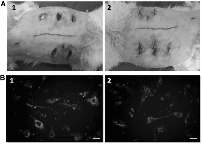
Each rabbit was anesthetized by an intramuscular injection of ketamine and xylazine, at dose of 60 and 6 mg/kg body weight, respectively. The surgical area was shaved with an electric razor, minute hairs were removed by hair remover cream and disinfected using 70% ethanol. After a deep surgical plane of general anaesthesia had been reached, 2 cm long incisional wounds were created in the dorsal skin of the animals. A grid line was made using a permanent marker, leaving a 3 cm margin between wounds, to avoid the risk of cell migration to adjacent wounds. Each animal received 5 wounds, with 3 wounds on one side and 2 wounds on opposite side of dorsal line (Fig. 2A). Both the epidermal and dermal layers were incised down to the subcutaneous connective tissue, creating a full-skin thickness wound. The wounds were then sutured (interrupted) using nylon threads (Fig. 2B). A 5 ml syringe with a 22 gauge needle was then used to administer PKH26 labelled caprine BM-MSCs suspended in approximately 500 µl of the sterile PBS to each wound (four wounds per animal) intra-dermally along the suture line. Sterile PBS alone was injected to the fifth wound which was maintained as a negative control. The wounds were left untouched for 5–10 min and allowed to dry.
Fig. 2.
A Induction of incisional wounds upon rabbits. (1) Five incisional wounds created on either side of the dorsal midline. (2) Wounds created were sutured (interrupted) by nylon threads. B PKH26 labeling of caprine BMMSCs in vitro. (1) Adherent labeled cells at first passage showing typical cell membrane distribution of the dye excluding the nucleus (Scale bar 50 μm). (2) Adherent labeled cells at third passage with comparatively less intense fluorescence due to dye dilution (Scale bar 50 μm). (Color figure online)
Collection of skin biopsy
The animals were re-anaesthetized using xylazine–ketamine cocktail at the aforementioned dosage. Full thickness skin specimens from the healing wound (one out of five wounds created on each animals) with a safety margin of 0.5 cm of healthy skin around the wound were excised, respectively, at the 3rd, 7th, 10th and 14th days post transplantation from each experimental animals (n = 4). The samples were then rapidly embedded in frozen section embedding medium (Fisher Scientific, Springfield, NJ, USA). Thin 6 µm sections were cut on a cryostat at −20 °C, mounted on glass microscope slides and protected from light until evaluation by fluorescence and light microscopy. The third day skin cryo-sections were maintained as positive control.
Result and discussion
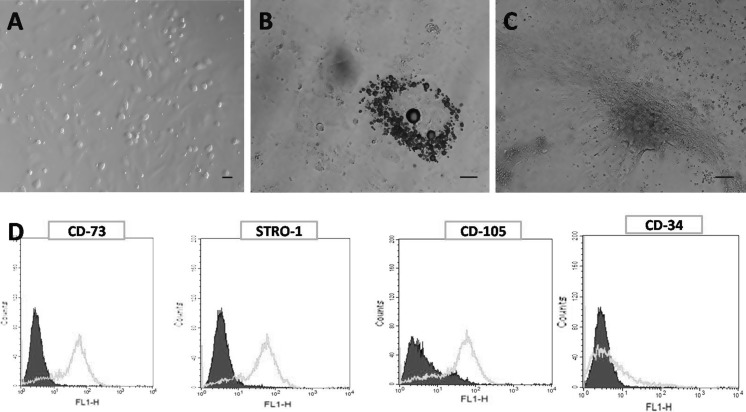
A density gradient technique was used to isolate caprine bone marrow derived mesenchymal stem cells (cBMMSCs) as established in humans (Juopperi et al. 2007) and in other animal species (Gade et al. 2013). After 24 h the cells were found attached to the plastic wall of cell culture dishes. These cells were fibroblastoid with spindle shaped or polymorphic morphology (Fig. 1A). Replacing the medium and subculturing, cBMMSCs were purified. The subcultured cells had identical morphology. Passaging was performed once in every 5 days and more than 4 times. Flow cytometry demonstrated that cBMMSCs did not express hematopoietic marker CD34, but had high expression of MSCs specific markers CD73, CD105 and Stro-1 (Fig. 1D). MSC identity of in vitro cultured caprine bone marrow derived cells was further proven by differentiation into adipogenic (Fig. 1B) and osteogenic cells (Fig. 1C) as demonstrated by Oil Red O and von Kossa staining respectively.
Fig. 1.
In vitro culture and characterization of caprine BM-MSCs. A Morphology of caprine bone marrow derived mesenchymal stem cells exhibiting typical fibroblastoid phenotype in primary culture (Scale bar 30 μm). B Adipogenesis induced lipid droplets observed in red colour after specific Oil Red O staining in in vitro expanded MSCs (Scale bar 50 μm). C Osteogenic differentiation of MSCs, brownish black coloured mineral deposition as demonstrated by von Kossa staining (Scale bar 50 μm). D Flowcytometric analysis of surface antigens in in vitro expanded caprine Bone marrow derived MSCs. Cells were stained with primary antibodies directed against CD-73, STRO-1, CD-105, CD-34 and counter stained by FITC conjugated secondary antibodies. Calibrated histogram represents the number of events on the Y-axis and FITC-fluorescent intensity (FLH-1) on X-axis. The shadowed histogram indicate negative controls. (Color figure online)
One classical method to label cells is using viral vectors to express fluorescent proteins, which has been a costly and complicated procedure and is associated with the toxicity problems (Tucker 2001). We have used red fluorescent lipophilic dye PKH26 that reportedly integrates into the cell membrane stably, without disturbing its surface marker expression (Parish 1999). In the present study, the labelling efficiency of PKH26 with cBMMSCs was analysed for pooled samples (n = 5) at 24 h post seeding and was found to be 98.5 ± 0.5% in adherent cells (Fig. 2C). Similarly high labelling efficiency has been reported by Shao-Fang et al. (2011) where they had used PKH26 to label human umbilical cord MSCs. The red fluorescence of PKH26 labelled cBMMSCs was observed even in 3rd passage adherent cells, but with reduced intensity than initial passages probably due to dye dilution with multiple cell divisions (Fig. 2D). Furthermore, the digital photographs of PKH26 labelled cells reveals that the dye probably stains the cell organelles and membranes excluding the nucleus and cytosol (Askenasy and Farkas 2002; Fig. 2C).
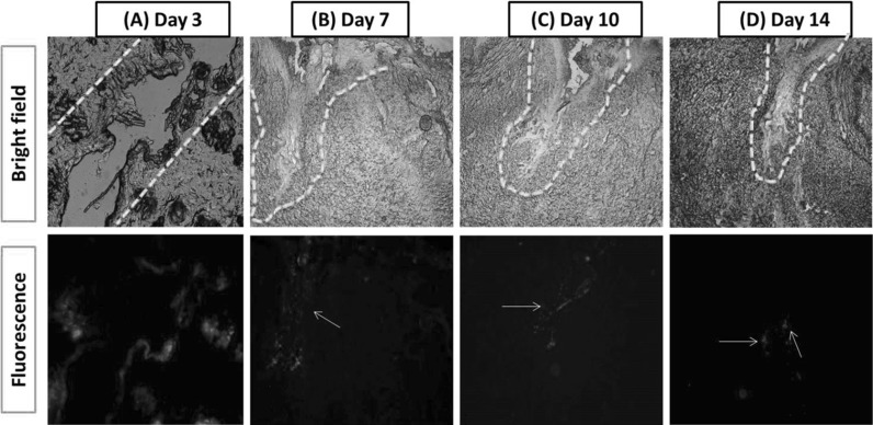
It has been reported that, following the systemic administration of MSCs, homing of MSCs at the site of injury is stated to be inefficient and many MSCs are trapped in the lungs thereby making it difficult to trace the fate of the injected cells (Hu et al. 2013). Most studies have used the technique of injecting MSCs intra-dermally into or around the wound area. Although this method has been shown to improve wound healing, there was always an apprehension about the engraftment efficiency and cell retention at the wound site (Wagner et al. 2009). Hence in the present study, authors attempted to exhibit the persistence/fate and migration pattern of transplanted cells in the site of injury when administered intra-dermally. To demonstrate that, incisional wounds created upon rabbits were treated with PKH26 labelled cBMMSCs and were imaged at day 3, 7, 10 and 14 post transplantation. The 3rd day skin cryosection which has been maintained as the positive control exhibited a diffuse dispersion pattern for the cBM-MSCs, which specifically found trapped within the hair follicles and injured tissues in the immediate wound area (Fig. 3A). At day 7, the labelled caprine stem cells were found to be concentrated at newly formed skin areas probably due to the high vascularity and angiogenesis (Fig. 3B). However, by day 10, the recruited caprine MSCs were observed primarily located around the edge of wound (Fig. 3C). It was hypothesised that the chemotaxis signals (SDF-1a) expressed by wound were mainly dispersed around the edge of incision, which promote the enrichment of cBM-MSCs in the region of the wound edge and the lack of cBM-MSCs in the centre of wound (Chen et al. 2015). Grossly the incisional wounds created on rabbits were healed up completely leaving a scar by 12–13 days post-surgery. Even then the skin sections collected at day 14 exhibited the red fluorescence at low intensity in the regenerated skin area (Fig. 3D). The presence of red fluorescence at the wound area even after the complete healing of wounds apparently shows that the stem cells have got an undeniable role in remodelling phase, which is the last stage of the wound healing process (Martin 1997). The progressive reduction in the intensity of the red fluorescence must be due to dilution of the membrane dye by multiple cell divisions. Similarly has been reported by Parish (1999) that, the fluorescent intensity of the PKH26 dye will decrease gradually after it is being distributed to their daughter cells. Although in the present study, MSCs have been shown to demonstrate long-term integration into healing wounds, a high number of research suggests that their therapeutic advantage is credited to their release of trophic mediators like stromal cell-derived factor-1, epidermal growth factor, vascular endothelial growth factor (VEGF), keratinocyte growth factor, insulin-like growth factor, and matrix metalloproteinase-9, rather than a direct structural contribution (Kirana et al. 2012). Although PKH26 has been proven to be a safe fluorescent marker with good biocompatibility, a recent study by Li et al. (2013) has indicated that PKH labeling was not limited to the transplanted cells. Later it has been contradicted by Nagyova et al. (2014) who has demonstrated that direct co-cultures of PKH labeled rat MSCs with unlabeled rat MSCs did not transfer dye from donor to unlabeled recipient cells apparently due to the lack of ‘Diluent C(R)’ which is an iso-osmotic, salt and solvent free staining vehicle which facilitates the instantaneous segregation of the dye into the lipid bilayer of cell membranes (Wallace et al. 2008). Furthermore, gradual reduction in the intensity of the red fluorescence as the day progresses authenticate our results in the present study.
Fig. 3.
Persistence of PKH26 labelled caprine BMMSCs at different time points post transplantation when observed under bright field and fluorescent microscopy. Dotted line represents the site of injury. White arrows points the distribution of PKH26 labelled cells segregated along the wound margin and within wound bed. (Color figure online)
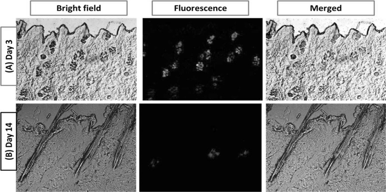
In addition to this, the current experiment demonstrated that the transplanted stem cells have a tendency to get localized in and around the hair follicle region (Fig. 4A) and which were retained there throughout the experimental period of fourteen days (Fig. 4B). This may be attributed to the involvement and contribution of exogenous stem cells in hair follicle development as described by Toyoshima et al. (2012). This observation certainly will provide a significant contribution to the further advancement of tissue engineering techniques that will facilitate future regenerative treatment for hair loss caused by trauma or by diseases such as alopecia and baldness.
Fig. 4.
A Localization of PKH26 labelled caprine BMMSCs in and around hair follicles on 3rd day post injection B which found persisting throughout the experimental period of 14 days even after the complete healing of wound. (Color figure online)
Conclusion
Herein we demonstrated that the transplanted stem cells and their daughter cells can persist at the wound site even after the completion of healing process suggesting their inevitable roles in all phases of wound healing mechanism. Also it was validated that, labelling cells with PKH26 may suggest a potential prospect for longitudinal tracking of MSC or other cell types without negotiating its cell migration ability. Additionally our observations significantly improve the technological progress of bioengineered hair follicles in regenerative therapy.
Electronic supplementary material
Below is the link to the electronic supplementary material.
Supplementary material (Figure)-Negative control wound cryo-sections administered with PBS alone when observed under bright field and fluorescent microscopy did not observe any red fluorescent signals at 3rd and 14th day post surgery (JPEG 172 kb)
Acknowledgements
Authors are thankful to Director, ICAR-IVRI for providing necessary financial assistance.
Compliance with ethical standards
Conflict of interest
None of the authors have any conflict of interest to declare.
Footnotes
Electronic supplementary material
The online version of this article (doi:10.1007/s10616-017-0097-0) contains supplementary material, which is available to authorized users.
Contributor Information
M. D. Pratheesh, Email: pratheesh@kvasu.ac.in
G. Taru Sharma, Email: gts553@ivri.res.in.
References
- Askenasy N, Farkas DL. Optical imaging of PKH-labeled hematopoietic cells in recipient bone marrow in vivo. Stem Cells. 2002;20:501–513. doi: 10.1634/stemcells.20-6-501. [DOI] [PubMed] [Google Scholar]
- Bantubungi K, Blum D, Cuvelier L, Wislet-Gendebien S, Rogister B, Brouillet E, Schiffmann SN. Stem cell factor and mesenchymal and neural stem cell transplantation in a rat model of Huntington’s disease. Mol Cell Neurosci. 2008;37:454–470. doi: 10.1016/j.mcn.2007.11.001. [DOI] [PubMed] [Google Scholar]
- Chen L, Tredget EE, Wu PY, Wu Y. Paracrine factors of mesenchymal stem cells recruit macrophages and endothelial lineage cells and enhance wound healing. PLoS ONE. 2008;3:e1886. doi: 10.1371/journal.pone.0001886. [DOI] [PMC free article] [PubMed] [Google Scholar]
- Chen G, Tian F, Li C, Zhang Y, Weng Z, Zhang Y, Peng R, Wang Q. In vivo real-time visualization of mesenchymal stem cells tropism for cutaneous regeneration using NIR-II fluorescence imaging. Biomaterials. 2015;53:265–273. doi: 10.1016/j.biomaterials.2015.02.090. [DOI] [PubMed] [Google Scholar]
- Dominici M, Le Blanc K, Mueller I, Slaper-Cortenbach I, Marini F, Krause D, Deans R, Keating A, Prockop DJ, Horwitz E. Minimal criteria for defining multipotentmesenchymal stromal cells. The International Society for Cellular Therapy position statement. Cytotherapy. 2006;8:315–317. doi: 10.1080/14653240600855905. [DOI] [PubMed] [Google Scholar]
- Friedenstein J, Chailakhyan RK, Latsinik NV. Stromal cells responsible for transferring the microenvironment of the hemopoietic tissues. Cloning in vitro and retransplantation in vivo. Transplantation. 1974;17:331–340. doi: 10.1097/00007890-197404000-00001. [DOI] [PubMed] [Google Scholar]
- Gade NE, Pratheesh MD, Nath A, Dubey PK, Amarpal S, Sharma B, Saikumar G, Sharma GT. Molecular and cellular characterization of buffalo bone marrow derived mesenchymal stem cells. Reprod Domest Anim. 2013;48:358–367. doi: 10.1111/j.1439-0531.2012.02156.x. [DOI] [PubMed] [Google Scholar]
- Henriksson HB, Svanvik T, Jonsson M, Hagman M, Horn M, Lindahl A, Brisby H. Transplantation of human mesenchymal stems cells into intervertebral discs in a xenogeneic porcine model. Spine. 2009;34:141–148. doi: 10.1097/BRS.0b013e31818f8c20. [DOI] [PubMed] [Google Scholar]
- Hu C, Yong X, Li C, Lü M, Liu D, Chen L, Hu J, Teng M, Zhang D, Fan Y, Liang G. CXCL12/CXCR4 axis promotes mesenchymal stem cell mobilization to burn wounds and contributes to wound repair. J Surg Res. 2013;183:427–434. doi: 10.1016/j.jss.2013.01.019. [DOI] [PubMed] [Google Scholar]
- Inoue Y, Iriyama A, Ueno S, Takahashi H, Kondo M, Tamaki Y, Araie M, Yanagi Y. Subretinal transplantation of bone marrow mesenchymal stem cells delays retinal degeneration in the RCS rat model of retinal degeneration. Exp Eye Res. 2007;85:234–241. doi: 10.1016/j.exer.2007.04.007. [DOI] [PubMed] [Google Scholar]
- Juopperi TA, Schuler W, Yuan X, Collector MI, Dang CV, Sharkis SJ. Isolation of bone marrow-derived stem cells using density-gradient separation. Exp Hematol. 2007;35:335–341. doi: 10.1016/j.exphem.2006.09.014. [DOI] [PubMed] [Google Scholar]
- Khong SML, Duscher D, Januszyk M, Than PA, Maan Z, Gurtner GC. Single cell transcriptomic analysis of human fetal bone marrow-derived mesenchymal stem cells in wound healing. Plast Reconstr Surg. 2015;135:123–124. doi: 10.1097/01.prs.0000465624.46646.20. [DOI] [Google Scholar]
- Kirana S, Stratmann B, Prante C, Prohaska W, Koerperich H, Lammers D, Gastens MH, Quast T, Negrean M, Stirban OA, Nandrean SG, Götting C, Minartz P, Kleesiek K, Tschoepe D. Autologous stem cell therapy in the treatment of limb ischaemia induced chronic tissue ulcers of diabetic foot patients. Int J Clin Pract. 2012;66:384–393. doi: 10.1111/j.1742-1241.2011.02886.x. [DOI] [PubMed] [Google Scholar]
- Lee JW, Fang X, Gupta N, Serikov V, Matthay MA. Allogeneic human mesenchymal stem cells for treatment of E. coli endotoxin-induced acute lung injury in the ex vivo perfused human lung. Proc Natl Acad Sci USA. 2009;106:16357–16362. doi: 10.1073/pnas.0907996106. [DOI] [PMC free article] [PubMed] [Google Scholar]
- Li P, Zhang R, Sun H, Chen L, Liu F, Yao C, Du M, Jiang X. PKH26 can transfer to host cells in vitro and vivo. Stem Cells Dev. 2013;22:340–344. doi: 10.1089/scd.2012.0357. [DOI] [PMC free article] [PubMed] [Google Scholar]
- Lyngbaek S, Ripa RS, Haack-Sørensen M, Cortsen A, Kragh L, Andersen CB, Jørgensen E, Kjaer A, Kastrup J, Hesse B. Serial in vivo imaging of the porcine heart after percutaneous, intramyocardially injected 111In-labeled human mesenchymal stromal cells. Int J Cardiovasc Imaging. 2010;26:273. doi: 10.1007/s10554-009-9532-4. [DOI] [PubMed] [Google Scholar]
- Mackenzie TC, Flake AW. Human mesenchymal stem cells persist, demonstrate site-specific multipotential differentiation, and are present in sites of wound healing and tissue regeneration after transplantation into fetal sheep. Blood Cells Mol Dis. 2001;27:601–604. doi: 10.1006/bcmd.2001.0424. [DOI] [PubMed] [Google Scholar]
- Martin P. Wound healing-aiming for perfect skin regeneration. Science. 1997;276:75–81. doi: 10.1126/science.276.5309.75. [DOI] [PubMed] [Google Scholar]
- Nagyova M, Slovinska L, Blasko J, Grulova I, Kuricova M, Cigankova V, Harvanova D, Cizkova DA. Comparative study of PKH67, DiI, and BrdU labeling techniques for tracing rat mesenchymal stem cells. In Vitro Cell Dev Biol Anim. 2014;50:656–663. doi: 10.1007/s11626-014-9750-5. [DOI] [PubMed] [Google Scholar]
- Parish CR. Fluorescent dyes for lymphocyte migration and proliferation studies. Immunol Cell Biol. 1999;77:499–508. doi: 10.1046/j.1440-1711.1999.00877.x. [DOI] [PubMed] [Google Scholar]
- Plotnikov AN, Shlapakova I, Szabolcs MJ, Danilo P, Jr, Lorell BH, Potapova IA, Lu Z, Rosen AB, Mathias RT, Brink PR, Robinson RB, Cohen IS, Rosen MR. Xenografted adult human mesenchymal stem cells provide a platform for sustained biological pacemaker function in canine heart. Circulation. 2007;116:706–713. doi: 10.1161/CIRCULATIONAHA.107.703231. [DOI] [PubMed] [Google Scholar]
- Pratheesh MD, Gade NE, Katiyar AN, Dubey PK, Sharma B, Saikumar G, Amarpal S, Sharma GT. Isolation, culture and characterization of caprine mesenchymal stem cells derived from amniotic fluid. Res Vet Sci. 2013;94:313–319. doi: 10.1016/j.rvsc.2012.08.002. [DOI] [PubMed] [Google Scholar]
- Pratheesh MD, Gade NE, Dubey PK, Nath A, Sivanarayanan TB, Madhu DN, Sharma B, Amarpal S, Saikumar G, Sharma GT. Molecular characterization and xenogenic application of Wharton’s jelly derived caprinemesenchymal stem cells. Vet Res Commun. 2014;38:139–148. doi: 10.1007/s11259-014-9597-y. [DOI] [PubMed] [Google Scholar]
- Ramasamy R, Tong CK, Seow HF, Vidyadaran S, Dazzi F. The immunosuppressive effects of human bone marrow-derived mesenchymal stem cells target T cell proliferation but not its effector function. Cell Immunol. 2008;251:131–136. doi: 10.1016/j.cellimm.2008.04.009. [DOI] [PubMed] [Google Scholar]
- Rodriguez-Menocal L, Shareef S, Salgado M, Shabbir A, Van Badiavas E. Role of whole bone marrow, whole bone marrow cultured cells, and mesenchymal stem cells in chronic wound healing. Stem Cell Res Ther. 2015;6:1–11. doi: 10.1186/s13287-015-0001-9. [DOI] [PMC free article] [PubMed] [Google Scholar]
- Schlosser S, Dennler C, Schweizer R, Eberli D, Stein JV, Enzmann V, Giovanoli P, Erni D, Plock JA. Paracrine effects of mesenchymal stem cells enhance vascular regeneration in ischemic murine skin. Microvasc Res. 2012;83:267–275. doi: 10.1016/j.mvr.2012.02.011. [DOI] [PubMed] [Google Scholar]
- Shao-Fang Z, Hong-Tian Z, Zhi-Nian Z, Yuan-Li H. PKH26 as a fluorescent label for live human umbilical mesenchymal stem cells. In Vitro Cell Dev Biol Anim. 2011;47:516–520. doi: 10.1007/s11626-011-9424-5. [DOI] [PubMed] [Google Scholar]
- Tang J, Wang J, Kong X, Yang J, Guo L, Zheng F, Zhang L, Huang Y, Wan Y. Vascular endothelial growth factor promotes cardiac stem cell migration via the PI3K/Akt pathway. Exp Cell Res. 2009;315:3521–3531. doi: 10.1016/j.yexcr.2009.09.026. [DOI] [PubMed] [Google Scholar]
- Toyoshima KE, Asakawa K, Ishibashi N, Toki H, Ogawa M, Hasegawa T, Irié T, Tachikawa T, Sato A, Takeda A, Tsuji T. Fully functional hair follicle regeneration through the rearrangement of stem cells and their niches. Nat Commun. 2012;3:784. doi: 10.1038/ncomms1784. [DOI] [PMC free article] [PubMed] [Google Scholar]
- Tucker KL. In vivo imaging of the mammalian nervous system using fluorescent proteins. Histochem Cell Biol. 2001;115:31–39. doi: 10.1007/s004180000213. [DOI] [PubMed] [Google Scholar]
- Wagner J, Kean T, Young R, Dennis JE, Caplan AI. Optimizing mesenchymal stem cell-based therapeutics. Curr Opin Biotechnol. 2009;20:531–536. doi: 10.1016/j.copbio.2009.08.009. [DOI] [PubMed] [Google Scholar]
- Wallace PK, Tario JD, Jr, Fisher JL, Wallace SS, Ernstoff MS, Muirhead KA. Tracking antigen-driven responses by flow cytometry: monitoring proliferation by dye dilution. Cytom A. 2008;73:1019–1034. doi: 10.1002/cyto.a.20619. [DOI] [PubMed] [Google Scholar]
- Yamagata K, Kumagai K. Experimental study of lymphogenous peritoneal cancer dissemination: migration of fluorescent-labelled tumor cells in a rat model of mesenteric lymph vessel obstruction. J Exp Clin Cancer Res. 2000;219:211–217. [PubMed] [Google Scholar]
- Zhao M, Amiel SA, Ajami S, Jiang J, Rela M, Heaton N, Huang GC. Amelioration of streptozotocin-induced diabetes in mice with cells derived from human marrow stromal cells. PLoS ONE. 2008;3:e2666. doi: 10.1371/journal.pone.0002666. [DOI] [PMC free article] [PubMed] [Google Scholar]
Associated Data
This section collects any data citations, data availability statements, or supplementary materials included in this article.
Supplementary Materials
Supplementary material (Figure)-Negative control wound cryo-sections administered with PBS alone when observed under bright field and fluorescent microscopy did not observe any red fluorescent signals at 3rd and 14th day post surgery (JPEG 172 kb)


