Abstract
Clostridium botulinum strains secrete their neurotoxins (BoNT) along with a group of neurotoxin-associated proteins (NAPs) that enhance the oral toxicity and provide protection to the neurotoxin against acidity, temperature and proteases in the G.I. tract. A major component of NAPs is Hn-33, a 33kDa protein, which is also protease resistant and strongly protects BoNT. The complex form of BoNT/A is used as a commercial therapeutic formulation against many neuromuscular disorders and for cosmetic purposes. Immune response against this formulation could hinder its long-term use; therefore, it is important to characterize the immunological properties of the associated proteins. This study aims to understand the immunological reactivity of BoNT/A complex, BoNT, NAPs, and Hn-33 through a series of competitive enzyme-linked immunosorbent assays (ELISA). The results indicated that BoNT/A complex competed 6 times more with complex antibodies compared to the neurotoxin confirming that the higher immunogenicity of BoNT/A complex was indeed a result of the associated proteins with the neurotoxin complex. While the nearly identical immuno-reactivity of BoNT/A complex and Hn-33 with Hn-33 antibodies indicated that the reactivity was due to the higher immunogenicity not the abundance of Hn-33 in the complex. Both the ELISA and immuno-blot results implied that Hn-33 is primarily responsible for eliciting the antibody response in BoNT/A complex, therefore it may be possible to employ Hn-33 as an adjuvant for development of vaccines against botulism.
Keywords: Botulinum, Clostridium, Complex, Hn-33, Immunogenicity, ELISA
1. Introduction
There are seven immunogenically distinct serotypes of botulinum neurotoxin (BoNT) A-G that are produced by various strains of Clostridum botulinum. BoNT acts on the neuromuscular junction blocking the release of the neurotransmitter acetylcholine thereby resulting in flaccid muscle paralysis (Cai et al., 2007). Clostridium botulinum strains secrete their neurotoxins along with a group of other proteins that exhibit hemagglutinin activity known as neurotoxin associated proteins (NAPs), which vary in number depending on the serotype and the form of progenitor toxin (Fu, 1998). The NAPs provide protection to the neurotoxin against environmental adversities such as acidity and temperature, and against proteolytic attack in gastric juice (Sakaguchi, 1983). The complex form of BoNT is currently used in therapeutic applications, but the role of NAPs in these formulations remains unclear.
BoNT has been used for over 20 years as therapeutic agent for treatment of various diseases and as a cosmetic enhancement tool to remove wrinkles. BoNT ability to paralyze or weaken the injected muscle over a certain period of time and leave other muscles unaffected makes its utility as a pharmaceutical ideal. Currently, the botulinum neurotoxin type A (BoNT/A) complex , commercially known as Botox™ (Allegran, Inc) and Dysport™, and type B complex, commercially known as Myobloc™ and Neurobloc™, are safe and effective for the treatment of numerous kinds of muscle disorders including dystonias, spasticity, tremors, and migraines (Schantz, 1992; Ting, 2004). Although the other serotypes of BoNT C, E, F are not commercially available they are under investigation for their therapeutic potential. There has been extensive research on NAPs in order to understand their biological and structural role in BoNT complexes and their interaction with the toxin, but there is limited research on understanding the immunological role of individual components of BoNT/A complex. Further research on the immunogenicity of BoNT/A complex proteins will help in understanding immuno-resistance to botulinum therapy and developing more efficient vaccines against botulism.
This research aimed to understand and characterize the immunological reactivity of BoNT/A and its associated proteins through a series of enzyme-linked immuno-sorbent assays (ELISA). Using ELISA technique to assess immuno-reactivity is at times marred with alterations in the conformation of the antigen upon binding with the plates; however, competitive ELISA provides a convenient avenue to distinguish immuno-reactivity of antigens in solution and bound state. Our findings with competitive ELISA confirmed that the Hn-33 component of BoNT/A complex accounts for most of the immuno-reactivity of BoNT/A complex, and that BoNT/A is accessible to antibodies even in its complex form.
2. Materials and Methods
2.1 Materials
96-well immuno micro-titer plates (Thermo Fisher Scientific, Pittsburg, PA), dry milk, rabbit anti-BoNT/A complex IgG (Merdian Life Science, Inc., Saco, ME), rabbit anti-BoNT/A IgG (Merdian Life Science, Inc., Saco, ME), rabbit anti-Hn-33 IgG (Merdian Life Science, Inc., Saco, ME), goat anti-rabbit IgG alkaline phosphatase antibody (Sigma Aldrich, St. Louis, MO), anti-rabbit IgG-HRP conjugated secondary antibody (Santa Cruz Biotechnology Inc., Santa Cruz, CA), 2,2′-Azino-bis(3-ethylbenzothiazoline-6-sulfonic acid) diammonium salt (ABTS) (Sigma Aldrich, St. Louis, MO), 3% hydrogen peroxide (H2O2), Immun-Blot PVDF membrane (BioRad, Hercules, CA) and, 5-bromo-4-chloro-3 –indoyl phosphate (BCIP) and nitro-blue tetrazolium chloride (NBT) (BioRad, Hercules, CA). Sodium Carbonate buffer (15mM Na2CO3 and 35mM NaHCO3, pH 9.6), phosphate buffered saline containing 0.05% Tween-20 (0.1M PBST, pH 7.4), phosphate-citrate buffer (0.1M PCB, pH 5.0), and tris buffered saline containing 0.05% Tween-20 (0.01M TBST, pH 7.4) were all prepared in de-ionized distilled water.
2.2 Protein purification
BoNT/A and BoNT/A complex were prepared according to previously established procedures (Dasgupta, 1984). During the purification of the neurotoxin, pool containing mostly NAPs with residual toxin was obtained. The NAPs were recovered by centrifugation and dialyzed against 20mM phosphate buffer pH 7.0 overnight at 4°C. Then the pool was loaded onto an SP-Sephadex C-50 column and equilibrated with the 20mM phosphate buffer pH 7.0 overnight. The residual neurotoxin remained bound to the SP-Sephadex C-50 column and needed a linear gradient of increasing Na+ for its elution (Kukreja et al., 2009). The recombinant Hn-33 was prepared as described previously (Zhou et al., 2007).
2.3 Indirect ELISA
Flat-bottom 96-well immuno micro-titer plates (Thermo Fisher Scientific, Pittsburg, PA) were coated with 100µL/well of 1 ng/µL of antigens (BoNT/A complex, BoNT/A, rHn-33, and NAPs) dissolved 15mM Na2CO3 and 35mM NaHCO3 pH 9.6 and allowed to adhere overnight at 4°C. The plates were washed thrice with PBST (200µL/well) to remove any unbound antigen. To prevent any non-specific binding the plates were subsequently blocked with 200µL of 3% dry milk PBST and incubated for 1hr. at 37 °C. Following a washing step as outlined above, a 2-fold serial dilutions of the antibodies (100µL/well) in 1% dry milk in PBST were performed and plates were incubated for 1 hr. at 37 °C. Following another washing step, the plates were incubated with anti-rabbit IgG-HRP conjugated secondary antibody (Santa Cruz Biotechnology Inc., Santa Cruz, CA) for 1 hr at 37°C. Another washing step, then 100µL of a substrate solution containing ABTS and 3% hydrogen peroxide in 0.1 M phosphate-citrate buffer pH 5.0 was added to each well for 30 min. at 37°C. After 30 min. the plate was read at 405nm on Molecular Devices Spectra Max M5 spectrophotometer using the Soft Max Pro Version 5.0.1 software. For controls, no antigen was incubated with the lowest dilution of the antibody (blank) and no primary antibody was incubated with 1% dry milk in PBST. All the absorbance values were subtracted from the blank. Each ELISA data set was performed in triplicate, normalized and averaged. The antibody titer was measured by plotting absorbance vs. log of antibody dilution using the mean and standard deviation data for each triplicate data set.
2.4 Competitive ELISA
Antigen coating and washing steps were the same as described for the indirect ELISA. Following a washing step as outlined above, 2-fold serial dilutions of the competition antigens (initial antigen concentration: 40 ng/µL) in 1% dry milk in PBST (75µL/well) were performed. Then 75 µL of 2X corresponding antibody titer was added. The wells containing the antigen and antibody solution were mixed thoroughly and the plates were incubated for 2 hr. at 37 °C. Following a washing step, plates were incubated with anti-rabbit IgG-HRP conjugated secondary antibody (Santa Cruz Biotechnology Inc., Santa Cruz, CA) for 1 hr at 37°C, and further development of the plates and recording of the data was carried out as described for indirect ELISA.
2.5 Western Blot Analysis
Western blot analysis was carried out using rabbit anti-BoNT/A complex IgG, anti-BoNT/A IgG, and anti-Hn33 IgG (Merdian Life Science, Inc., Saco, ME). SDS- PAGE was run using a Mini Protean III system (BioRad, Hercules, CA) on a 12% polyacrylamide gel at voltage of 60V for stacking and 120V for resolving at room temperature. The proteins were transferred to Immun-Blot PVDF membrane (BioRad, Hercules, CA) using Mini TransBlot system (BioRad, Hercules, CA) at constant voltage of 100V for 90min with Towbin buffer pH 8.6 (25mM, 192mM glycine, 0.1% SDS, 20% methanol). The membrane was incubated in blocking buffer consisting of 3% dry milk in 0.01 M TBS, pH 7.4 with gentle shaking for 1hr. at room temperature. Next the membrane was washed with TBST thrice for 5 min. duration. The membrane was incubated with the antibodies in 1% dry milk in 0.01 M TBST pH 7.4 for 1hr. at room temperature with gentle shaking. Following the washing step as outlined above, the membrane was incubated with goat anti-rabbit IgG alkaline phosphatase antibody (Sigma Aldrich, St. Louis, MO) in 1% dry milk in 0.01 M TBST pH 7.4 for 1 hr. at room temperature with gentle shaking. After the membrane was washed thrice with TBST, colorimetric detection was carried out using BCIP and NBT (BioRad, Hercules, CA) as substrate. The membrane was scanned on Odyssey Li-cor Version 3.0 Infrared Imaging System.
3. Results and Discussion
Understanding the immunological properties of BoNT and its associated proteins is critical for designing detection systems against botulism and developing more efficient vaccines. Previous studies provided information on the immunogenicity of BoNT/A complex and its neurotoxin associated proteins, but it was unclear whether the immuno-reactivity was from the abundance of associated proteins in the complex or due to higher immunogenicity of these proteins. The ELISA experiments in previous studies involved testing antigens adsorbed to microplate surface, which could introduce conformational changes thus compromising immuno-reactivity results. In order to provide a better understanding on the immuno-reactivity of BoNT/A complex proteins two different types of ELISA were employed, indirect and competitive ELISA. Indirect ELISA involved surface absorption interaction of BoNT/A and its associated proteins with antibodies, while competitive ELISA involved a solution phase interaction.
3.1 Indirect ELISA Analysis of BoNT/A and its associated proteins
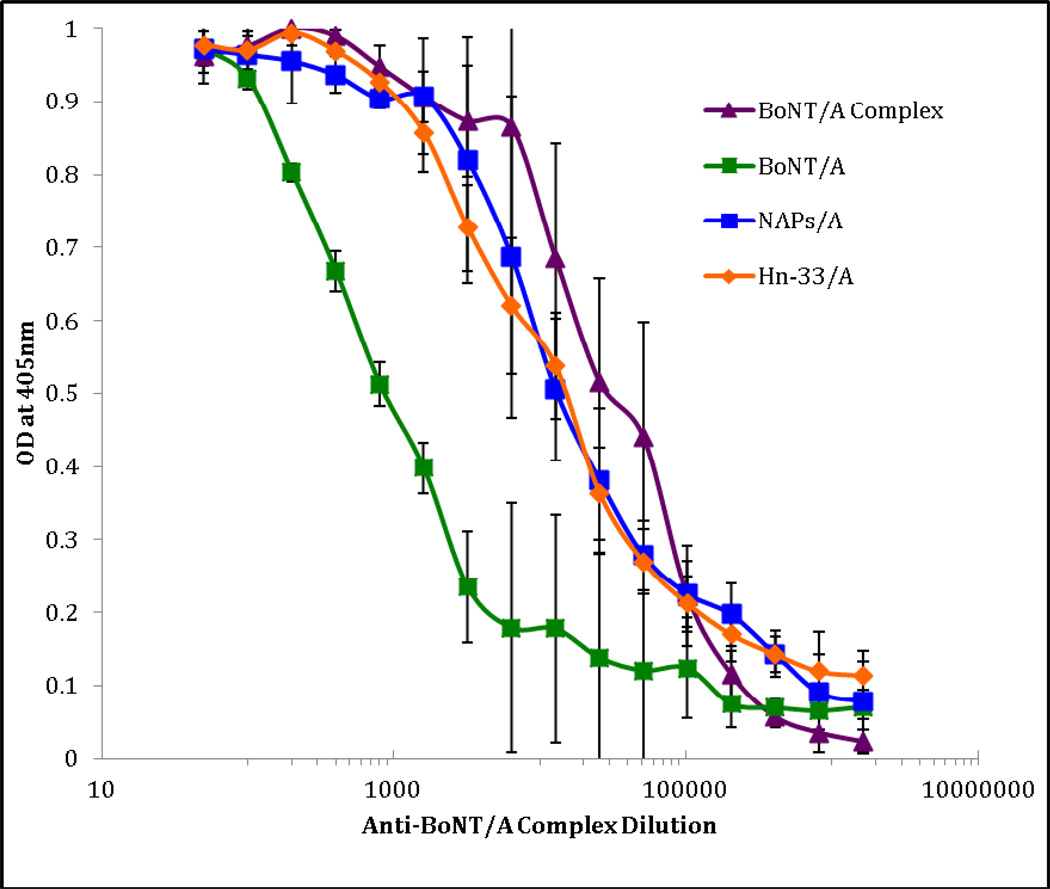
To have a better understanding on the immunological role of BoNT/A complex it is important to characterize the immunological properties of NAPs. Using indirect ELISA, antibodies against BoNT/A complex were examined for their interaction with BoNT/A complex, BoNT/A, NAPs, and Hn-33 (Fig. 1). Based on the results, the BoNT/A complex antigen had a 14-fold higher titer than BoNT/A antigen. These observations confirmed previous results that showed that BoNT/A complex antibodies had a much stronger reactivity with the complex than the pure neurotoxin (Kukreja et al., 2009; Singh, 1995). The neurotoxin was responsible for eliciting some of the BoNT/A complex antibody immune response, but the NAPs appear to have the more significant immunogenic role. This agrees with previous suggestions that BoNT/A detection in food and other environmental samples may be more sensitive with antibodies against the whole complex rather than only the neurotoxin (Singh, 1995). However, since there is an uncertainty of NAPs composition in the BoNT/A complex, a major question remains to be answered on the effectiveness of the NAPs in the detection of BoNT.
Fig. 1.
Indirect ELISA titration curve of anti-BoNT/A complex binding to BoNT/A complex, BoNT/A, NAPs/A, and Hn-33/A. The 96 well immuno-plate was coated with 1ng/µL of corresponding antigen followed by a 2-fold serial dilution of anti-rabbit BoNT/A complex IgG and developed with anti-rabbit HRP conjugated IgG. The error bars represented the standard deviation from three independent experiments.
The anti-BoNT/A complex had a higher reactivity towards the NAPs compared to BoNT (Fig. 1). Individually, both the NAPs and Hn-33 showed higher reactivity than BoNT towards BoNT/A complex antibodies indicating that NAPs and Hn-33 are more immunogenic. Interestingly, the NAPs and Hn-33 did not result in equal immunogenicity, given that the NAPs exhibited a titer about 4-fold higher and the Hn-33 exhibited a 7-fold higher titer than the BoNT. It is not clear what accounts for the immuno-reactivity to be higher with the Hn-33 alone rather than with the NAPs. Since current results have indicated that Hn-33 has a greater molecular ratio of 3 as compared to the other NAPs components with a ratio of 2 (Bryant, 2012), its abundance could have in part contributed to the higher immuno-reactivity. Another possibility for the difference in the immuno-reactivity could be a result of the accessibility of Hn-33 to interact with the complex antibodies. Hn-33 as a part of BoNT/A complex and purified NAPs may exhibit different structural changes affecting the accessibility of the Hn-33 to the antibodies. To answer such questions further research should be preformed on the structural aspects of BoNT/A complex, NAPs and Hn-33 and their influence on immunological characteristics.
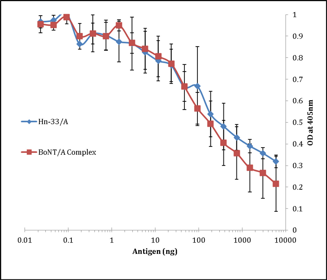
To provide further insight into the immunogenic role of Hn-33, the antibodies raised against purified type A Hn-33 were tested for their interaction with BoNT/A complex and Hn-33. The results indicated nearly identical reactivity with BoNT/A complex and Hn-33 (Fig. 2), which suggested that the reactivity was due to the higher immunogenicity, not the abundance of the proteins in the complex. It also suggested that Hn-33 is primarily responsible for eliciting the antibody response in the BoNT/A complex.
Fig. 2.
Indirect ELISA titration curve of anti-Hn-33/A binding to rHn-33/A and BoNT/A complex. The 96 well immuno-plate was coated with 1ng/µL of corresponding antigen followed by a 2-fold serial dilution of anti-rabbit Hn-33/A IgG and developed with antirabbit HRP conjugated IgG. The error bars represented the standard deviation from three independent experiments.
3.2 Competitive ELISA Analysis of BoNT/A and its associated proteins
The indirect ELISA results provided some additional insight on the immunogenicity of botulinum neurotoxin and its associated proteins, but still left questions about the immunogenicity of individual components of NAPs unanswered. This study of understanding the role of the BoNT/A complex components was limited, Furthermore, since the indirect assay’s measurement relied on the interaction of soluble antibodies with solid-phase antigens, it was not possible to evaluate comparative immuno-reactivity accurately as the surface interaction could mask epitopes or reorient them due to conformational changes. Use of competitive ELISA allowed the interactions of the antibodies with the BoNT/A, BoNT/A complex, and Hn-33 to be investigated in a solution phase, mimicking the actual physiological environment.
Firstly, the antigenic nature of BoNT/A complex compared to neurotoxin was analyzed for its reaction against both the complex and the neurotoxin antibodies. Previous results of BoNT/A antibody reaction with BoNT/A complex had suggested that the neurotoxin was less accessible to its antibodies in the complex form (Singh, 1995). The competitive ELISA results also indicated that BoNT/A was slightly less reactive to its antibodies in the complex form implying that the NAPs did hinder the immuno-accessibility of the neurotoxin in the complex. Thus, the results based on immuno-reactivity of surface adsorbed BoNT/A and BoNT/A in solution are compatible, and the differences reflect the reactivity due to molecular structure of the BoNT/A complex.
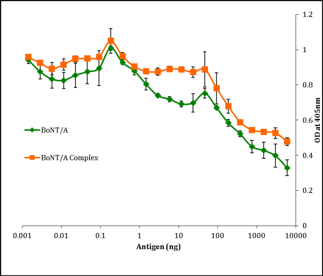
Interaction of anti-BoNT/A complex with BoNT/A and BoNT/A complex was further investigated with competitive ELISA (Fig. 4). The indirect ELISA data indicated that complex antibodies reacted 14 times better with the complex than the pure neurotoxin, which suggested that a majority of complex antibodies were produced against NAPs. In the competitive ELISA analysis, the 50% inhibition of the binding of antibodies against the BoNT/A complex was observed at approximately 8ng of BoNT/A complex, while BoNT/A was at approximately 50ng. Thus the BoNT/A complex competed 6 times more with complex antibodies compared to the BoNT/A, confirming that higher immunogenicity of BoNT/A complex was indeed due to the NAPs.
Fig. 4.
Competitive ELISA titration curve of anti-BoNT/A complex competing with BoNT/A or BoNT/A complex. The 96 well immuno-plate was coated with 1ng/µL of BoNT/A, followed by a 2-fold serial dilution of BoNT/A or BoNT/A complex with 2X titer of anti-rabbit BoNT/A IgG and developed with anti-rabbit HRP conjugated IgG. The error bars represented the standard deviation from three independent experiments.
To understand the immunological role of Hn-33 in NAPs, the immuno-reactivity of Hn-33 against anti-BoNT/A complex (Fig. 5) and anti-Hn-33 (Fig. 6) were examined. Based on previous results NAPs are composed of NAP-53 (50-kDa), Hn-33 (36-kDa), NAP-22 (24-kDa), NAP-17 (17-kDa) with a molecular ratio of 2:3:2:2, respectively in comparison to BoNT/A (Bryant, 2012). Both BoNT/A complex and Hn-33 competed nearly identically for the binding of anti-BoNT/A complex and anti-Hn33 antibodies. Similarly, previous competitive ELISA results indicated that Hn-33 competed for 78% of complex antibodies (Sharma, 2000). The difference between our current data and previously published data was likely the source of the antibodies. The antibodies used for the previous study were obtained from the US Army and were raised in horses (Sharma, 2000). The nature of the BoNT/A complex used for immunization of the horses may result in a slightly different antibody response. Nevertheless, our current observations and previous results, suggested that Hn-33 competed just as well as BoNT/A complex with the BoNT/A complex antibodies. The immunogenicity of Hn-33 was effectively the same when it was part of the BoNT/A complex or alone. Indirect ELISA showing a slightly greater reactivity with BoNT/A complex than Hn-33 was probably due to the presence and accessibility of NAPs. The solution phase binding of antibodies with BoNT/A complex, NAPs, and Hn-33 in competitive ELISA is likely to allow more effective interactions without any interference of surface adsorption that may block certain epitopes on these proteins. These results confirmed that Hn-33 is the major immuno-reactive protein of NAPs and its utilization in vaccines may stimulate the immune system and increase the immunity to botulism disease. Since there is a need to improve the current formulation of the botulism vaccine, Hn-33 should be further investigated as an adjuvant for its ability to accelerate, prolong, and enhance the immune responses against BoNTs.
Fig. 5.
Competitive ELISA titration curve of anti-Complex/A competing with Hn-33/A or BoNT/A Complex. The 96 well immuno-plate was coated with 1ng/µL of BoNT/A complex, followed by a 2-fold serial dilution of BoNT/A Complex or rHn-33/A with 2X titer of anti-rabbit BoNT/A complex IgG and developed with anti-rabbit HRP conjugated IgG . The error bars represented the standard deviation from three independent experiments.
Fig. 6.
Competitive ELISA titration curve of anti-Hn-33/A competing with Hn-33/A or BoNT/A complex. The 96 well immuno-plate was coated with 1ng/µL of BoNT/A complex, followed by a 2-fold serial dilution of BoNT/A complex or rHn-33/A with 2X titer of anti-rabbit Hn-33 IgG and developed with anti-rabbit HRP conjugated IgG. The error bars represent the standard deviation from three independent experiments.
Botulinum in its complex form is commonly used in therapeutic and cosmetic formulations. With high doses and long-term treatment the development of neutralizing antibodies and secondary non-responsiveness to treatment becomes a huge issue in its effectiveness. Previous results (Kukreja et al., 2009) and current ELISA results indicate that pure neurotoxin generates significantly less antibody response as compared with BoNT/A complex. Subsequently, the pure neurotoxin may be less likely to induce antibodies that lead to non-responsiveness of treatment. It has been shown that BoNT/A free from associated proteins was successful in the treatment of cervical dystonia (Benecke, 2005), but further studies are needed on its ability to treat other neuromuscular disorders and any problems it may create in the absence of NAPs.
3.3 Immunoblot Analysis of BoNT/A and its associated proteins
The immunoblotting was performed to further characterize the immunogenicity of BoNT/A and its associated proteins. BoNT/A complex, BoNT/A, NAPs, and Hn-33 were examined for their immunological interaction with antibodies raised against BoNT/A, BoNT/A complex, and Hn-33. The Western blot analysis of anti-BoNT/A exhibited a reaction with the neurotoxin components, heavy chain and light chain, in BoNT/A complex and purified BoNT/A, but there was a slight reaction with NAPs preparation as well (Fig. 7, Panel A. This reaction suggested that the NAPs preparation had some residual toxin left over from the purification process. The reaction of anti-complex immuno-reactivity separately with BoNT/A complex, NAPs, and Hn-33 showed Hn-33 as the highest immunogenic protein (Fig. 7, Panel B). In BoNT/A complex, the neurotoxin showed less immunogenicity than Hn-33, reinforcing the ELISA data that the presence of Hn-33 in the BoNT/A complex might be responsible for its higher immunogenicity. Equal immuno-reactivity was observed with anti-Hn 33 against Hn-33 and Hn-33 when part of BoNT/A complex and NAPs, confirming the indirect and competitive ELISA results (Fig. 7, Panel C).
Fig. 7.
Western Blot Analysis of 2µg of rHn-33, NAPs, BoNT/A, and BoNT/A complex under reducing conditions. Lane 1: rHn-33, Lane 2: NAPs, Lane 3: BoNT/A, Lane 4: BoNT/A complex, Lane 5:Thermo Scientific Page Ruler™ Plus Prestained Ladder. Panel A: Probed with rabbit anti-BoNT/A IgG Panel B: Probed with rabbit anti-BoNT/A complex IgG Panel C: Probed with rabbit anti-Hn-33 IgG.
3.4 Conclusion
A similar immuno-reactivity of BoNT/A and its associated proteins was observed in the surface adsorbed interaction of indirect ELISA and solution phase interaction of competitive ELISA. The results confirmed past observations that BoNT/A complex had a higher immunogencity than BoNT/A and suggested that the higher immuno-reactivity was not from the abundance of the associated proteins in the complex. Based on the indirect ELISA, competitive ELISA and immuno-blot results, Hn-33 was primarily responsible for eliciting a majority of the antibody response in the BoNT/A complex. These observations provide a better understanding on the immunogenicity of BoNT/A complex and its associated proteins for developing a more efficient vaccine, improving the current therapeutic formulation, and designing a detection system against botulism.
Fig. 3.
Competitive ELISA titration curve of anti-BoNT/A competing with BoNT/A or BoNT/A complex. The 96 well immuno-plate was coated with 1ng/µL of BoNT/A, followed by a 2-fold serial dilution of BoNT/A or BoNT/A complex with 2X titer of anti-rabbit BoNT/A IgG and developed with anti-rabbit HRP conjugated IgG.
Highlights.
BoNT complex immunogenicity primarily from its associated proteins (NAPs)
Hn-33 component of NAPs accounts for most of the immunogenicity of NAPs
Direct, competitive, and Western blot results provide consistent results
Acknowledgements
The authors would like to thank Paul Lindo, Stephen Riding, and Jenny Davis for BoNT/A complex, BoNT/A, NAPs, and Hn-33 purification. This work was supported by NIH Grant 1U01A1078070-02 and a Department of Homeland Security
Contract - HSHQDC-11-C-00064.
Abbreviations
- BoNT
botulinum neurotoxin
- BoNT/A
botulinum neurotoxin type A
- NAPs
neurotoxin-associated proteins
- NBP
neurotoxin-binding protein
- ELISA
enzyme-linked immunosorbent assay
- PBS
phosphate buffered saline
- PBST
phosphate buffered saline containing 0.05% Tween-20
- TBST
Tris buffered saline containing 0.05% Tween-20
- ABTS
2,2′-Azino-bis (3-ethylbenzothiazoline-6-sulfonic acid) diammonium salt
- IgG
immunoglobulin G
Footnotes
Publisher's Disclaimer: This is a PDF file of an unedited manuscript that has been accepted for publication. As a service to our customers we are providing this early version of the manuscript. The manuscript will undergo copyediting, typesetting, and review of the resulting proof before it is published in its final citable form. Please note that during the production process errors may be discovered which could affect the content, and all legal disclaimers that apply to the journal pertain.
Conflict of Interest
The authors declare that there are no conflicts of interest.
References
- Benecke R, Jost W, Kanovsky P, Ruzicka E, Comes G, Stat D, Grafe S. A new botulinum toxin type A free of complexing proteins for treatment of cervical dystonia. Neurology. 2005;64:1949–1951. doi: 10.1212/01.WNL.0000163767.99354.C3. [DOI] [PubMed] [Google Scholar]
- Bryant A, Davis J, Cai S, Singh BR. Molecular composition and extinction coefficient of native botulinum neurotoxin complex produced by clostridium botulinum hall A strain. 2012 doi: 10.1007/s10930-013-9465-6. Submitted for publication. [DOI] [PubMed] [Google Scholar]
- Cai S, Singh BR, Sharma S. Botulism diagnostics: from clinical symptoms to in vitro assays. Critical reviews in microbiology. 2007;33:109–125. doi: 10.1080/10408410701364562. [DOI] [PubMed] [Google Scholar]
- Dasgupta B, Sathyamoorthy V. Purfication and amino acid composition of type A botulinum neurotoxin. Toxicon. 1984;22:415–424. doi: 10.1016/0041-0101(84)90085-0. [DOI] [PubMed] [Google Scholar]
- Fu F, Sharma S, Singh BR. A protease-resistant novel hemagglutinin purified from type A clostridium botulinum. Journal of Protein Chemistry. 1998;7:53–60. doi: 10.1023/a:1022590514771. [DOI] [PubMed] [Google Scholar]
- Kukreja R, Chang TW, Cai S, Lindo P, Riding S, Zhou Y, Ravichandran E, Singh BR. Immunological characterization of the subunits of type A botulinum neurotoxin and different components of its associated proteins. Toxicon. 2009;53:616–624. doi: 10.1016/j.toxicon.2009.01.017. [DOI] [PubMed] [Google Scholar]
- Sakaguchi G. Clostridium botulinum toxins Pharmac. Ther. 1983;19:165–194. doi: 10.1016/0163-7258(82)90061-4. [DOI] [PubMed] [Google Scholar]
- Schantz E, Johnson E. Properties and use of botulinum toxin and other microbial neurotoxins in medicine. Microbiological Reviews. 1992;56:80–99. doi: 10.1128/mr.56.1.80-99.1992. [DOI] [PMC free article] [PubMed] [Google Scholar]
- Sharma S, Singh BR. Immunological properties of HN-33 purified from type A clostridium botulinum. Journal of Natural Toxins. 2000;9:357–362. [PubMed] [Google Scholar]
- Singh BR, Lopes T, Silvia M. Immunological characterization of type A botulinum neurotoxin in its purified and complexed forms. Toxicon. 1995;34:267–275. doi: 10.1016/0041-0101(95)00113-1. [DOI] [PubMed] [Google Scholar]
- Ting P, Freiman A. The story of clostridium botulinum- from food poisoning to botox. Clinical Medicine. 2004;4:258–261. doi: 10.7861/clinmedicine.4-3-258. [DOI] [PMC free article] [PubMed] [Google Scholar]
- Zhou Y, Paturi S, Lindo P, Shoesmith SM, Singh BR. Cloning, expression, purification, and characterization of biologically active recombinant hemagglutinin-33, type A botulinum neurotoxin associated protein. The Protein Journal. 2007;26:29–37. doi: 10.1007/s10930-006-9041-4. [DOI] [PubMed] [Google Scholar]


