



Abstract
A patient presented for an elective transcatheter aortic valve replacement with temporary transvenous pacing (TVP) wires placement per protocol. On postoperative day 1, the patient remained stable, so the wires were subsequently removed, after which the patient acutely decompensated, with transthoracic echocardiography revealing pericardial effusion. Emergent pericardiocentesis was performed, and a pericardial drain was placed. Three days later, the drain was removed; again, the patient acutely decompensated, requiring another emergent pericardiocentesis. Despite the relatively benign nature of TVP wires and pericardial drains, the possibility of cardiac tamponade should be kept in mind as a potential complication when they are being removed.
Keywords: Cardiac tamponade, pericardial drain, pericardial effusion, transthoracic echocardiogram, transvenous pacing wires
Introduction
Cardiac tamponade is a life-threatening emergency, defined as slow or rapid cardiac compression caused by pericardial fluid – blood, pus, clots or effusion or air, trauma or rupture of the heart.[1,2] Prompt recognition and intervention are essential to avert potentially fatal consequences. One rare etiology for cardiac tamponade is myocardial perforation from pacing wires placement. We present a case report of two separate episodes of cardiac tamponade in a patient with the first occurring after removal of transvenous pacing (TVP) wires and the second occurring after removal of the pericardial drain in place to treat the first episode.
Case Report
A 79-year-old female with a medical history of hypertension, severe aortic stenosis, and chronic heart failure presented for an elective transcatheter aortic valve replacement (TAVR). TAVR with 26 mm CoreValve™ was successfully performed under general anesthesia. Temporary TVP wires were placed in the right ventricle (RV) under fluoroscopic guidance through the left common femoral vein without any difficulty, and she was subsequently transferred to the Intensive Care Unit (ICU), extubated and in stable condition. The temporary pacemaker was set as VVI, the rate of 50 beats/min, and at the threshold of 10 mA.
Two-dimensional transthoracic echocardiography (TTE) performed on the postoperative day (POD) 1 was unremarkable [Figure 1], so decision was made to remove the pacer wires. Thirty minutes after removal, the patient became diaphoretic and hypotensive (blood pressure [BP] 40/25 mmHg). The patient was immediately resuscitated with intravenous fluid boluses and started on a norepinephrine drip, after which the BP increased to 100/60 mmHg. A stat TTE was performed at bedside showed moderate pericardial effusion around RV/right atrium, excessive respiratory variation of the mitral inflow velocities, and mild RV collapse during diastole indicative of cardiac tamponade physiology [Figure 2]. The patient underwent emergency pericardiocentesis, 180 ml of sanguineous fluid was drained and a pericardial drain was left in place. A presumed diagnosis of RV rupture by temporary pacing wires was made which became evident after the removal of wires.
Figure 1.
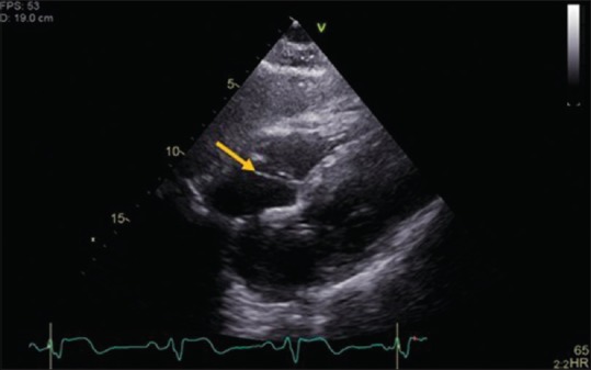
Transthoracic echocardiogram showing temporary pacing wire in the right ventricle
Figure 2.
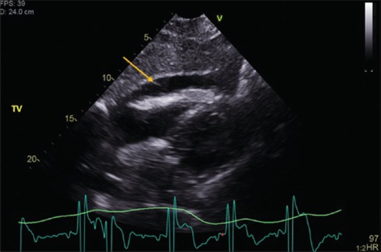
Transthoracic echocardiogram after removal of pacing wires, yellow arrow shows pericardial effusion around the right ventricle
On POD 3, the patient remained stable, draining minimal amount of fluid (approximately 15 ml in 24 h). Repeat TTE showed no new pericardial effusion, so a decision was made to remove the pericardial drain. Twenty minutes after, again, the patient developed similar acute symptoms of diaphoresis and became hemodynamically unstable. Emergent TTE revealed reaccumulation of pericardial fluid. Another pericardiocentesis was done with the draining of 200 ml of frank blood. Coronary angiogram revealed no obstruction, and an aortic root aortogram showed no leakage or tear. A transesophageal echocardiography confirmed good valve function.
The patient was transferred back to the ICU with the plan to continue conservative management and leave the drain in place for 72 h as per cardiothoracic surgeon's recommendations. The patient remained stable and follow-up TTE and computed tomography of the chest was negative for any fluid reaccumulation. On the 4th day after redrainage (POD 7), pericardial catheter was clamped and TTE was performed 2 h postclamping showing no accumulation of pericardial fluid. The drain was removed, vital signs were monitored continuously for 4 h, and after 24 h, the patient was transferred from the ICU to step-down.
Discussion
Temporary pacing wire implantation is indicated for all TAVR procedures because of potential risk of atrioventricular (AV) conduction block postprocedure due to proximity of AV node and left bundle branch to the noncoronary cusp of the aortic valve.[3] Temporary pacing wire placement is associated with a wide range of complications, including failure to secure the venous access, failure to implant the leads correctly, sepsis, life-threatening arrhythmias, and puncture of arteries, lungs, or myocardium.[4] The potentially significant acute complications of permanent pacemaker placement included lead displacement (0.5%–4.8%), pneumothorax (1.9%–3.7%), and myocardial perforation (0.37%–1%).[5]
In this particular case, the patient had presumed RV tear. Myocardial perforation can occur in any part of the heart that comes in contact with leads, but majority of perforations occur at the apex of RV. The RV is the most common site for lead deployment, and myocardial wall is thinner at the apex than at the septum or RV outflow tract. In spite of its rarity, it is imperative to be cognizant of this complication when removing TVP wires at the bedside as early recognition and treatment is the key to prevent fatal outcome. Threshold of the pacemaker does not play a role in potential tear when removing the TVP wires.[6] The patient's initial signs and symptoms may include nausea, fatigue, lightheadedness, dyspnea, retrosternal chest pain, tachycardia, cyanosis, neck venous engorgement, pulsus paradoxus, and confusion.[7] The current guidelines recommend frequent measurements of vital signs during first 4 h after the removal of the pacing wires.[8]
After early recognition, prompt treatment and appropriate diagnostic studies are the key. Electrocardiogram (EKG) and chest X-ray (CXR) may not always aid in the diagnosis. CXR may not show changes until large amount of fluid collection in pericardial sac. Furthermore, low voltage QRS or electrical alterns may not always present on EKG.[9] Echocardiography remains the diagnostic imaging modality of choice and allows quantification of pericardial effusion and tamponade.[7] The patient should be resuscitated with fluids to facilitate the maximum filling of heart. Ionotropic agents help increase the stroke volume and increase systemic vascular resistance.[10] Definitive treatment includes pericardiocentesis or thoracotomy.[7,10] Similar to the removal of TVP wires, caution should be used in removal of pericardial drain as well.
In our case, the patient developed reaccumulation of blood in pericardial sac after removal of drain. A possible etiology may be a small thrombus attached to epicardium, which could have been dislodged along with the removal of drain. This could have potentially caused that same unhealed RV tear to reaccumulate the fluid in the pericardial sac. There is different consensus on timing of pericardial drain removal in different studies, ranged from an average of 2–4.8 days. Sagristà-Sauleda et al. recommended that catheter should be managed as long as drainage is >25 cc/day.[11,12] There have been few case reports of developing hydropneumopericardium after removal of pericardial drain, which were believed to be secondary to leakage of drainage system or communications between pleura and pericardium.[13,14,15] Other rare complications include breakage of catheter in the pericardium, knotting of catheter around the pulmonary artery, and impossible drainage due to loop around the heart.[16]
Despite the relatively benign nature of TVP wires, the possibility of cardiac tamponade should be kept in mind as a potential complication when they are being removed. Prompt recognition and early intervention are imperative to prevent complications. We recommend applying clinical judgment based on clinical improvement, amount of fluid drainage, and drain clamping during deciding when to remove the pericardial drain as pericardial effusion can be a complication from removal as well. In addition, proper precautions should be used including monitoring vital signs closely and recognizing the signs/symptoms of tamponade as mentioned earlier and start resuscitation efforts if needed after the drain removal.
Declaration of patient consent
The authors certify that they have obtained all appropriate patient consent forms. In the form the patient(s) has/have given his/her/their consent for his/her/their images and other clinical information to be reported in the journal. The patients understand that their names and initials will not be published and due efforts will be made to conceal their identity, but anonymity cannot be guaranteed.
Financial support and sponsorship
Nil.
Conflicts of interest
There are no conflicts of interest.
References
- 1.Spodick DH. Pathophysiology of cardiac tamponade. Chest. 1998;113:1372–8. doi: 10.1378/chest.113.5.1372. [DOI] [PubMed] [Google Scholar]
- 2.Costa IV, Soto B, Diethelm L, Zarco P. Air pericardial tamponade. Am J Cardiol. 1987;60:1421–2. doi: 10.1016/0002-9149(87)90639-4. [DOI] [PubMed] [Google Scholar]
- 3.Piazza N, de Jaegere P, Schultz C, Becker AE, Serruys PW, Anderson RH. Anatomy of the aortic valvar complex and its implications for transcatheter implantation of the aortic valve. Circ Cardiovasc Interv. 2008;1:74–81. doi: 10.1161/CIRCINTERVENTIONS.108.780858. [DOI] [PubMed] [Google Scholar]
- 4.McCann P. A review of temporary cardiac pacing wires. Indian Pacing Electrophysiol J. 2007;7:40–9. [PMC free article] [PubMed] [Google Scholar]
- 5.Vanezis AP, Prasad R, Andrews R. Pacemaker leads and cardiac perforation. JRSM Open. 2017;8:1–3. doi: 10.1177/2054270416681432. [DOI] [PMC free article] [PubMed] [Google Scholar]
- 6.Banaszewski M, Stepinska J. Right heart perforation by pacemaker leads. Arch Med Sci. 2012;8:11–3. doi: 10.5114/aoms.2012.27273. [DOI] [PMC free article] [PubMed] [Google Scholar]
- 7.Ristic AD, Imazio M, Adler Y, Anastasakis A, Badano LP, Brucato A, et al. Triage strategy for urgent management of cardiac tamponade: A position statement of the European Society of Cardiology Working Group on Myocardial and Pericardial Diseases. Eur Heart J. 2014;35:2279–84. doi: 10.1093/eurheartj/ehu217. [DOI] [PubMed] [Google Scholar]
- 8.Mahon L, Bena JF, Morrison SM, Albert NM. Cardiac tamponade after removal of temporary pacer wires. Am J Crit Care. 2012;21:432–40. doi: 10.4037/ajcc2012585. [DOI] [PubMed] [Google Scholar]
- 9.Little WC, Freeman GL. Pericardial disease. Circulation. 2006;113:1622–32. doi: 10.1161/CIRCULATIONAHA.105.561514. [DOI] [PubMed] [Google Scholar]
- 10.Collins D. Aetiology and management of acute cardiac tamponade. Crit Care Resusc. 2004;6:54–8. [PubMed] [Google Scholar]
- 11.Gibbs CR, Watson RD, Singh SP, Lip GY. Management of pericardial effusion by drainage: A survey of 10 years’ experience in a city centre general hospital serving a multiracial population. Postgrad Med J. 2000;76:809–13. doi: 10.1136/pmj.76.902.809. [DOI] [PMC free article] [PubMed] [Google Scholar]
- 12.Sagristà-Sauleda J, Mercé AS, Soler-Soler J. Diagnosis and management of pericardial effusion. World J Cardiol. 2011;3:135–43. doi: 10.4330/wjc.v3.i5.135. [DOI] [PMC free article] [PubMed] [Google Scholar]
- 13.Dregoesc I, Iancu A, Stef A, Molnar A. Massive hydropneumopericardium after pericardial drain removal. Eur Heart J. 2016;37:3617–8. doi: 10.1093/eurheartj/ehw407. [DOI] [PubMed] [Google Scholar]
- 14.Lee SH, Kim WH, Lee SR, Rhee KS, Chae JK, Ko JK. Cardiac tamponade by iatrogenic penumopericardium. J Cardiovasc Ultrasound. 2008;16:26–8. [Google Scholar]
- 15.Attar A, Sharifkazemi MB, Moaref AR. Bubbles in pericardium fluid: Multimodality imaging in iatrogenic hydropericardium. Arch Cardiovasc Imaging. 2015;3:e22787. [Google Scholar]
- 16.Kraus MB, Spitznagel RA, Kugler JA. Cardiovascular collapse with attempted pericardial drain withdrawal. Ann Pediatr Cardiol. 2016;9:241–3. doi: 10.4103/0974-2069.189118. [DOI] [PMC free article] [PubMed] [Google Scholar]