Abstract
Thymocyte differentiation antigen 1 (Thy-1) is a glycosylphosphatidyl inositol (GPI)-linked cell surface glycoprotein expressed on numerous cell types, which regulates signals affecting cell adhesion, migration, differentiation, and survival. In addition, Thy-1 has been detected in serum, cerebral spinal fluid, wound fluid from venous ulcers, synovial fluid from joints in rheumatoid arthritis and more recently urine. We previously detected Thy-1 in the conditioned media of cytokine-stimulated lung fibroblasts, suggesting that Thy-1 shedding may be a response to cellular stress. Soluble and membrane bound forms of Thy-1 from in vivo sources have been shown to be identical in size when deglycosylated, suggesting that soluble Thy-1 is separated from the diacyl glycerol portion of its GPI-anchor by hydrolysis within the GPI moiety. For Thy-1 and other GPI-anchored proteins, delipidation induces a stable change in conformation that manifests itself in a change in antibody affinity for soluble forms. Using epitope tagged recombinant soluble Thy-1, we report that widely available monoclonal antibodies to human Thy-1 are unable to detect soluble Thy-1 by immunoblotting. We reevaluated the Thy-1 that we previously reported in the conditioned media of normal human lung fibroblasts and found it to be entirely insoluble. These findings suggest that most Thy-1 reported in body fluids retains its GPI anchor and may be associated with membrane fragments or vesicles. This phenomenon should be considered in the generation of antibodies and controls for Thy-1 bioassays. Furthermore, the changes in Thy-1 conformation with delipidation, beyond affecting antibody affinity, likely affect the ligand affinity and biological function of soluble vs. released membrane-associated forms.
Keywords: Antibody Recognition, Delipidated THY1, Fibroblast, GPI Anchor, Recombinant THY1
Thy-1 cell surface antigen (also Thymocyte differentiation antigen 1, CD90 or Thy-1), a glycosylphosphatidyl inositol (GPI)-linked cell surface glycoprotein on stem cells and multiple mature cell types, was originally discovered in an attempt to raise antiserum against leukemia-specific antigens from the C3H mouse strain in the AKR mouse strain and vice versa. The antibodies were found to strongly label thymoctyes as well as peripheral T cells.1 For this reason, the original designation for the antigen changed from Theta to Thy-1.2 The cell types expressing Thy-1 in normal and pathological conditions, and the immunologic and non-immunologic roles of Thy-1 have been reviewed elsewhere.3–5
Our lab previously reported that normal human lung fibroblasts (NHLF) treated with several different pro-inflammatory cytokines undergo a decrease in the level of cell surface Thy-1 expression. This decrease coincides with an increase of detectable Thy-1 in the conditioned media (CM), suggesting release of Thy-1 from the cell surface. From these observations, we concluded that Thy-1 is likely separated (shed) from the diacyl glycerol portion of its GPI-anchor by a “GPIase” hydrolyzing a bond within the GPI moiety.6 Others have demonstrated Thy-1 detectable in serum, wound fluid, urine and cerebral spinal fluid in a variety of normal and pathological conditions.7–9 Taken together, these findings suggested that released Thy-1 may serve as a useful biomarker for certain pathological conditions. In addition, recent studies have shown that a soluble recombinant Thy-1 in which the GPI-attachment signal is replaced by the Fc fragment of human IgG1 (Thy-1-Fc) alters activation of latent Transforming Growth Factor-β and cell phenotype in lung fibroblasts,10 indicating that Thy-1 may have a role as a soluble mediator in addition to its function on the cell surface. For these reasons, antibody detection of Thy-1 in biological fluids is important in clinical and research applications.
However, antibody mediated detection or purification of Thy-1 is known to be problematic for several reasons. Many of the monoclonal antibodies employed to detect Thy-1 in western blots require that it be prepared under non-reducing (NR) conditions, including the clones OX7, G7, HO-13-4, 5E10, and ASO2.8,11–13 For many antibodies that recognize GPI-anchored proteins at the cell surface, their affinity is lost or greatly diminished if that same protein is delipidated. This phenomenon has been reviewed elsewhere.14 For Thy-1 in particular, several monoclonal and polyclonal antibodies to mouse Thy-1 have been shown not to react with delipidated, soluble Thy-1.11,12 Though mostly demonstrated for antibodies to mouse Thy-1, a group reported the reactivity of an antiserum raised against membrane-bound human Thy-1 failed to recognize hydrophilic human Thy-1 purified from cerebral spinal fluid. For Thy-1 and other GPI-anchored proteins, the general consensus is that delipidation induces a stable change in conformation that manifests itself in a change in antibody affinity.14 Antibodies that recognize human Thy-1 with a GPI anchor are predicted to have lower affinity for Thy-1 if it is delipidated. Therefore we reasoned that the Thy-1 we detected in fibroblast supernatants and others detected in several body fluids using antibodies to membrane-bound Thy-1 may not be a truly soluble, cleaved form.
The widely-used monoclonal antibodies K117 (American Type Culture Collection Number: HB-8553), 5E10 (STEMCELL Technologies 01437), and AS02 (Millipore CP28) were generated from mice immunized with a human astrocytoma cell line, a human erythroleukemia cell line, and human dermal fibroblasts, respectively. The antigen recognized at the cell surface by K117, 5E10, and AS02 is Thy-1. We performed the following studies to characterize recognition of soluble and GPI-anchored forms of Thy-1 by antibodies from these clones.
Materials and Methods
Recombinant Constructs of THY1
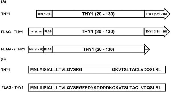
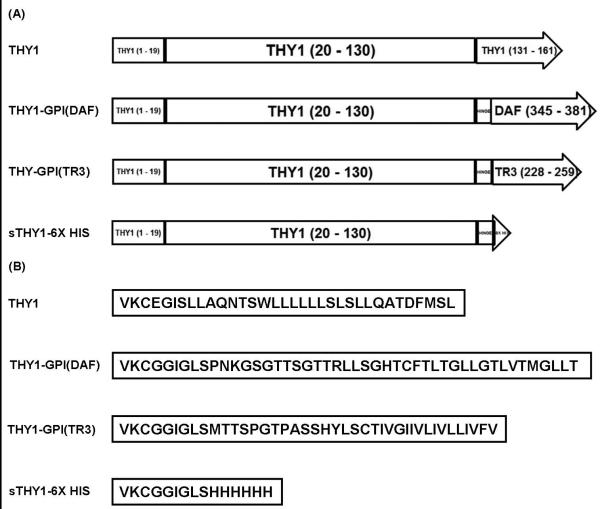
For expression of wild type (WT) human Thy-1, the complete cDNA of human THY1 (AAH65559.1) was ligated into the mammalian expression vector pcDNA3.1/Zeo (+) (Invitrogen V860-20) with the Kozak sequence GCCGCC15 just upstream of the start codon. The restriction sites EcoRI and NotI were used at the 5′ and 3′ end, respectively [Figure 1. A and B]. To express mature Thy-1 with an N-terminal FLAG tag, a coding sequence for the FLAG epitope16,17 was cloned immediately downstream of that of the ER localization signal18 and just upstream of the first codon for mature Thy-1 [Figure 1. A and B]. To express Thy-1 with its GPI-attachment signal replaced with that of another glycoprotein, coding sequences for the foreign GPI-attachment signal were cloned downstream of a “hinge” region. This “hinge” region comprises the 6 amino acids (AA) downstream of mature Thy-1, EGISLL, changed to GGIGLS as was previously shown to successfully add the transmembrane domains of Cluster of Differentiation 8 and Neural Cell Adhesion Molecule to mouse Thy-1Thy119. Using site-directed mutagenesis, an intermediate construct was made first by changing the sequence just downstream of the codons for mature Thy-1 to code for GGIG followed by the BstBI restriction site. Expression vectors containing the cDNA of the GPI-attachment signals for Decay-Accelerating Factor (DAF) and TNF-related Apoptosis-inducing Ligand Receptor 3 (TRAIL-R3) were kindly provided by Daniel F. Legler (University of Konstanz, Konstanz, Germany). The cDNA of TRAIL-R3 contains multiple threonine, alanine, proline, and glutamine-rich repeats that present multiple annealing sites for the 5′ cloning primer. To avoid this possibility, the TRAIL-R3 containing expression vector was cut with PvuII and NotI producing a 115 bp fragment. This fragment was used as the template for PCR. PCR products of TRAIL-R3 and DAF had the restriction sites of BstBI and NotI introduced 24 base pairs upstream of the final codon in the mature protein and after the stop codon, respectively. These PCR products were cloned into the intermediate construct with BstBI and NotI. Finally, the BstBI sites in both were converted to codons for LS by site directed mutagenesis [Figure 2. A and B]. To express sThy-1 with a C-terminal histidine tag, an intermediate construct was made introducing half the changes required for six histidines then a stop codon to follow the “hinge” region by site-directed mutagenesis. The intermediate was then used as the template in another round of site directed mutagenesis to complete the required changes [Figure 2. A and B]. To express soluble Thy-1 with an N-terminal FLAG tag, the “hinge” region was introduced followed by a stop codon into FLAG – THY1 by site directed mutagenesis [Figure 1. A and B]. See Figures 1. and 2. for schematics of these constructs.
FIGURE 1. Primary Structures of and N-terminally tagged recombinant Thy-1.
(A) Full length diagrams of primary structures for WT Thy-1 and N-terminally tagged recombinant Thy-1. (B) Alignment of the AA sequences at the N-terminus of N-terminally tagged recombinant Thy-1 with WT Thy-1.
FIGURE 2. Primary Structures of recombinant Thy-1 with modifications to the C-terminus.
(A) Full length diagrams of primary structures for WT Thy-1 and recombinant THY1 with modifications to the C-terminus. (B) Alignment of the AA sequences at the C-terminus of recombinant Thy-1 with modifications to the C-terminus with WT Thy-1.
Cell Culture of RFL-6 and CCL-210
Rat fetal lung fibroblasts (RFL-6) (American Type Culture Collection) were maintained in F-12K supplemented with 10% FBS, 1% penicillin, and 1% streptomycin with medium exchanged every 2 days. CCL-210 (American Type Culture Collection), NHLF, were maintained in MEM supplemented with 10% FBS, 1% penicillin, and 1% streptomycin with medium exchanged every 2 days. Both were passaged using 0.25% trypsin. Cell extracts (CE) and CM were collected in the following manner. Adherent cells were washed twice with PBS then harvested by scraping into ice cold IP lysis buffer (Pierce 87788). Cell lysates were incubated on ice for 5 min with periodic vortexing then centrifuged at 13,000 × g and 4°C for 10 min. The supernatant containing the CE was stored at -80°C prior to use. CM from adherent cells was cleared of cellular debris by centrifugation at 1,200 × g and 4°C for 5 min. The supernatant was either immediately concentrated using Amicon's Ultra-2 Centrifugal Filter Unit with a 3kDa retention membrane, precipitated with methanol, subjected to differential centrifugation, or stored at -80°C. For cytokine treatment, cells at 90% confluency were washed with serum free media (SFM) then serum starved in SFM for 24 hrs, followed by culture in either MEM alone or 20ng/mL each TNFα (GIBCO PHC3015L) and IL-1β (GIBCO PHC815) in MEM for 24 hrs to 48 hrs.
Stable Expression of Recombinant Thy-1 in RFL-6
RFL-6 were transfected using Lipofectamine 2000 (Invitrogen 11668) according to the manufacturer's protocol. After 24 hrs, cells were subcultured in growth media so as to be near 10% confluent the following day, then washed with SFM and cultured in growth media supplemented with 500μg/mL Zeocin (Invitrogen). Stably transfected cells were selected after several passages showed no visual indication of cell death.
Deglycosylation of Thy-1 with PNGase F
CEs or CM were diluted in water and brought to 1% SDS with a 10% stock solution then incubated at 100°C for 10 min., following the manufacturer's standard protocol for Peptide: N-Glycosidase F (PNGase F) (New England BioLabs P0704) except for omission of dithiothreitol from the denaturing buffer. An aliquot was taken as the control for Thy-1 prior to deglycosylation. G7 reaction buffer and NP40 were added to a final concentration of 1X and 1%, respectively. After adding PNGase F, this reaction mixture was incubated at 37°C for 1 hour then diluted with PBS to the same total protein concentration.
Release of Thy-1 from Cell Surfaces with PI-PLC
Cells expressing Thy-1 were cultured in growth media to near 90% confluency, washed with SFM, then incubated with 0.1U/mL Phosphatidylinositol-Specific Phospholipase C (PI-PLC) (Invitrogen P-6466) in SFM for 24 hrs.
Analysis of Cell Surface Thy-1 Expression by Flow Cytometry
Flow cytometry was performed as previously described6 using the following antibodies: anti-human Thy-1 and mouse IgG1 κ isotype control conjugated to FITC (BD Pharmingen 555595 and 555748, respectively).
Western Blot Analysis
Unless otherwise specified, all samples were prepared in NR SDS loading buffer (50mM Tris-HCl pH 6.8, 2% SDS, and 10% glycerol). Sample preparations were electrophoresed in 10% polyacrylamide gels then transferred onto PVDF membranes. Depending on the primary antibody, membranes were either blocked in 5% Milk in TBST or TBST alone. The primary antibodies for detecting the mature Thy-1 polypeptide, K117, 5E10, and AS02 were diluted to 0.1μg/mL in TBST. The primary antibodies for detecting the FLAG (Sigma-Aldrich M2 F1804) and 6 histidine epitopes (Rockland 600-401-382) were diluted to 1.0μg/mL in 5% Milk in TBST. All primary antibodies were incubated with membranes overnight at 4°C. Antibodies bound to antigen were visualized with secondary antibodies conjugated to horseradish peroxidase in conjunction with a chemiluminescent substrate. The secondary antibodies used were goat anti-mouse (H+L) (Bio-Rad 172-1011) and goat anti-rabbit (H+L) (Thermo Scientific 32460) diluted 1/200,000 and 1/40,000 in blocking buffer, respectively. When required, membranes were stripped with Pierce's Restore western blot stripping buffer following the manufacturer's protocol.
Partitioning of Conditioned Media using Triton X-114
Triton X-114 (Sigma-Aldrich 93422) was added to concentrated CM to a final concentration of 2%. An aliquot was taken as the control for total Thy-1 prior to partitioning. With periodic vortexing throughout, the remainder was kept on ice for 10 min, then another 10 min at 37°C. Following this, the sample was immediately centrifuged at 21,000 × g for 10 min at room temperature. Centrifuging produced a clear partition between the insoluble and soluble phases. Prior to western blot analysis, volumes were adjusted to represent equal fractions of starting volume of CM.
Differential centrifugation and Methanol Precipitation of Conditioned Media
Two 150μL aliquots were taken from the supernatant of CM centrifuged at 1,200 × g and 4°C for 5 min. One aliquot was centrifuged again at 21,000 × g for 5 min at 4°C. After this, the supernatant was removed. All samples were precipitated by adding 1,350μL of methanol, then leaving them at -20°C overnight. The following day all were centrifuged at 21,000 × g for 30 min at 4°C. The supernatant was poured off and the precipitant was allowed to air dry before being resuspended in 35μL of 1X SDS loading buffer then submitted to western blot analysis.
Results
Anti-human Thy-1 monoclonal antibodies which react with Thy-1 at the cell surface do not recognize delipidated Thy-1
Human Thy-1 from CE is readily detected by western blot in NR conditions using either K117, 5E10, or AS02 monoclonal antibody [Figure 3. A and B and Supplementary Figure 1.]. The NHLF cell line CCL-210 was treated with PI-PLC as described in Methods. Analysis by flow cytometry showed Thy-1 was completely removed by PI-PLC (data not shown). However, Thy-1 was not detected in the CM of PI-PLC treated CCL-210 using the K117 monoclonal antibody, despite being easily detected in CEs diluted to the same volume as the CM (data not shown).
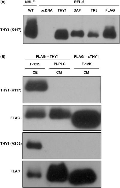
FIGURE 3. Complete GPI anchor of Thy-1 required for recognition by monoclonal antibodies raised against membrane bound form.
(A) Mature Thy-1 detected by western blot in the CEs of NHLF and RFL-6 stably transfected with pcDNA3.1/Zeo(+) (pcDNA) or pcDNA3.1/Zeo (+) with a recombinant form of Thy-1 cloned in: WT THY1 (THY1), THY1 – GPI(DAF) (DAF), THY1 – GPI(TR3) (TR3), or FLAG – THY1 (FLAG). (B) Mature Thy-1 detected by western blot in CE containing FLAG – Thy-1 or CM containing PI-PLC released FLAG – Thy-1 or FLAG – sThy-1 using K117 and AS02. After stripping the membranes of antibodies for detecting mature Thy-1, FLAG was detected using M2.
In order to confirm the presence of Thy-1 released by PI-PLC into CM, a recombinant form was engineered to be expressed at the cell surface with an N-terminal FLAG tag. This was accomplished by introducing the FLAG epitope16,17 downstream of the N-terminal ER localization signal18 and just upstream of the first residue of mature Thy-1, designated FLAG – THY1 [Figure 1 A and B]. The size of FLAG – THY1 stably expressed by RLF-6 relative to WT Thy-1 is in keeping with the predicted polypeptide mass difference of ~1.3kDa and with being correctly glycosylated [Figure 3A]. CE from RFL-6 stably expressing FLAG – THY1 and the CM of the same cell line treated with PI-PLC were compared in western blots with anti-FLAG as the primary antibody (data not shown). Using FLAG band intensity to control for loading of Thy-1, CM from PI-PLC treated fibroblasts were used in western blots with clone K117 5E10, and AS02 as the primary antibodies. Each western blot showed a band in the lane corresponding to CE but not the CM [Figure 3. B and Supplementary Figure 1.] After stripping the membranes of primary and secondary antibodies for detecting Thy-1, each was re-probed with FLAG antibody. Unlike with clones K117, 5E10, and AS02, the immunoblot for FLAG revealed a band in each lane equal in size. However, the band appearing in the lane in which PI-PLC released FLAG – THY1 was run was equal or greater in intensity as predicted [Figure 3.B]. Thus, sufficient FLAG – THY1 was present for clones K117, 5E10, and AS02 to detect Thy-1 were it not delipidated.
Monoclonal antibodies from clones K117 and 5E10 recognize epitopes on Thy-1 independent of its glycosylation, but are abolished under reducing conditions
Glycosylation of Thy-1, which is exclusively N-linked, can account for more than 50% of its total mass. Between different cell and tissue types, the carbohydrate moiety composition may vary dramatically. As a consequence, the molecular mass of Thy-1 ranges from 25 to 37kDa.7,12,13,20 Of the anti-mouse monoclonal antibodies that have affinity for non-delipidated Thy-1, OX7, H140-150, H154-177,12 and AS0213 have been demonstrated to recognize their respective epitopes independent of glycosylation. Therefore, the epitopes lost with delipidation are likely confined to a region of the Thy-1 polypeptide. The mature Thy-1 polypeptide has four cysteine residues. As a member of the immunoglobulin superfamily, each cysteine is predicted to form a disulfide bond with one of the other under oxidizing conditions.21 Western blots to detect Thy-1 that utilize antibodies from clones HO-13-4, G7,11 OX7,12 5E10,8 or AS0213 as the primary are conducted under NR conditions. NR conditions are used with clone K117 as well [Figure 3].
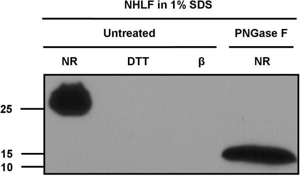
To determine if recognition by clones K117 and 5E10 is contingent on glycosylation or disulfide bonds, both were used as the primary antibodies in western blots of native, reduced and completely deglycosylated Thy-1 in CCL-210 extract. PNGase F cleaves between the innermost GlcNAc and asparagine of oligosaccharides from N-linked glycoproteins and was shown to completely remove the glycans from human Thy-1.7 With clone K117 [Figure 4] and 5E10 (data not shown), a single band of a size corresponding to fully glycosyated Thy-1 was detected in the lane containing non-PNGase F treated CE prepared under NR conditions. To circumvent possible diffusion of the reducing agent into adjacent lanes, samples were run in western blots with an empty lane between those with reducing agents and those without [Figure 4]. No bands were detected in the lanes containing non-PNGase F treated cell lysate prepared under reducing conditions. A single band of a size corresponding to completely deglycosyated Thy-1, ~14kDa, was detected in the lane containing PNGase F treated CE prepared under NR conditions. Moreover, the bands were equal in intensity and relative size [Figure 4]. As is the case with clones OX7, H140-150, H154-177, and AS02, the epitopes recognized by K117 and 5E10 on human Thy-1, which are lost with delipidation, remain with deglycosylation. Additionally, breaking disulfide bonds with reducing agents completely abolishes epitope recognition by these antibodies.
FIGURE 4. Monoclonal antibodies from clones K117 and 5E10 recognize epitopes on Thy-1 independent of its glycosylation, but are abolished under reducing conditions.
Mature Thy-1 detected by western blot in equal dilutions of untreated or PNGase F treated NHLF extract prepared as indicated under NR conditions or with 100mM DTT (DTT) or 2.5% β-mercaptoethanol (β).
Substitution of the native GPI anchor attachment signal of THY1 with the GPI-attachment signals of DAF or TRAIL-R3 does not alter antibody reactivity despite the presence of an intervening 15 amino acid region
Recombinant THY1 hybrids were engineered to have GPI anchors attach using the DAF and TRAIL-R3 GPI attachment signals, designated THY1 – GPI(DAF) and THY1 – GPI(TR3). To accomplish this, a “hinge” region was placed between mature THY1 and the foreign GPI-attachment sequences. The hinge region is based on prior publication by a group that successfully replaced the GPI-attachment signal of mouse Thy-1 with the transmembrane domain of cluster of differentiation 8 and Neural Cell Adhesion Molecule.19 Both GPI attachment signals consist of the C-terminus 8 residues upstream of the GPI anchorage sites of DAF and TRAIL-R3 [Figure 2. A and B].
Thy-1 is detected in RFL-6 cells transfected with expression vectors for THY1 – GPI(DAF) or THY1 – GPI(TR3) as assessed by western blot using clone K117 [Figure 3. A]. The bands for each are the same size and in keeping with the predicted polypeptide mass difference of ~1.3kDa relative to the band for WT Thy-1 and are correctly glycosylated. Identical results were obtained using clone 5E10 (not shown). After 24 hr incubation with PI-PLC, cells expressing THY1 – hinge – GPI(DAF) and – GPI(TR3) are completely negative for cell surface Thy-1 as assessed by flow cytometry. This confirms that both are GPI anchored and neither GPI-anchor possesses an additional palmitoyl group on the inositol. PI-PLC hydrolyzes the bond in the GPI anchor that liberates the diacylglycerol but not the palmitoyl group from the inositol.22 For both, the GPI anchor is predicted to attach 15 AA downstream of the WT attachment site of Thy-1 [Figure 2. B]. This is confirmed by the size of each relative to native Thy-1 and recombinant WT Thy-1 [Figure 3. A]. Thus, GPI anchors preserve the conformation required for recognition of Thy-1 by K117 and 5E10 even when they are up to 15 AA removed from the WT attachment site. Moreover, a GPI anchor attached to Thy-1 by a non-endogenous GPI anchor attachment signal can confer a conformation recognizable by K117 and 5E10.
Recombinant soluble Thy-1 is not recognized by the anti-human Thy-1 monoclonal antibodies from clones K117, 5E10, and AS02
Recombinant soluble Thy-1 without a GPI anchor can be expressed in mammalian cells by introducing a stop codon downstream or in place of the codon for CYS 130 and upstream of the GPI anchor attachment signal.9,18 Without a tether to the inner leaflet, recombinant Thy-1 in the ER traffics to the Golgi, then into the CM. This approach was taken in the design of recombinant sThy-1 [Figure 1. A-B and 2. A-B].
Two bands, approximately 25 and 20kDa in size, are detected in the CM of RFL-6 transfected with expression vectors for FLAG – sTHY1 and sTHY1 – 6X HIS. The slower migrating band is always a greater intensity than the smaller band and in keeping with the predicted polypeptide mass difference, lacking a GPI anchor, and being correctly glycosylated [Figure 5. A and B]. To determine if the two bands represent differentially glycosylated recombinant sThy-1, western blots were performed using sThy-1 in CM treated with PNGase F. With PNGase F treatment, the CM no longer contained the ~25 and ~20kDa bands but rather a single band between 15 and 10kDa [Figure 5. B]. The predicted molecular mass of the polypeptide alone is ~13.9kDa.
FIGURE 5. Characterization of soluble recombinant Thy-1 with 6 histidine epitope tag at C-terminus.
(A) Mature Thy-1 detected by western blot in the CE of NHLF or CM containing sTHY1 – 6X HIS prepared as indicated under NR conditions or with 2.5% β-mercaptoethanol (β). After stripping the membranes of antibodies for detecting mature Thy-1, the 6X HIS epitope tag was probed for. (B) 6X HIS epitope detected by western blot in equal dilutions of untreated or PNGase F treated CM containing sThy-1 – 6X HIS.
N-terminally tagged sThy-1 was used to confirm that sufficient recombinant sThy-1 is present in CM of transfected cells for K117, 5E10, and AS02 to be able to detect it, if it maintained the correct conformation without a complete GPI anchor. CEs from RFL-6 cells expressing FLAG – THY1- and FLAG – sTHY1-containing CM were run in western blots with anti-FLAG as the primary antibody. The relative concentration of Thy-1 in these preparations was determined in the same manner as before by assuming FLAG band intensity is directly proportional to the concentration of Thy-1. Sample preparations of CE and CM were loaded so a greater amount of FLAG – sTHY1 was run in western blots with clone K117, 5E10, and AS02 as the primary antibody. As with FLAG – THY1 delipidated by PI-PLC, both western blots showed a band in the lane corresponding to the cell lysates but not the CM [Figure 3. B]. After stripping the membranes of primary and secondary antibodies for detecting Thy-1, each was re-probed with FLAG antibody. As anticipated, there was more FLAG – sTHY1 in the lanes containing the CM [Figure 3. B].
Unlike the case with anti-human Thy-1 monoclonal antibodies, recognition of the histidine epitope at the C-terminus of sTHY1 – 6X HIS requires or at least is greatly facilitated with reducing conditions [Figure 5. A]. Reduced and non-reduced sTHY1 – 6X HIS run in adjacent lanes reveals a ~25 kDa band that spans the entire lane of the former and at the edge of the latter [Figure 5. A]. Even after concentrating several fold, non-reduced sTHY1 – 6X HIS was never detected by western blot with K117 (data not shown).
The increased Thy-1 in the Conditioned Media of normal human fibroblasts treated with pro-inflammatory cytokines, that coincides with a decrease at the cell surface, is insoluble
The monoclonal antibody from clone K117 detects basal and cytokine induced increases in levels of Thy-1 in the CM of CCL-210. However, as demonstrated in Figures 3. B, K117 does not recognize sThy-1 expressed without a GPI-anchor or Thy-1 delipidated with PI-PLC. This suggests that Thy-1 detected in the CM of CCL-210 retains a complete GPI-anchor and is therefore insoluble.
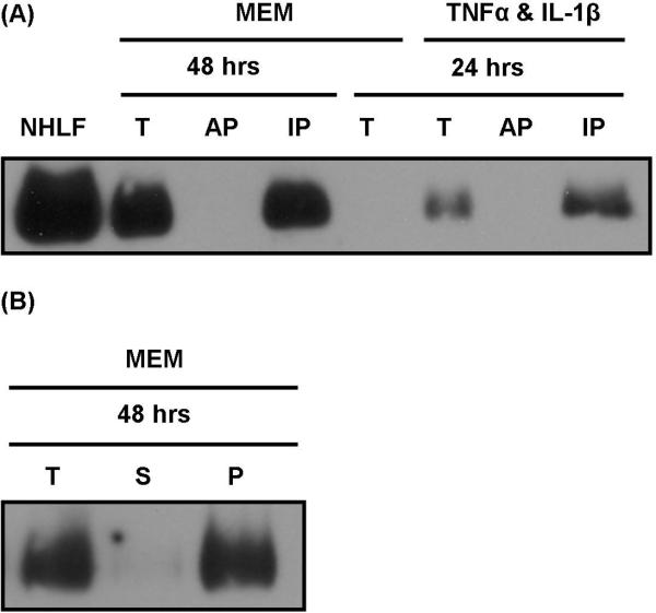
CCL-210 CM was partitioned into soluble and insoluble phases using the non-ionic detergent Triton X-114 to assess the solubility of Thy-1 detected in it. Insoluble phase and soluble phase were submitted to western blot analysis using K117 as the primary. None of the Thy-1 detected in the CM of CCL-210 was retained in the soluble phase. Rather, Thy-1 detected in the CM of either cytokine stimulated or un-stimulated NHLF partitioned exclusively into the insoluble phase [Figure 6. A]. Moreover, the vast majority of Thy-1 detected in the pre-concentrated CM, retained following centrifugation at 1,200 × g, was removed following centrifugation at ≥ 21,000 × g [Figure 6. B].
FIGURE 6. Thy-1 in the Conditioned Media of Normal Human Lung Fibroblasts is insoluble.
(A) CM was collected from CCL-210 provided either MEM or 20ng/mL TNFα and IL-1β in MEM for 24 to 48 hrs. Using Triton X-114, concentrated CM was partitioned into an aqueous (AP) and insoluble phase (IP). Mature Thy-1 detected by western blot in the CE of NHLF or partitioned and pre-partitioned CM by western blot using K117. (B) CM was collected from CCL-210 provided MEM 48 hrs. CM was submitted to differential centrifugation of 1,200 × g and 21,000 × g then precipitated with methanol. Mature Thy-1 was detected in the precipitated material of total (T) prior to, supernatant (S) after, and pellet (P) after 21,000 × g by western blot using K117.
Discussion
The monoclonal antibodies from clones K117, 5E10, and AS02 recognize human Thy-1 at the surface of cells. Thus, these antibodies recognize epitopes displayed by Thy-1 while in a native conformation. Additionally, all three detect Thy-1 in western blots, but not under reducing conditions. This suggests that these antibodies recognize epitopes comprised of segments in the polypeptide held in close proximity by disulphide bonds. The mature Thy-1 polypeptide has four cysteine residues. As Thy-1 is a member of the immunoglobulin superfamily, each cysteine is predicted to form a disulfide bond with one of the other under oxidizing conditions.21 The complete deglycosylation of Thy-1 does not affect the affinities these antibodies have for it. Therefore, the epitopes are likely confined to a region of the Thy-1 polypeptide. Taken together, recognition of Thy-1 by K117, 5E10, and AS02 requires the THY1 polypeptide be in a conformation it assumes at the surface of cell membranes.
Molecular dynamic models comparing rodent Thy-1 inserted into a lipid monolayer, vs. delipidated Thy-1 as if by GPI specific phospholipase D1 (GPLD1), vs. without any portion of a GPI anchor, demonstrate distinct conformational differences. The conformation of rodent Thy-1 without any portion of a GPI anchor is intermediate between the other two but closer to the GPLD1 delipidated model. Interestingly, these same studies suggest the glycan chain of the GPI anchor is tightly folded on itself, bringing the protein in close proximity to the cell surface. A bulk of it may even fit into a lectin-type binding site on the adjacent surface of Thy-1.12,23 The conformational differences are appreciable on the surface of Thy-1 opposite the GPI anchor; known epitopes in these areas become compromised,12 consistent with altered antibody affinity. For Thy-1 and GPI-anchored proteins, the general consensus is that delipidation induces a stable change in conformation that manifests itself in a change in antibody affinity.14 The positions of TYR residues in human Thy-1 are better suited than those in rodent Thy-1 for using circular dichroism spectra to detect conformational changes. A discernable and stable shift in the circular dichroism spectrum of human Thy-1 occurs within an hour of its delipidation. Taken all together, antibodies that recognize human Thy-1 with a GPI anchor are predicted to have lower affinity for Thy-1 if it is delipidated.
We evaluated the relative affinity of three widely available monoclonal Thy-1 antibodies, K117, 5E10, and AS02, for the mature Thy-1 polypeptide with a complete GPI anchor, delipidated by PI-PLC, and expressed as a soluble recombinant protein by omitting the GPI attachment signal. Of the three antibodies, AS02 was shown by another group to detect an increase of Thy-1 in the supernatant of PI-PLC treated human fibroblast. Thy-1 was also detected in the supernatant of untreated cells, however.13 In order to detect the mature Thy-1 polypeptide independent of conformation, recombinant forms were engineered to be expressed with an N-terminal FLAG tag. Our findings demonstrate that recognition of Thy-1 by monoclonal antibodies from clones K117, 5E10, and AS02 in western blots is abolished or greatly diminished if Thy-1 is made soluble by PI-PLC or expressed without a GPI anchor attachment signal. Remarkably, GPI anchors can mediate the conformation of Thy-1 required for recognition by K117 and 5E10 from as far away as 15 AA and attached by a non-endogenous GPI anchor attachment signal. Moreover, detection of THY1 – GPI(DAF) and – GPI(TR3) suggest that the epitopes these antibodies bind do not encompass both the polypeptide and the glycan core of the GPI anchor.
Recombinant soluble Thy-1 has been designed in a number of different ways. In one design, three AA followed by a stop codon were introduced immediately following CYS 130 of rat Thy1.1.18 The additional AA, GGS, were included to allow it to be purified by affinity chromatography using OX-7,18 shown to lose affinity for Thy-1 if delipidated.12 Though our FLAG – sTHY1 has a 6 AA extension, it was not detected in western blots using K117, 5E10, or AS02 [Figure 3B and Supplementary Figure 1]. Additionally, sTHY1 – 6X HIS, with a 12 AA extension, was not detected in western blots using K117. In a second design, six histidines followed by a stop codon were introduced immediately following LYS 129 of human Thy-1, thereby omitting CYS 130. Interestingly, this form of recombinant soluble Thy-1 was detected in western blots by AS02. Also, it was used to establish the standard curve in a sandwich ELISA in which AS02 and 5E10 were the detection antibodies.9 Two potential explanations could account for why these antibodies detect this form but not ours. One, the disulfide bond formed with CYS 130 may place a constraint at the C-terminus that sequesters elements downstream of it. This is supported by reducing conditions exposing the histidine epitope at the C-terminus of sTHY1– 6X HIS in western blots. Two, detection by AS02 and 5E10 may require a greater amount of a soluble Thy-1 relative to the GPI-anchored form, in which case absolute values for each would not be comparable.
The integrin and syndecan-4 binding motifs RLD and RETKK, respectively, were indentified and characterized using a recombinant hybrid of Thy-1 in which the GPI-attachment signal is replaced by the Fc fragment of human IgG1. Though soluble, the Fc fragment molecular mass at ~25.6kDa is twice the mature Thy-1 polypeptide. The relatively large size could presumably supply the constraint at the carboxyl terminus for Thy-1 antibody recognition. Thy-1-Fc forms a dimer24 through the Fc fragments making it further unsuited as a surrogate for WT soluble Thy-1.25 We tested whether commercially available Thy-1-Fc (ALX-522-091) is recognized by K117. A band just over 100kDa was detected suggesting Thy-1-Fc has a conformation more akin to GPI anchored Thy-1 (data not shown). Although without an ability to detect Thy-1-Fc by western blot independent of its conformation, the possibility cannot be discounted that the recognition is non-specific. It is not known whether the binding motifs RLD or RETKK are altered in the soluble Thy-1 conformation in a manner that would influence affinity for integrins or syndecan-4. Potential affinity differences aside, the bivalent nature of Thy-1-Fc likely cross links cell surface receptors that native sThy-1 would not. Absent a transmembrane domain, clustering is an important mechanism by which Thy-1 signals.19,26 Thus, soluble Thy-1-Fc most likely better approximates WT Thy-1 at the cell surface.
There are only two known mammalian enzymes that release GPI-anchored proteins by cutting within the GPI moiety, GPLD127–29 and Angiotensin-converting Enzyme (ACE).30–32 Both have been shown to release Thy-1 from cell surfaces.31–33 ACE cleaves between the first and second mannose of the GPI moiety leaving ethanolamine-phosphate-mannose.31,32 GPLD1 cleaves the inositol phosphate linkage within the GPI moiety so the phosphate is retained by the diacylglycerol.14,29,34 Compared with the remnant GPLD1 leaves behind, ACE leaves behind one more similar in size to a small AA extension and less similar to the one PI-PLC generates. However, it is unknown if Thy-1 released by ACE is recognized by antibodies demonstrated to be deficient in recognizing other forms of soluble Thy-1. The GPI anchor remnant, ethanolamine-phosphate-mannose, could act in a similar manner as the three AA at the C-terminus of sTHY1.1 – GGS.
Despite the inability to recognize soluble Thy-1, K117 detects basal levels of Thy-1 in the CM of NHLF and the increase of Thy-1 that comes with pro-inflammatory cytokine treatment. These incongruous results led to experiments to assess the solubility of Thy-1 detected in CM. As suggested, Thy-1 detected in the CM of CCL-210 is entirely insoluble. Differential centrifugation revealed the Thy-1 to be associated with a rather large particle as it was cleared from the CM at speeds as low as 21,000 × g. Microparticles released from cell membranes are enriched in GPI-linked proteins, and are increasingly recognized to play an important role in intracellular communication in a number of physiological processes and pathological conditions.35–39
A group recently published detection of elevated Thy-1 in the cell-free post-digestion supernatant, a by-product in processing prostate tissue for cell sorting by collagenase digestion. The presence of Thy-1 in the supernatant was attributed to either “cell shedding, secretion, or enzymatic release.” However, this was done by western blot using 5E10 as the primary antibody.8 Based on the results reported here, the Thy-1 thus detected likely retains a complete GPI anchor. On the other hand, mass spectrometry, but not western blot, analysis of urine from patients with prostate cancer identified Thy-1; conversely, Thy-1 was not detected in the urine of post-prostatectomy patients.8 It may be that the Thy-1 in the urine in prostate cancer is a truly soluble, delipidated form.
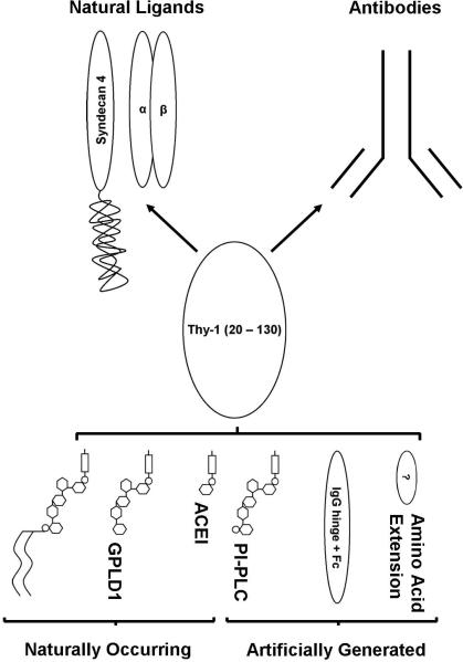
We report that K117, 5E10, and AS02 are deficient in detecting soluble forms of Thy-1 by western blot. Thus, there is a need to develop antibodies to Thy-1 that take into consideration the conformation of delipidated Thy-1. Additionally, the conformation of recombinant forms of Thy-1 used as controls in assays that rely on antibody recognition must be evaluated carefully. Beyond antibody recognition, the conformation of recombinant forms of Thy-1 used to elucidate its function should also be evaluated. The aforementioned considerations are summarized and depicted in figure 7. Furthermore, the nature of Thy-1 released by cytokines, which appears to maintain its membrane-bound conformation and may have important biological significance, needs to be evaluated further.
FIGURE 7. Diagram of GPI anchored, delipidated, and recombinant Thy-1 depicted with relative affinity to antibodies raised against GPI-anchored and known ligands of Thy-1.
Delipidation induces a change in the conformation of Thy-1 causing it to lose affinity for antibodies that recognize the native conformation of THY1 with a complete GPI-anchor. Recombinant soluble Thy-1, i.e. expressed without a GPI-attachment signal, also has a conformation that is not recognized by these same antibodies. However, some forms of recombinant soluble Thy-1 with either a specific AA extension or possibly omission of certain AAs, i.e. CYS 130, may restore affinity. This phenomenon is depicted in the upper half of the diagram, in which white ovals represent the mature Thy-1 polypeptide and narrow black ovals are AA extensions off CYS 130. The conformation assumed by Thy-1 released from cell surfaces with GPLD1 and ACE with respect to antibody affinity, is not known. Moreover, the effect conformational differences may have on the affinity for known natural ligands or whether new ones are gained is not known. All are important to consider when raising antibodies to, using a recombinant form of, or examining the function of Thy-1.
Supplementary Material
SUPPLEMENTARY FIGURE 1. Complete GPI anchor of Thy-1 required for recognition by the monoclonal antibody 5E10. Mature Thy-1 detected by western blot in CE containing FLAG – THY1 or CM containing PI-PLC released FLAG – THY1 or FLAG – sTHY1 using 5E10. After stripping the membranes of antibodies for detecting mature Thy-1, FLAG was detected using M2.
Acknowledgments
We thank Drs. Celia Espinoza, Kornelia Kis, Wendy Liu, and Simon Wong for their helpful suggestions and discussions. We also thank Elizabeth Boyd for all her administrative services and Dr. Daniel F. Legler for his gift of expression vectors containing the cDNA of the GPI-attachment signals for DAF and TRAIL-R3. We acknowledge the support of the UCSD Center for AIDS Research (NIH/DAIDS, P30 AI36214, PI D. Richman) Molecular Biology Core and Flow Cytometry Research Core Facilities at the VA San Diego Healthcare System.
Supported by National Institutes of Health Grant R01HL082818 (JSH)
Abbreviations
- AA
amino acids
- ACE
Angiotensin-converting Enzyme
- CE
Cell extracts
- CM
conditioned media
- DAF
Decay-Accelerating Factor
- GPI
glycosylphosphatidyl inositol
- GPLD1
GPI specific phospholipase D1
- NHLF
normal human lung fibroblasts
- NR
non-reducing
- PI-PLC
Phosphatidylinositol-Specific Phospholipase C
- PNGase F
Peptide: N-Glycosidase F
- RFL-6
Rat fetal lung fibroblasts
- SFM
serum free media
- Thy-1
Thy-1 cell surface antigen
- THY1
Human gene symbol for Thy-1
- Thy1
Mouse gene symbol for Thy-1
- TRAIL-R3
TNF-related Apoptosis-inducing Ligand Receptor 3
- WT
wild type
Contributor Information
John E. Bradley, University of Alabama at Birmingham, Rady Children's Hospital of San Diego 9500 Gilman Drive, MC 0731 La Jolla, CA 92093-0731
Joy M. Chan, University of California-San Diego, Rady Children's Hospital of San Diego 9500 Gilman Drive, MC 0731 La Jolla, CA 92093-0731
James S. Hagood, Pediatric Respiratory Medicine, University of California- San Diego and Rady Children's Hospital of San Diego 9500 Gilman Drive, MC 0731 La Jolla, CA 92093-0731.
References
- 1.Reif AE, Allen JM. Specificity of isoantisera against leukaemic and thymic lymphocytes. Nature. 1963 Dec 28;200:1332–1333. doi: 10.1038/2001332b0. [DOI] [PubMed] [Google Scholar]
- 2.Reif AE, Allen JM. The AKR thymic antigen and its distribution in leukemias and nervous tissues. J Exp Med. 1964 Sep 1;120:413–433. doi: 10.1084/jem.120.3.413. [DOI] [PMC free article] [PubMed] [Google Scholar]
- 3.Rege TA, Hagood JS. Thy-1 as a regulator of cell-cell and cell-matrix interactions in axon regeneration, apoptosis, adhesion, migration, cancer, and fibrosis. FASEB J. 2006 Jun;20(8):1045–1054. doi: 10.1096/fj.05-5460rev. [DOI] [PubMed] [Google Scholar]
- 4.Rege TA, Hagood JS. Thy-1, a versatile modulator of signaling affecting cellular adhesion, proliferation, survival, and cytokine/growth factor responses. Biochim Biophys Acta. 2006 Oct;1763(10):991–999. doi: 10.1016/j.bbamcr.2006.08.008. [DOI] [PMC free article] [PubMed] [Google Scholar]
- 5.Bradley JE, Ramirez G, Hagood JS. Roles and regulation of Thy-1, a context-dependent modulator of cell phenotype. Biofactors. 2009 Jun;35(3):258–265. doi: 10.1002/biof.41. [DOI] [PMC free article] [PubMed] [Google Scholar]
- 6.Hagood JS, Prabhakaran P, Kumbla P, Salazar L, MacEwen MW, Barker TH, et al. Loss of fibroblast Thy-1 expression correlates with lung fibrogenesis. Am J Pathol. 2005 Aug;167(2):365–379. doi: 10.1016/S0002-9440(10)62982-3. [DOI] [PMC free article] [PubMed] [Google Scholar]
- 7.Almqvist P, Carlsson SR. Characterization of a hydrophilic form of Thy-1 purified from human cerebrospinal fluid. J Biol Chem. 1988 Sep 5;263(25):12709–12715. [PubMed] [Google Scholar]
- 8.True LD, Zhang H, Ye M, Huang C-Y, Nelson PS, von Haller PD, et al. CD90/THY1 is overexpressed in prostate cancer-associated fibroblasts and could serve as a cancer biomarker. Mod Pathol. 2010 Oct;23(10):1346–1356. doi: 10.1038/modpathol.2010.122. [DOI] [PMC free article] [PubMed] [Google Scholar]
- 9.Saalbach A, Wetzig T, Haustein UF, Anderegg U. Detection of human soluble Thy-1 in serum by ELISA. Fibroblasts and activated endothelial cells are a possible source of soluble Thy-1 in serum. Cell Tissue Res. 1999 Nov;298(2):307–315. doi: 10.1007/s004419900079. [DOI] [PubMed] [Google Scholar]
- 10.Zhou Y, Hagood JS, Lu B, Merryman WD, Murphy-Ullrich JE. Thy-1-integrin alphav beta5 interactions inhibit lung fibroblast contraction-induced latent transforming growth factor-beta1 activation and myofibroblast differentiation. J Biol Chem. 2010 Jul 16;285(29):22382–22393. doi: 10.1074/jbc.M110.126227. [DOI] [PMC free article] [PubMed] [Google Scholar]
- 11.Kukulansky T, Abramovitch S, Hollander N. Cleavage of the glycosylphosphatidylinositol anchor affects the reactivity of thy-1 with antibodies. J Immunol. 1999 May 15;162(10):5993–5997. [PubMed] [Google Scholar]
- 12.Barboni E, Rivero BP, George AJ, Martin SR, Renoup DV, Hounsell EF, et al. The glycophosphatidylinositol anchor affects the conformation of Thy-1 protein. J Cell Sci. 1995 Feb;108(Pt 2):487–497. doi: 10.1242/jcs.108.2.487. [DOI] [PubMed] [Google Scholar]
- 13.Saalbach A, Kraft R, Herrmann K, Haustein UF, Anderegg U. The monoclonal antibody AS02 recognizes a protein on human fibroblasts being highly homologous to Thy-1. Arch Dermatol Res. 1998 Jul;290(7):360–366. doi: 10.1007/s004030050318. [DOI] [PubMed] [Google Scholar]
- 14.Bütikofer P, Malherbe T, Boschung M, Roditi I. GPI-anchored proteins: now you see “em, now you don”t. FASEB J. 2001 Feb;15(2):545–548. doi: 10.1096/fj.00-0415hyp. [DOI] [PubMed] [Google Scholar]
- 15.Kozak M. An analysis of 5’-noncoding sequences from 699 vertebrate messenger RNAs. Nucleic Acids Res. 1987 Oct 26;15(20):8125–8148. doi: 10.1093/nar/15.20.8125. [DOI] [PMC free article] [PubMed] [Google Scholar]
- 16.Einhauer A, Jungbauer A. The FLAG peptide, a versatile fusion tag for the purification of recombinant proteins. J Biochem Biophys Methods. 2001 Oct 30;49(1-3):455–465. doi: 10.1016/s0165-022x(01)00213-5. [DOI] [PubMed] [Google Scholar]
- 17.Hopp TP, Prickett KS, Price VL, Libby RT, March CJ, Cerretti DP, et al. A short polypeptide marker sequence useful for recombinant protein identification and purification. Nature Biotechnology. 1988 Oct 1;6(10):1204–1210. [Google Scholar]
- 18.Devasahayam M, Catalino PD, Rudd PM, Dwek RA, Barclay AN. The glycan processing and site occupancy of recombinant Thy-1 is markedly affected by the presence of a glycosylphosphatidylinositol anchor. Glycobiology. 1999 Dec;9(12):1381–1387. doi: 10.1093/glycob/9.12.1381. [DOI] [PubMed] [Google Scholar]
- 19.Tiveron MC, Nosten-Bertrand M, Jani H, Garnett D, Hirst EM, Grosveld F, et al. The mode of anchorage to the cell surface determines both the function and the membrane location of Thy-1 glycoprotein. J Cell Sci. 1994 Jul;107(Pt 7):1783–1796. doi: 10.1242/jcs.107.7.1783. [DOI] [PubMed] [Google Scholar]
- 20.Hoessli D, Bron C, Pink JR. T-lymphocyte differentiation is accompanied by increase in sialic acid content of Thy-1 antigen. Nature. 1980 Feb 7;283(5747):576–578. doi: 10.1038/283576a0. [DOI] [PubMed] [Google Scholar]
- 21.Perkins SJ, Williams AF, Rademacher TW, Dwek RA. The Thy-1 glycoprotein: a three-dimensional model. Trends Biochem Sci. 1988 Aug;13(8):302–303. doi: 10.1016/0968-0004(88)90124-7. [DOI] [PubMed] [Google Scholar]
- 22.Roberts WL, Myher JJ, Kuksis A, Low MG, Rosenberry TL. Lipid analysis of the glycoinositol phospholipid membrane anchor of human erythrocyte acetylcholinesterase. Palmitoylation of inositol results in resistance to phosphatidylinositol-specific phospholipase C. J Biol Chem. 1988 Dec 15;263(35):18766–18775. [PubMed] [Google Scholar]
- 23.Rademacher TW, Edge CJ, Dwek RA. Dropping anchor with the lipophosphoglycans. Curr Biol. 1991 Feb;1(1):41–42. doi: 10.1016/0960-9822(91)90123-e. [DOI] [PubMed] [Google Scholar]
- 24.Leyton L, Schneider P, Labra CV, Rüegg C, Hetz CA, Quest AFG, et al. Thy-1 binds to integrin β3 on astrocytes and triggers formation of focal contact sites. Current Biology. 2001 Jul 10;11(13):1028–1038. doi: 10.1016/s0960-9822(01)00262-7. [DOI] [PubMed] [Google Scholar]
- 25.Avalos AM, Valdivia AD, Muñoz N, Herrera-Molina R, Tapia JC, Lavandero S, et al. Neuronal Thy-1 induces astrocyte adhesion by engaging syndecan-4 in a cooperative interaction with alphavbeta3 integrin that activates PKCalpha and RhoA. J Cell Sci. 2009 Oct 1;122(Pt 19):3462–3471. doi: 10.1242/jcs.034827. [DOI] [PMC free article] [PubMed] [Google Scholar]
- 26.Mayor S, Rothberg KG, Maxfield FR. Sequestration of GPI-anchored proteins in caveolae triggered by cross-linking. Science. 1994 Jun 24;264(5167):1948–1951. doi: 10.1126/science.7516582. [DOI] [PubMed] [Google Scholar]
- 27.Low MG, Prasad AR. A phospholipase D specific for the phosphatidylinositol anchor of cell-surface proteins is abundant in plasma. Proc Natl Acad Sci USA. 1988 Feb;85(4):980–984. doi: 10.1073/pnas.85.4.980. [DOI] [PMC free article] [PubMed] [Google Scholar]
- 28.Davitz MA, Hom J, Schenkman S. Purification of a glycosyl-phosphatidylinositol-specific phospholipase D from human plasma. J Biol Chem. 1989 Aug 15;264(23):13760–13764. [PubMed] [Google Scholar]
- 29.Scallon BJ, Fung WJ, Tsang TC, Li S, Kado-Fong H, Huang KS, et al. Primary structure and functional activity of a phosphatidylinositol-glycan-specific phospholipase D. Science. 1991 Apr 19;252(5004):446–448. doi: 10.1126/science.2017684. [DOI] [PubMed] [Google Scholar]
- 30.Kondoh G, Watanabe H, Tashima Y, Maeda Y, Kinoshita T. Testicular Angiotensin-converting enzyme with different glycan modification: characterization on glycosylphosphatidylinositol-anchored protein releasing and dipeptidase activities. J Biochem. 2009 Jan;145(1):115–121. doi: 10.1093/jb/mvn148. [DOI] [PubMed] [Google Scholar]
- 31.Kondoh G, Tojo H, Nakatani Y, Komazawa N, Murata C, Yamagata K, et al. Angiotensin-converting enzyme is a GPI-anchored protein releasing factor crucial for fertilization. Nat Med. 2005 Feb;11(2):160–166. doi: 10.1038/nm1179. [DOI] [PMC free article] [PubMed] [Google Scholar]
- 32.Mayor S. ACEing GPI release. Nat Struct Mol Biol. 2005 Feb;12(2):107–108. doi: 10.1038/nsmb0205-107. [DOI] [PubMed] [Google Scholar]
- 33.Bergman AS, Carlsson SR. Saponin-induced release of cell-surface-anchored Thy-1 by serum glycosylphosphatidylinositol-specific phospholipase D. Biochem J. 1994 Mar 15;298(Pt 3):661–668. doi: 10.1042/bj2980661. [DOI] [PMC free article] [PubMed] [Google Scholar]
- 34.Metz CN, Brunner G, Choi-Muira NH, Nguyen H, Gabrilove J, Caras IW, et al. Release of GPI-anchored membrane proteins by a cell-associated GPI-specific phospholipase D. EMBO J. 1994 Apr 1;13(7):1741–1751. doi: 10.1002/j.1460-2075.1994.tb06438.x. [DOI] [PMC free article] [PubMed] [Google Scholar]
- 35.Tetta C, Bruno S, Fonsato V, Deregibus MC, Camussi G. The role of microvesicles in tissue repair. Organogenesis. 2011 Apr 1;7(2):105–115. doi: 10.4161/org.7.2.15782. [DOI] [PMC free article] [PubMed] [Google Scholar]
- 36.Muralidharan-Chari V, Clancy JW, Sedgwick A, D'Souza-Schorey C. Microvesicles: Mediators of Extracellular Communication During Cancer Progression. J Cell Sci. 2010 May 15;123(10):1603–1611. doi: 10.1242/jcs.064386. [DOI] [PMC free article] [PubMed] [Google Scholar]
- 37.Butikofer P, Kuypers FA, Xu CM, Chiu DT, Lubin B. Enrichment of two glycosylphosphatidylinositol-anchored proteins, acetylcholinesterase and decay accelerating factor, in vesicles released from human red blood cells. Blood. 1989 Oct 1;74(5):1481–1485. [PubMed] [Google Scholar]
- 38.Kim H-S, Choi D-Y, Yun SJ, Choi S-M, Kang JW, Jung JW, et al. Proteomic Analysis of Microvesicles Derived from Human Mesenchymal Stem Cells. J Proteome Res. 2011;11(2):839–849. doi: 10.1021/pr200682z. [DOI] [PubMed] [Google Scholar]
- 39.Anderson HC, Mulhall D, Garimella R. Role of extracellular membrane vesicles in the pathogenesis of various diseases, including cancer, renal diseases, atherosclerosis, and arthritis. Laboratory Investigation. 2010 Aug 30;90(11):1549–1557. doi: 10.1038/labinvest.2010.152. [DOI] [PubMed] [Google Scholar]
Associated Data
This section collects any data citations, data availability statements, or supplementary materials included in this article.
Supplementary Materials
SUPPLEMENTARY FIGURE 1. Complete GPI anchor of Thy-1 required for recognition by the monoclonal antibody 5E10. Mature Thy-1 detected by western blot in CE containing FLAG – THY1 or CM containing PI-PLC released FLAG – THY1 or FLAG – sTHY1 using 5E10. After stripping the membranes of antibodies for detecting mature Thy-1, FLAG was detected using M2.


