Abstract
We describe a burn patient who developed skin lesions on her skin-graft harvest and skin-graft recipient (burn) sites. Orf virus infection was confirmed by a combination of diagnostic assays, including molecular tests, immunohistochemical analysis, pathologic analysis, and electron microscopy. DNA sequence analysis grouped this orf virus isolate among isolates from India. Although no definitive source of infection was determined from this case, this is the first reported case of orf virus infection in a skin graft harvest. Skin graft recipients with exposures to animals may be at risk for this viral infection.
Keywords: Orf virus, skin graft, thermal burn, Parapoxvirus
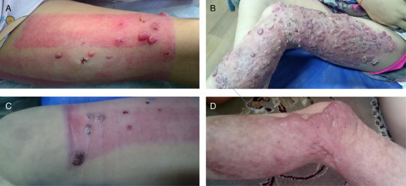
A previously healthy 4-year-old girl from a rural area of Gonbad-e-Kavus, Golestan Province, Iran, was playing in her family’s kitchen when she sustained third-degree skin burns, affecting 35% of her body, from hot water in December 2015. The majority of the burns were on her trunk and left upper and lower extremities. Seven days after the incident, the patient received a skin graft harvested from her right thigh and grafted to the burn areas on her left arm and leg. Four days later, while she was still at the hospital, she developed 8–10 red, nodular lesions measuring 3–8 mm in diameter at the skin-graft harvest site (right thigh; Figure 1A) and approximately 100 pustular/papular lesions in the graft recipient sites (trunk and left upper/lower extremities; Figure 1B). Complete blood count and human immunodeficiency virus tests confirmed she was immunocompetent. She also had no known underlying medical conditions, and skin conditions such as atopy or eczema were not present. No pruritus was ever reported by the patient prior to appearance of lesions. The patient’s father was a livestock breeder of sheep and cattle, in which animals were housed in a structure near the household; however, there was no reported history of sick animals, and the family reported that the patient had no history of contact with these animals. No cloth wrappings nor salves were ever applied by the family to the wounds. One week after the skin graft, 2 biopsy specimens from 2 individual lesions were collected from the affected skin-graft harvest site. Biopsy specimens were not acquired from the skin-graft recipient sites because the burn sites were under special postoperative dressing, and the clinical and morphological similarities of the lesions strongly indicated that both harvest and recipient sites were of similar cause. Because of the clinical presentation and environmental history, the 2 formalin-fixed paraffin-embedded (FFPE) skin biopsy specimens were sent to the Poxvirus and Infectious Disease Pathology Laboratories at the Centers for Disease Control and Prevention (CDC) in Atlanta, Georgia, and subjected to histopathologic analysis, immunohistochemical analysis, transmission electron microscopy (TEM), and molecular analysis.
Figure 1.
Lesion progression over skin graft harvest site and burn site. (A) Papules/nodules in skin-graft harvest site in right thigh 11 days after burn, (B) papules/pustules within the burn site of left thigh after skin graft 11 days after burn; (C) resolution of infection in skin graft harvest approximately four weeks after burn; (D) resolution of burn site (left thigh) approximately four weeks after burn.
METHODS
Histopathologic and Immunohistochemical Analysis of Paraffin-Fixed Biopsy Specimens and TEM
Routine hematoxylin and eosin–stained sections were prepared using standard histologic methods. Immunohistochemical analysis was performed using a colorimetric, indirect, polymer-based immunoalkaline phosphatase technique (Biocare Medical, Concord, California). Polyclonal sheep anti–orf virus and bovine anti–bovine papular stomatitis virus antibodies known to detect parapoxviruses, including orf virus, and a polyclonal rabbit anti–monkeypox virus antibody known to detect orthopoxviruses were used as previously described [1]. On-slide embedding and TEM were performed as previously described [2].
DNA Extraction From Paraffin-Fixed Biopsy Specimens
DNA was extracted from the formalin-fixed biopsy specimens as previously described [3] by the QIAamp DNA mini kit (Qiagen, Valencia, California).
Orf Virus–Specific Real-Time Polymerase Chain Reaction (PCR) Analysis and Phylogenetic Analysis
A real-time PCR assay that targets the orf virus gene J6R was performed on DNA [4]. Reactions were performed in 25-µL volumes on an ABI7900 instrument (Applied Biosystems, Foster City, California).
Sequence analysis assays have been previously described [5]. Briefly, PCR amplicons generated from each tissue specimen were sequenced for analysis. Phylogenetic analyses were performed using the Bayesian analysis software package (v1.83; available at: http://beast.bio.ed.ac.uk), BEAST, BEAUti, and Tracer [6]. The analyses used a Markov chain Monte Carlo chain length of 8 000 000 with an HKY nucleotide rate substitution model, strict molecular settings, and sampling of every 1000 states. The DNA sequences were aligned using BioEdit (available at: http://www.mbio.ncsu.edu/BioEdit/BioEdit.html) and Clustal alignment programs.
Data supporting the results of this article are available in GenBank (accession numbers are included). The sequences used in the phylogenetic analysis of the B2L amplicon were selected from available parapoxviruses or other high G + C content poxviruses in GenBank, including the following isolates: orf viruses ORFV_IND 67_04 (GenBank accession no. DQ263305), ORFV_FIN_F07_3748S (JN773702), ORFV_CHN_Gansu388 (KC485343), ORFV_NZ2 (U06671), ORFVIA82 (AY386263), ORFV_SA00 (AY386264), ORFV_D1701 (HM133903), and ORFV_VA0910054 (KF830857); pseudocowpox viruses PCPV_F05_990C (JF773694), PCPV_VR634 (GQ329670), PCPV_IT_1303 (JN171852), PCPV_GE3_07 (KF478804), PCPV_JP_IW2010H (AB921003), PCPV_BR_ SV721 (KC896641), PCPV_BSH07012 (KF830854), PCPV_ BSH07013 (KF830855), and PCPV_VA0904 (KF830856); sealpox virus SEAV_V842 (AY952943); and bovine papular stomatitis viruses BPSV_VA0982 (KF830859), BPSV_VA09186 (KF830860), BPSV_BSH07005 (KF830858), and BPSV_AR02 (AY386265). 2016_001B2l is the isolate from this study. The sequences used in the phylogenetic analysis of the J6R amplicon included the following isolates: molluscum contagiosum viruses MOCV_UK (GenBank accession no. JQ269324) and MOCV_2008_031 (GQ902057); the uncharacterized crocodile poxvirus CROV_Nile (DQ356948); pseudocowpox viruses PCPV_BSH07012 (GQ902051), PCPV_BSH07013 (GQ902052), PCPV_MD06025 (GQ902049), PCPV_FIN00_120R (GQ329669), PCPV_VA0904J (KF830862), and PCPV_GA08024J (KF830863); squirrel poxvirus SQRV_UK (HE601899), orf virus ORFV_VA2010910054J (KF830861); and bovine papular stomatitis viruses BPSV_ BSH07005 (GQ902054), BPSV_WA07058 (GQ902053), BPSV_VA0982 (KF830861), and BPSV_VA09186J (KF830864). Both amplicons from the patient will be submitted to GenBank.
RESULTS
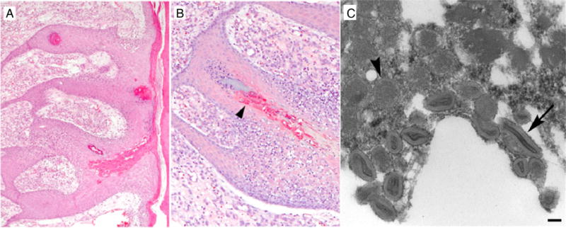
The 2 biopsy specimens from the skin-graft harvest site exhibited epidermal hyperplasia, superficial dermal edema, and vascularization with focal keratinocyte ballooning degeneration, pallor of superficial keratinocytes, and rare eosinophilic cytoplasmic inclusions, all of which are consistent with a Parapoxvirus infection (Figure 2A). Immunohistochemical staining was positive for Parapoxvirus, with antigen localized to keratinocytes (Figure 2B), and negative for Orthopoxvirus. TEM captured images of mature Parapoxvirus particles from the FFPE specimens (Figure 2C). Orf virus DNA signatures were amplified by real-time PCR from the FFPE biopsy specimens.
Figure 2.
Histopathology (A) and immunohistochemistry (B) of formalin-fixed paraffin-embedded skin biopsy from the patient’s affected skin harvest site and (C) transmission electron microscopy image. (A) Epidermal hyperplasia, vascularization and keratinocyte degeneration; (B) arrow indicates parapoxvirus antigen localization (red) to keratinocytes; (C) immature (arrowhead), spherical particles with a homogenous core and mature (long arrow), oval parapoxvirus particles with a dense core from epidermis skin graft harvest site lesion. Bar, 100 nm.
Two amplicon sequences (high G + C and B2L) were used for the phylogenetic analysis of this isolate. The high G + C PCR amplicon is 620 base pairs in size and highly conserved among poxviruses with high G + C content, including pseudocowpox viruses, bovine papular stomatitis viruses, molluscum contagiosum viruses, and crocodile poxviruses. The sequence isolated from the burn patient biopsy specimen (2016_001GC) grouped closely to other known orf viruses (Supplementary Figure 1A). The 590–base pair B2L amplicon sequence (2016_001B2L) also grouped with the orf virus clade and was closely related to an isolate from India (Supplementary Figure 1B).
DISCUSSION
Parapoxviruses are a genus of poxviruses whose natural host are ungulates; however, transmission to other mammals, such as humans, has been documented [7]. In humans, orf virus infections, or contagious ecthyma, are commonly associated with contact with infected animals, usually goats and sheep, or infected fomites [7]. The virus infects epidermal keratinocytes through breaks in the skin barrier, such as a cut or burn, resulting in pustular lesions that then ulcerate. The typical clinical presentation in immunocompetent individuals is one or a few characteristic localized, self-limiting lesions that often appear approximately 1 week following exposure. Lesions often last for 1–4 weeks before resolving spontaneously, but it is not uncommon for lesions to persist for significantly longer before full resolution. Although there is no established treatment for orf virus infections, topical imiquimod, excision, and cryotherapy have been options for immunocompromised patients [8, 9]. In the thermal-burn patient presented in this report, the majority of the lesions for both the skin-graft harvest and burn sites resolved spontaneously 3 weeks after initial presentation, with no complications. Cryotherapy was, however, used on 3 remaining lesions 1 week after most [10] lesions resolved, and no scarring was noted (Figure 1C and 1D).
In this report, the isolate from Iran is most related to an isolate from India isolated in 2006 [10]. Varying regional practices in the trade of ungulates may impact the spread of orf viruses; however, the significance of the orf virus isolated from the patient (2016_001GC) grouping with an isolate from India is not clear.
The pathogenesis, resulting in the characteristic capillary proliferation, is associated with the viral-encoded protein vascular endothelial growth factor (VEGF) [11]. Viral VEGF promotes angiogenesis and epidermal regeneration, which is believed to be important in maintaining viral growth as the virus establishes an infection in the skin [12]. The viral VEGF also is believed to be involved in crust or scab formation, characteristics of healing orf virus lesions [11]. The shed scabs are important for viral transmission because they contain a significant amount of infectious viral particles and are highly resistant to environmental degradation [12]. VEGF, therefore, appears to be important during both replication and transmission of orf virus. VEGF is also produced in normal host cells to promote healing through re-epithelization. There is some evidence that thermal burns are associated with an upregulation of VEGF production by the host [12]; however, it is unknown whether or how host VEGF upregulation in burns play a role in orf virus pathogenesis in conjunction with viral VEGF.
This is the first documented report of orf virus infection in a thermal-burn patient at the skin-graft harvest site. Although no biopsy specimen was collected from the affected recipient’s burn site, orf virus infection was strongly suggested because of the timing and characteristic dermatologic presentation occurring concurrently with the orf virus infection at the skin-graft harvest site. Both sites exhibited almost complete lesion resolution approximately 3 weeks after initial presentation. The use of lanolin-based emollients, such as wool-derived fats, were ruled out as a possible source, based on the patient’s history and medical management. The patient’s history of residence near sheep and cows is a suspicious exposure risk; however, there were no reports of any animals that exhibited clinical signs or symptoms suggestive of orf virus infection in the weeks prior to illness in the patient. Transmission through confirmed infectious fomites has been a cause in previous documented orf virus infection cases associated with burn patients [13]; however, in the case of this burn patient, it is unknown whether the infection of the harvest and burn sites were due to concurrent infection from a common animal or fomite source or whether virus transmission was the direct result of the grafting process (ie, transmission of orf virus from the infected skin-graft harvest site to the susceptible skin-graft recipient site).
Limitations of this investigation included a lack of environmental specimen collection (via swabs) from the patient’s home to test fomites; a thorough investigation of possible exposures from infected animals, including a thorough evaluation and sample collection from farm animals; and a reliance of only dermatologic presentation of affected burn sites.
Parapoxvirus infections are generally self-limiting; however, burn patients are uniquely at risk for complicated orf virus infections because of the breakdown of the epidermal barrier [13, 14]. In rare instances, lesions can be disseminated and have a complicated recovery compounded by secondary skin infections requiring medical intervention [13]. Orf virus lesions can develop into large, exophytic lesions (giant orf) that can spread beyond the initial sites of inoculation in immunocompromised hosts [9]. This could explain the differential lesion distribution between the graft harvest site and the burn sites. The orf virus infection at the skin-graft harvest site was limited to isolated lesions. In contrast, the orf virus infection in the burn sites appeared more evenly distributed across the affected tissue, which could be due to the localized immunocompromised environment of burn sites [15]. Despite the patient’s immunocompetence, skin burns create anatomical regions that can be immunologically distinct from the rest of the host. It should be noted that burns can also impair systemic immunity, as well as skin immunity, and therefore may also contribute to more-diffuse lesions observed in the burn sites. Finally, graft harvest sites will have an absence of epidermis, which may have limited the ability of orf virus to invade and multiply efficiently. Ultimately, the anatomical differences and immunological status in this patient did not affect the outcome—the patient’s infection at the skin-graft harvest and recipient sites have fully resolved with no recurrence.
Although the occurrence may be rare, thermal-burn patients may be a unique category of patients at particular risk of acquiring various skin infections, such as orf virus infection [14]. Because of this patient’s unique orf virus infection at the site of the skin-graft harvest, we propose that, in addition to thermal-skin-burn patients, skin-graft donors and recipients may be at risk for orf virus infection, particularly if the donor site is not properly disinfected. Although person-to-person transmission of orf virus is not known to occur, early recognition of orf virus can guide the management of patients and make identifying an infection source a priority. In the hospital setting, a contaminated fomite source could infect multiple susceptible patients with orf virus in a burn ward, as documented by Midili et al [13]. This case exemplifies not only the importance of early orf virus diagnosis in a hospital setting, but also the importance of interagency and interdisciplinary collaboration to achieve a positive patient outcome.
Supplementary Material
Footnotes
Supplementary materials are available at http://jid.oxfordjournals.org. Consisting of data provided by the author to benefit the reader, the posted materials are not copyedited and are the sole responsibility of the author, so questions or comments should be addressed to the author.
Potential conflicts of interest. All authors: No reported conflicts. All authors have submitted the ICMJE Form for Disclosure of Potential Conflicts of Interest. Conflicts that the editors consider relevant to the content of the manuscript have been disclosed.
References
- 1.Osadebe LU, Manthiram K, McCollum AM, et al. Novel poxvirus infection in 2 patients from the United States. Clin Infect Dis. 2015;60:195–202. doi: 10.1093/cid/ciu790. [DOI] [PMC free article] [PubMed] [Google Scholar]
- 2.Hayat MA. Principles and techniques of electron microscopy : biological applications. 4. Cambridge, UK, and New York: Cambridge University Press; 2000. [Google Scholar]
- 3.Guarner J, Bhatnagar J, Shieh WJ, et al. Histopathologic, immunohistochemical, and polymerase chain reaction assays in the study of cases with fatal sporadic myocarditis. Hum Pathol. 2007;38:1412–9. doi: 10.1016/j.humpath.2007.02.012. [DOI] [PubMed] [Google Scholar]
- 4.Zhao H, Wilkins K, Damon IK, Li Y. Specific qPCR assays for the detection of orf virus, pseudocowpox virus and bovine papular stomatitis virus. J Virol Methods. 2013;194:229–34. doi: 10.1016/j.jviromet.2013.08.027. [DOI] [PubMed] [Google Scholar]
- 5.Li Y, Meyer H, Zhao H, Damon IK. GC content-based pan-pox universal PCR assays for poxvirus detection. J Clin Microbiol. 2010;48:268–76. doi: 10.1128/JCM.01697-09. [DOI] [PMC free article] [PubMed] [Google Scholar]
- 6.Drummond AJ, Rambaut A. BEAST: Bayesian evolutionary analysis by sampling trees. BMC Evol Biol. 2007;7:214. doi: 10.1186/1471-2148-7-214. [DOI] [PMC free article] [PubMed] [Google Scholar]
- 7.Centers for Disease Control Prevention. Human Orf virus infection from household exposures - United States, 2009–2011. MMWR. 2012;61:245–8. [PubMed] [Google Scholar]
- 8.Lederman ER, Green GM, DeGroot HE, et al. Progressive ORF virus infection in a patient with lymphoma: successful treatment using imiquimod. Clin Infect Dis. 2007;44:e100–3. doi: 10.1086/517509. [DOI] [PubMed] [Google Scholar]
- 9.Degraeve C, De Coninck A, Senneseael J, Roseeuw D. Recurrent contagious ecthyma (Orf) in an immunocompromised host successfully treated with cryotherapy. Dermatology. 1999;198:162–3. doi: 10.1159/000018095. [DOI] [PubMed] [Google Scholar]
- 10.Hosamani M, Bhanuprakash V, Scagliarini A, Singh RK. Comparative sequence analysis of major envelope protein gene (B2L) of Indian orf viruses isolated from sheep and goats. Vet Microbiol. 2006;116:317–24. doi: 10.1016/j.vetmic.2006.04.028. [DOI] [PubMed] [Google Scholar]
- 11.Savory LJ, Stacker SA, Fleming SB, Niven BE, Mercer AA. Viral vascular endothelial growth factor plays a critical role in orf virus infection. J Virol. 2000;74:10699–706. doi: 10.1128/jvi.74.22.10699-10706.2000. [DOI] [PMC free article] [PubMed] [Google Scholar]
- 12.Wise LM, Inder MK, Real NC, Stuart GS, Fleming SB, Mercer AA. The vascular endothelial growth factor (VEGF)-E encoded by orf virus regulates keratinocyte proliferation and migration and promotes epidermal regeneration. Cell Microbiol. 2012;14:1376–90. doi: 10.1111/j.1462-5822.2012.01802.x. [DOI] [PubMed] [Google Scholar]
- 13.Midilli K, Erkilic A, Kuskucu M, et al. Nosocomial outbreak of disseminated orf infection in a burn unit, Gaziantep, Turkey, October to December 2012. Euro Surveill. 2013;18:20425. doi: 10.2807/ese.18.11.20425-en. [DOI] [PubMed] [Google Scholar]
- 14.Biyik Ozkaya D, Taskin B, Tas B, et al. Poxvirus-induced angiogenesis after a thermal burn. J Dermatol. 2014;41:830–3. doi: 10.1111/1346-8138.12589. [DOI] [PubMed] [Google Scholar]
- 15.Ruocco V, Ruocco E, Piccolo V, Brunetti G, Guerrera LP, Wolf R. The immunocompromised district in dermatology: a unifying pathogenic view of the regional immune dysregulation. Clin Dermatol. 2014;32:569–76. doi: 10.1016/j.clindermatol.2014.04.004. [DOI] [PubMed] [Google Scholar]
Associated Data
This section collects any data citations, data availability statements, or supplementary materials included in this article.


