Abstract
Human P-glycoprotein (P-gp) controls drugs bioavailability by pumping out of the cells many structurally-unrelated drugs. The x-ray structure of the mouse P-gp ortholog was solved with two SSS- and one RRR-enantiomers of the selenohexapeptide inhibitor QZ59, found within the putative drug-binding pocket of the membrane domain outer leaflet. This offered the first opportunity to localize the well-known H- and R- drug-substrate sites in light of QZ59 inhibition mechanisms that were characterized here in cellulo and modelled towards Hoechst 33342 and daunorubicin transport. We found that QZ59-SSS competes efficiently with both substrates, displaying KI,app values of 0.15 and 0.3 μM, respectively 13 and 2 times lower than corresponding Km,app. In contrast, QZ59-RRR non-competitively inhibited daunorubicin transport with moderate efficacy (KI,app = 1.9 μM) and displayed a mixed-type inhibition towards Hoechst 33342 transport, resulting from a mainly non-competitive (Ki2,app = 1.6 μM) and a poor but significant competitive tendency (Ki1,app = 5 μM). These results suppose a positional overlap of QZ59 – drug-transport sites, total for the SSS enantiomer and partial for the RRR one. Crystal structures analysis suggests that the H site overlaps both QZ59-SSS locations while the R-site overlaps the most embedded one.
Keywords: P-glycoprotein, multidrug efflux ABC transporters, drug efflux mechanism, drug-binding sites, cancer, drug resistance
Introduction
The human P-glycoprotein (P-gp) is an integral membrane protein that actively pumps endo/exogenous compounds out of cells using the energy derived from ATP binding and hydrolysis [1]. P-gp is up-regulated in many cancer cells, where it reduces the intra-cellular concentrations of many chemotherapeutic drugs, thereby imparting multidrug resistance (MDR) [2]. P-gp is one of the best-known pumps in this context, in addition to Multidrug Resistance Protein 1 (MRP1/ABCC1) and Breast Cancer Resistance Protein (BCRP/ABCG2); all these proteins belong to the same ATP-binding cassette (ABC) transporter superfamily [3]. The implication of such pumps in chemoresistance necessitates the elucidation of the mechanisms of multidrug export and its selective inhibition. P-gp transports a broad spectrum of molecules sharing a marked hydrophobicity but structurally divergent [4, 5]. This polyspecificity is partly explained by the existence of at least three predicted drug-binding sites, termed H-, R-, and P-site. The H-site binds Hoechst 33342 and quercetin, the R-site preferentially binds rhodamine 123 and anthracyclins such as daunorubicin and doxorubicin, while the P-site binds the likes of prazosin and progesterone [6, 7]. A fourth site has also been suggested, which may bind non-transported modulators like GF120918 or nicardipine [8].
Among the different drug-binding sites of P-gp, the H- and R- sites have been more rigorously investigated. The use of thiol-reactive substrates in competition with drugs coupled to cysteine scanning mutagenesis has contributed to identify several amino acid residues from transmembrane domain close, or participating to the drug binding sites (reviewed in [9]). Notably, I340(TM6), A841(TM9), L975-V981-V982(TM12) were mapped in the R-site [10]. Location of the H-site is more controversial since fluorescence resonance energy transfer investigations using Hoechst 33342 suggested a location in the cytoplasmic/inner leaflet side of the transporter [11] while the use of a pharmacophore pattern for the same dye suggested a location within the outer leaflet [12].
The recent mouse P-gp (Abcb1a) x-ray structures revealed a large, internal drug-binding cavity enclosed between the two ‘halves’ of the ‘full’ transporter [13, 14]. For the first time the pump was co-crystallized with the RRR and SSS enantiomers of the cyclic selenohexapeptide, QZ59 [13]. One molecule of QZ59-RRR, and two of QZ59-SSS were identified in their respective co-crystal structures. As not uncommon for membrane proteins, these 3D-structures were obtained at a rather modest resolution of 3.8 – 4.35 Å but, thanks to the 3 Selenium atoms they included, QZ59s could be unambiguously located, within the outer leaflet interface of the drug-binding cavity, QZ59s were found to inhibit the mouse P-gp-mediated efflux of drugs such as colchicine and calcein, and the verapamil-stimulation component of mouse P-gp’s ATP hydrolysis activity [13]. Until now, no other compound, as either inhibitor or substrate drug, has ever been co-crystallized, making QZ59 enantiomers unique tools for localizing more precisely the drug-binding sites, at least inside the co-crystallized pump conformation. A detailed characterization of their inhibition mechanisms, competitive, non-competitive, mixed…, would let us know if they share, none, one or even several drug transport sites, which ones, and to what extent. Such information is crucial to understand the transport mechanism mediated by ABC pumps; it is also critical to guide the design of compounds acting at the molecular level with the highest efficiency.
We addressed this question in the present study. Our results revealed distinct inhibitory mechanisms of the QZ59 enantiomers toward drug-substrates binding to the H and R sites. As expected, such mechanisms allowed us to localize these substrate-binding sites within the transporter’s binding cavity.
Results
In order to test the effects of QZ59 inhibitors on the transport by P-gp of substrates binding to the H- and R-sites, as detailed in the experimental section, we used whole cells in order to provide a native environment for the pump [15] in terms of membrane lipid composition, substrate partitioning and bioavailability, protein trafficking, and membrane potential. Concentrations and kinetic parameters estimated under these conditions remain only apparent but, most importantly for the present study, fully comparable.
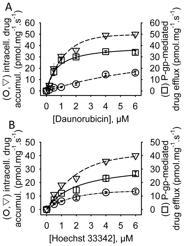
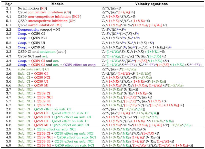
We measured the transport of daunorubicin and Hoechst 33342 mediated by P-gp (Fig. 1), from which we estimated with equation 2.1 (Table 1) maximal rates, Vm, of 41 ± 3 and 34 ± 3 pmol.s−1.mg−1 (of cell protein content) and Michaelis constants, Km of 0.7 ± 0.2 and 2 ± 0.4 μM for daunorubicin and Hoechst 33342, respectively. These values are in the same range that those previously reported for daunorubicin [15] and Hoechst 33342 [16] showing that the system set up here allows to characterize the QZ59 inhibition towards these substrates. For the further experiments, data sets were subjected to a detailed enzymatic analysis (fully displayed in Table S1) using non-linear regressions, and modeled using the equations displayed in Table 1. We then evaluated the models by statistical tools such as Goodness-Of-Fit (GOF) and corrected Akaike Information Criterion (AICc). As classically carried out, the choice of the best model was guided either by looking for the largest drop in AICc or, equally, the largest ΔAICc when using the basal Michaelis-Menten equation as reference (eq. 2.1).
Figure 1. Intracellular accumulations and P-gp-mediated transports of daunorubicin and Hoechst 33342 in control and P-gp expressing NIH3T3 cells.
Daunorubicin (A) and Hoechst 33342 (B) were added to NIH3T3 (triangles) and NIH3T3-P-gp (circles) cells and quantified as described in Methods. The net drug or dye accumulations corresponding to the difference in accumulation between both cell types are shown by squares. Data in triplicates were fitted as detailed in Methods with equations 1.1 (triangles and circles) and 2.1 (squares).
Table 1. Enzyme inhibition models used in this study.
Equations were used as detailed in Methods. S = substrate concentration, μM; I = QZ59 concentration, μM; Vm = maximal substrate efflux rate, in fmol of transported drug•cell−1•h−1; Km = Michaelis constant, μM; KI = inhibition constant, μM; Ki1 = inhibition constant for the E+I -> E•I partial reaction, μM; Ki2 = inhibition constant for the E•S+I->E•S•I partial reaction, μM; h = Hill number, KA = activation constant, μM; KSI = substrate inhibition constant, μM.
Eq. = equation;
NI= no inhibition;
CI = competitive inhibition;
NCI = non-competitive inhibition;
UI = uncompetitive inhibition;
MI = mixed inhibition;
coop. = cooperativity;
act. = activation;
sub = substrate.
Mechanism of drug efflux inhibition by QZ59-RRR
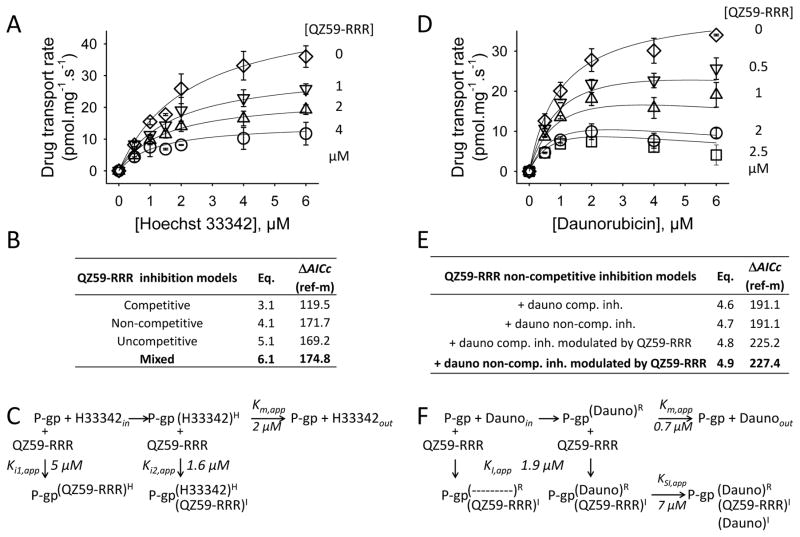
We first measured the QZ59-RRR-affected inhibition of Hoechst 33342 (used to probe the H-site) and daunorubicin (used to probe the R-site) efflux by human P-gp. Transport data in the absence or presence of increasing concentrations of the drug-substrate and QZ59-RRR were collected using the cell line NIH3T3-P-gp over-expressing human P-gp, compared to non-expressing NIH3T3 cells as control (Fig. 2A–F).
Figure 2. QZ59-RRR effect on P-gp-mediated drugs transport.
A, D. Plots of Hoechst 33342 (A) and daunorubicin (D) transport rates as a function of drug and QZ59-RRR concentrations. The experiments were done in triplicate generating 84 (A) and 90 (D) measures respectively. Traces correspond to the best fit obtained with eq. 6.1 (A) or eq. 4.9 (D), Table 1. B, E. ΔAICc scores of the tested models in respect of the reference model (eq. 2.1). C, F. Reaction schemes of substrates transport and QZ59-RRR effects with the estimated constants. H33342 = Hoechst 33342; Dauno = daunorubicin; R, H = R- or H-transport sites; I= inhibition site. Ki1, Ki2, KI, KSI and Km correspond to the inhibition and Michaelis constants of the reactions, as detailed in the text.
The mixed inhibition model (eq. 6.1) best fitted the data set for the inhibition of Hoechst 33342 export (Fig. 2A-C), leading to the largest ΔAICc (174.8, Fig. 2B). A detailed analysis of model fitting and scoring is displayed in Figure S1A, legend and comments. This mixed inhibition has a strong non-competitive contribution as suggested by the inhibition constants Ki1,app (for P-gp alone) of 5 ± 2 μM and Ki2,app (for the P-gp·Hoechst 33342 complex) of 1.6 ± 0.2 μM (panel C; Table S1). Indeed, the non-competitive inhibition model (eq. 4.1) confirmed this tendency, giving a comparably close ΔAICc (171.7, panel B). As drawn in panel C, this non-competitive mixed inhibition suggests that at the Ki2,app concentration (1.6 μM) QZ59-RRR binds to an inhibitory site, distinct to the H- site, while at high concentrations, i.e. in the range of Ki1,app (≥5 μM), QZ59-RRR tends to share the H- site.
The inhibition of P-gp-mediated daunorubicin efflux by QZ59-RRR is displayed in Fig. 2D-F and detailed in Fig. S1B. The initial kinetic analysis suggested a non-competitive model (Table S1). Additionally, we observed an inhibition mediated by daunorubicin itself upon its own efflux, which is clearly visible at high daunorubicin and QZ59-RRR concentrations in Fig. 2D. Regression analyses carried out on these data with equations 4.6 and 4.7 including these effects gave the same ΔAICc scores (191.1) for the competitive (eq. 4.6) and non-competitive (eq. 4.7) models (Fig. 2E). These models however did not correctly fit the data obtained at high daunorubicin and QZ59-RRR concentrations (Fig. 2E, S1B). Such data suggested that QZ59-RRR may enhance the daunorubicin-mediated self-inhibition. Indeed, introducing this effect into equations 4.8 and 4.9 led to the largest increase of ΔAICc values, 225.2 and 227.4 for models including competitive and non-competitive inhibition by daunorubicin (panel E). Although these models are fairly close in their fits (Fig. S1B), the latter model (eq. 4.9) with the significantly higher ΔAICc is more likely to be true. Thus, the QZ59-RRR-mediated inhibition of daunorubicin transport is non-competitive (KI,app = 1.9 ± 0.4 μM), added of a daunorubicin-mediated self-inhibition enhanced by QZ59-RRR (KSI,app = 7 ± 3 μM).
Taken together, our data suggest that QZ59-RRR inhibits drug efflux mainly through an inhibitory site, distinct to the R- and H- transport sites. At high concentrations however, QZ59-RRR partially shares or overlaps with the H-site, and also enhances the self-inhibitory action of R-site drug-substrates.
Mechanism of drug efflux inhibition by QZ59-SSS
The mouse P-gp-QZ59 co-crystal structures reveal distinct binding sites for the RRR and SSS enantiomers of QZ59, the latter being present in 2 different locations. We tested whether this distinction has a bearing on the mechanism of drug efflux inhibition.
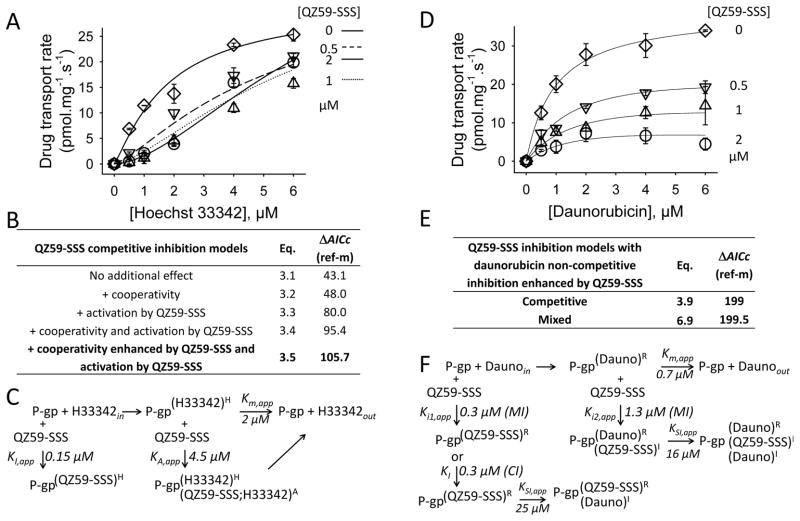
The inhibition P-gp-mediated Hoechst 33342 efflux by QZ59-SSS is presented in Fig. 3A–C.
Figure 3. QZ59-SSS effects on P-gp-mediated drugs transport.
A, D. Plots of Hoechst 33342 (A) and daunorubicin (D) transport rates as a function of drug and QZ59-SSS concentrations. The experiments were done in triplicate generating 72 data in both cases. Traces correspond to the best fits obtained with eq. 3.5 (A) or eq. 6.9 (D), Table 1. For clarity, traces in panel a have been drawn in solid lines for [QZ59-SSS] = 0 and 2 μM, and in dashed and dotted lines for [QZ59-SSS] = 0.5 and 1 μM. B, E. ΔAICc scores of the tested models in respect of the reference one (eq. 2.1). C, F. Reaction schemes of substrates transport and QZ59-SSS effects with the estimated constants. H33342 = Hoechst 33342; Dauno = daunorubicin; QZ59S = QZ59-SSS; R, H = R- or H-transport sites; I= inhibition sites; A= activation site. Ki1, Ki2, KI, KSI, KA and Km correspond to the inhibition, activation and Michaelis constants of the reactions, as detailed in the text.
The data at different concentrations of QZ59-SSS revealed a sigmoidal distribution, suggesting a cooperative behavior of the pump triggered by QZ59-SSS binding. Moreover, data at higher inhibitor concentrations revealed an activation effect by QZ59-SSS on the efflux of Hoechst 33342. We carried out the simulations accordingly, exploring the effects of cooperativity, inhibition and activation independently or together using equations 2.1 to 3.5. A detailed analysis is displayed in Figure S2A, legend and comments. Among these models, competitive inhibition models best fitted the data set (eq. 3.1–3.5, panel B). Furthermore, the models that included activation and cooperativity together (eq. 3.4, 3.5) led to the largest ΔAICc (95.4 and 105.7), in contrast to the models that included them separately (eq. 3.2, 3.3: 47.9 and 79.9). The best model (eq. 3.5, GOF displayed in Fig. S2A) suggests that QZ59-SSS competes with Hoechst 33342 transport (panel C), doing this with a very good efficiency as evidenced by the low value of the inhibition constant, KI,app, of 0.15 ± 0.04 μM, which is 13 times lower than the Km,app (2 μM). At high QZ59-SSS and Hoechst 33342 concentrations, QZ59-SSS tends to activate the Hoechst 33342 efflux, in a cooperative manner. The estimated activation constant KA,app of 4.5 μM suggests that this effect is mediated either through binding to additional low-affinity sites and/or with a poor efficacy.
The effects of QZ59-SSS on P-gp-mediated daunorubicin efflux are displayed in Fig. 3D–F. A detailed analysis is displayed in Figure S2B, legend and comments. The distribution of the values suggested an inhibition by daunorubicin at high substrate and QZ59-SSS concentrations as observed with QZ59-RRR, although to a lower extent. Competitive and mixed inhibition models (eq. 3.9 and 6.9, panel E) gave the largest ΔAICc, 199 and 199.5, respectively. The mixed inhibition model had a marginally better fit, as displayed in panel D and confirmed by the corresponding GOFs (Fig. S2B). As drawn in panel F, the mixed inhibition model suggests that QZ59-SSS competes with daunorubicin at the R transport site. This effect occurs with an inhibition constant Ki1,app of 0.35 ± 0.08 μM, which is ~2 times lower than the Km of daunorubicin transport. According to this model, QZ59-SSS also binds to the P-gp-daunorubicin complex on an inhibitory site, with an inhibition constant Ki2,app of 1.3 ± 0.9 μM. Ki2,app is ~4 times higher than Ki1,app indicating a marked competitive inhibition tendency. This is confirmed by the close ΔAICc obtained with the competitive model (eq. 3.9), leading to a KI,app of 0.28 ± 0.05 μM. At high concentrations, daunorubicin reaches its self-inhibitory site similar to previously described for QZ59-RRR on daunorubicin efflux. This effect is limited since the inhibition constant KSI,app is estimated to 16 μM (panel F).
Taken together, these data suggest that QZ59-SSS competes with drug substrates, which are historically known to bind the H- and R- sites, with a positional substrate overlap much more marked for the H-site than for the R-site. Additionally, at higher concentrations this inhibitor tends to activate the efflux of drugs that bind the H-site.
Discussion
Owing to its significant role in imparting multidrug resistance properties to cancer cells, P-gp is a validated therapeutic target. The inhibition of its drug export function is critical in order to improve the efficacy of many clinically important chemotherapeutic drugs. Thus, understanding the inhibitory mechanisms pertinent to this pump helps for developing more potent and selective inhibitors. The system set up here allowed to estimate the apparent transport and inhibition constants; we found them from 0.1 to 25 μM, values which correlate well with KD recently estimated for compounds interacting with P-gp [17, 18].
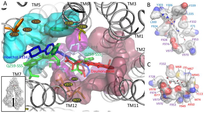
QZ59-RRR and -SSS enantiomers are two inhibitors that were co-crystallized with mouse P-gp [13], each in one of the 6 inward-facing conformations resolved to date [19]. The mouse P-gp is 87% identical in protein sequence to human P-gp making that the 3D structural information obtained with the mouse P-gp can be reasonably correlated to the inhibitory mechanism of QZ59 compounds towards the human P-gp. We found that in contrast to RRR, the SSS enantiomer competes with drugs binding to canonical H- and R-sites. This result is intriguing because QZ59-RRR and -SSS are distributed in a common groove in the outer leaflet of the membrane domain (Fig. 4A) while R- and H-drugs-binding sites are distinct, as previously reported [7].
Figure 4. Putative H and R drug-binding site locations in the (mouse) P-gp in respect to QZ59 inhibitors.
A. QZ59 binding region of the mouse P-gp x-ray structure (PDB code 3G61) as shown in the lower-left inset. QZ59-SSS1 and QZ59-SSS2 as resolved in 3G61 are in green. QZ59-RRR as resolved in the mouse P-gp x-ray structure (PDB code 3G60) is included in this view in light blue. Hoechst 33342 (blue) and daunorubicin (red) correspond each to the best rated molecule location from independent docking simulations as detailed in Fig. S3A-B (−9.1 ± 0.3 and −10.4 ± 0.3 kcal/mol). Orange residues correspond to those reported to interact with Hoechst 33342 [12]. Yellow residues are those which, when replaced by a cysteine, are protected by rhodamine from thiol modification by MTS-rhodamine [10]. Areas surrounding the H- and R-sites and those being common to both are in blue, red and purple respectively. B, C. Residues forming the putative H- (B) and R- (C) drug-binding sites in the mouse P-gp inward-facing conformation in which QZ59-SSS were co-crystallized. Drawn with the same color code as in panel A. Drawn with Pymol 1.6.
This contradiction is only apparent. Indeed, the KI,app for QZ59-SSS competitive inhibition toward Hoechst 33342 and daunorubicin, 0.15 and 0.3 μM respectively, let us suppose an overlap between drugs and QZ59-SSS, as the corresponding Km,app are 13 and 2 times higher. In addition, QZ59-RRR displays a moderate competitive tendency toward Hoechst 33342 while it acts strictly non-competitively toward daunorubicin. This suggests that the H site broadly overlaps the QZ59-binding groove, contrarily to the R site which shares it partially. A docking simulation carried out with Hoechst 33342 in the mouse QZ59-SSS·P-gp x-ray structure indeed gave the 3 best locations along the groove (Fig. S3A). Notably, as defined the H site is bordered by residues F299, Y303 and Y306 (Fig. 4A) that have been proposed to be part of it [12]. Another docking simulation done with daunorubicin suggested that the 4 best locations overlap the most embedded QZ59-SSS (Fig. S3B). We got exactly the same result by docking the thiol-reactive analog of rhodamine, methanethiosulfonate (MTS)-rhodamine (Fig. S3C) used previously by Loo and Clarke for mapping cysteine-mutagenized residues protected from its modification by rhodamine B [10]. Interestingly, docking showed that while the rhodamine moiety binds to the same R-site, the MTS moiety occupies a much larger space indeed surrounded by the cysteine-mutagenized residues protected by rhodamine B. As positioned, the R site overlaps the most embedded QZ59-SSS location and almost not that of QZ59-RRR (Fig. 4A), an observation fully consistent with the QZ59 enantiomers inhibition patterns. When looking at residues forming each H- and R- pocket (Fig. 4B–C), about 2/3 are distinct and 1/3 common, coherent with the competition between each drug above 2 μM early reported [7]. Note that Pajeva et al. in a recent study in which they tested elacridar and tariquidar-based inhibitors of P-gp indirectly reached to the same conclusions [20].
The location overlap between Hoechst 33342 and QZ59-RRR suggested in Fig. 4A reflects rather well the mixed non-competitive nature of the inhibition pattern resulting from a poor competitive component (Ki1,app of 5 μM) and a non-competitive one (Ki2,app of 1.6 μM), close to the Km,app (2 μM). Such a modest range of inhibition constants suggests that QZ59-RRR may shift in the presence of Hoechst 33342, similarly to the situation observed with proflavin and ethidium bound to the multidrug-binding transcription repressor QacR [21]. Putting together inward-facing conformations resolved to date [13, 14] (video S1) shows that the R and H drug-binding site locations that we propose here (Fig. 4) only exist at a given time, a situation which seems reasonable in a context of drug translocation. Experimentally determined structures of P-gp with bound drug substrates and inhibitors, along with computational simulations, will help in getting a better understanding of this fascinating membrane protein pump.
Materials and Methods
Reagents
Daunorubicin and Hoechst 33342 were from Sigma Aldrich. QZ59-RRR and QZ59-SSS cyclic hexapeptides were synthesized according to Tao et al. [22]. Compounds were dissolved in 100 % DMSO at 20 mM stock concentration and stored at − 20° C.
Cell Culture
The NIH3T3 parental cell line and NIH3T3-P-gp drug-resistant cell line transfected with human MDR1/A-G185 [23], were from ATCC and used as previously described [24]. Cells were grown in a cell growth medium (CGM) containing Dulbecco’s modified Eagle’s medium (PAA laboratories), 10% fetal bovine serum (PAA laboratories) and 1% penicillin/streptomycin (PAA Laboratories). The NIH3T3-P-gp growth medium was also supplemented with 60 ng/mL colchicine. Cells were incubated at 37° C in humidified 5% CO2.
Drug transport set up
We used settings of drug transport described by Spoelstra et al. [15], who fully characterized the cell P-gp-mediated daunorubicin efflux. Steady state conditions were used as described in this study, close to those described by Litman et al. [25], by incubating control cells and P-gp-expressing cells with up to 6 μM daunorubicin (instead of the 5 μM described in the reference study) for one hour in the culture medium. The same settings were used for Hoechst 33342: 5.104 NIH3T3 or NIH3T3-P-gp cells were incubated as above for 24 h in CGM and then 1 h in CGM with or without 0–6 μM daunorubicin or Hoechst 33342 and with or without 0–4 μM QZ59s. After incubation, cells were washed with Phosphate Buffered Saline (PBS, PAA laboratories), trypsinised, and stored on ice for approximately 30 min before quantification of their intracellular drug amount by flow cytometry. We checked by flow cytometry that virtually no cell lysis occurred during the overall treatment since cell fragments and dead cells corresponded to less than 0.1%. Flow cytometry was carried out with a FACS Calibur cytometer or FACS LSR II from BD Biosciences. Daunorubicin and Hoechst 33342 were excited with 488 and 355 nm lasers; the corresponding emissions were recorded with a 530/30 band pass filter and 450/50 band pass filter, respectively. All experiments were standardized with mid-range FL1 fluorescence beads (BD Biosciences). Data was collected with CellQuest Pro 4.0 or FACSDiva 6.1.2 softwares and then exported to FlowJo (TreeStar) for analysis. To facilitate the kinetic analysis, arbitrary fluorescence values were converted to apparent intracellular drug concentrations (nmoles drug•mg−1) by quantifying the intracellular drug amounts spectrophotometrically (see details in Fig. S4). This led to reasonable estimations, close to those reported earlier [15]. Under these conditions, drug range concentrations were then optimized, showing that a maximal accumulation was reached close to 6 μM drug, with or without QZ59 inhibitors (see Fig. S5) [15]. Although these drug/dye partly are well known to accumulate into the nucleus, this effect had a limited impact since we only considered the difference in accumulation between control NIH3T3 and NIH3T3-P-gp cells incubated in comparable conditions [25]. Intracellular drug accumulation was fitted using equations 1.1 or 1.2,
| (1.1) |
| (1.2) |
Regression analyses
Equations 2.1 to 7.1 used or built to fit P-gp-mediated drug efflux are detailed in Table 1. Regressions were performed using nls function in the statistical software R (2.14.1 version). Scripts are available upon request. Model validation and selection was achieved firstly by evaluating each model by Goodness-Of-Fit (GOF) diagnostics (Lowess function in R) comparing predictions to observations, and secondly by calculating the Akaike’s Information Criterion (AIC) score (equation 8, AIC function in R)),
| (8) |
In which, npar represents the number of parameters in the fitted model and k the penalty per parameter to be used (default k = 2). The log likelihood term is a measure of the fit between predicted and observed values (the lower the deviance value, the better the model); the k*npar term is a penalty for over-fitting by increasing the number of parameters [26]. Since in this study the data points/parameters ratio was within 14–40, we used the corrected AIC score, AICc (equation 9), which gives a more accurate score by taking into account the number of points and parameters:
| (9) |
Where N is the number of data points and K is the number of parameters +1. The simplest model with the smallest loss of information due to the prediction gives the smallest AICc score [27]. Models were compared by calculating the difference of AICc between a reference model and the tested one (m), using as reference the Michaelis and Menten equation 2.1, without inhibition (Table 1). The choice of the best model was guided by the largest drop in ΔAICc.
| (10) |
Akaike’s weights were calculated for models with close AICc, typically giving a difference of ~3 and lower using equation 11,
| (11) |
A ΔAICc(m-ref) of 0 (reference compared to itself) will give a probability of 0.5, thus 100% of chance to be correct; a ΔAICc(m-ref) of 2, which corresponds to the default value of k in eq. 8, will give a probability of 0.27, thus 73% of chance that the reference model is correct and 73/(100−73)=2.7 times more probable than the model m; in the same way, a ΔAICc(m-ref) of 6 will give a probability of 0.05, thus 95/(100−95)=20 times more probable than the model m, etc… [28].
Docking
Simulations were carried out with autodock vina 1.1.2 [29], preparing with autodocktools 4 [30] the Apo-mouse P-gp from the x-ray structure in which two QZ59-SSS molecules were resolved (PDB code 3G61) and ligands (QZR59-SSS, QZ59-RRR, Hoechst 33342, daunorubicin, MTS rhodamine). The docking box was designed to cover all the membrane region and using the following parameters: points in x-, y- and z-dimensions (50, 66, 60), spacing of 1 Å and a center grid box x, y and z of 20, 50 and 0. Exhaustivity was set up to the default value (8, testing 15 and 50 gave same results). These settings were tested by docking QZ59-SSS and QZ59-RRR on their corresponding P-gp 3D-structures, 3G61 and 3G60 (Fig. S6A–B). Scripts and 3D data are available upon request.
Supplementary Material
Figure S1A. Detailed process for fitting the inhibition by QZ59-RRR of Hoechst 33342 efflux.
Figure S1B. Fitting of the inhibition by QZ59-RRR of daunorubicin efflux.
Figure S2A. Fitting of the inhibition by QZ59-SSS of Hoechst 33342 efflux.
Figure S2B. Fitting of the inhibition by QZ59-SSS of daunorubicin efflux.
Figure S3A. Hoechst 33342 docking on the mouse P-gp x-ray structure 3G61.
Figure S3B. Daunorubicin docking on the mouse P-gp x-ray structure 3G61.
Figure S3C. MTS-Rhodamine docking on the mouse P-gp x-ray structure 3G61.
Figure S4. Intracellular drugs quantitations.
Figure S5. Intracellular drug-substrate saturation.
Figure S6A. QZ59-SSS docking on the mouse P-gp x-ray structure 3G61.
Figure S6B. QZ59-RRR docking on the mouse P-gp x-ray structure 3G60.
Detailed results of the regression analyses.
Conformational changes of the mouse P-gp visualized by morphing.
Acknowledgments
The authors thank Dr. Hélène Cortay for useful comments on the manuscript. This work was funded by NIH to RD and GC (RO1 GM94367). LM was supported by the Rhône-Alpes region (Cluster 10-ARC1 santé, Explora’doc). PF, VC, LM and ADP were supported by the Ligue Contre le Cancer, PF was supported by ANR [ANR-09-PIRI-0002-01, [ANR-EMMA-10-049-01] and Lyon Science Transfert (AAP 2009 L616).
Abbreviations
- ABC
ATP-binding cassette
- P-gp
Pleiotropic glycoprotein
- TM
Transmembrane
- CGM
cell growth medium (Dulbecco’s modified Eagle’s medium + 10% fetal bovine serum + 1% penicillin/streptomycin)
- MTT
3-(4,5-dimethylthiazol-2-yl)-2,5-diphenyltetrazolium bromide
- PBS
Phosphate Buffer Saline
- AIC
AKAIKE Index Criterion
- AICc
corrected AIC
- GOF
Goodness-Of-Fit
- MTS-rhodamine
methanethiosulfonate-rhodamine
Footnotes
The authors declare no conflict of interest.
References
- 1.Juliano RL, Ling V. A surface glycoprotein modulating drug permeability in Chinese hamster ovary cell mutants. Biochim Biophys Acta. 1976;455:152–62. doi: 10.1016/0005-2736(76)90160-7. [DOI] [PubMed] [Google Scholar]
- 2.Gottesman MM, Fojo T, Bates SE. Multidrug resistance in cancer: role of ATP-dependent transporters. Nat Rev Cancer. 2002;2:48–58. doi: 10.1038/nrc706. [DOI] [PubMed] [Google Scholar]
- 3.Dean M, Hamon Y, Chimini G. The human ATP-binding cassette (ABC) transporter superfamily. J Lipid Res. 2001;42:1007–17. [PubMed] [Google Scholar]
- 4.Szakacs G, Paterson JK, Ludwig JA, Booth-Genthe C, Gottesman MM. Targeting multidrug resistance in cancer. Nat Rev Drug Discov. 2006;5:219–234. doi: 10.1038/nrd1984. [DOI] [PubMed] [Google Scholar]
- 5.Eckford PDW, Sharom FJ. ABC Efflux Pump-Based Resistance to Chemotherapy Drugs. Chemical Reviews. 2009;109:2989–3011. doi: 10.1021/cr9000226. [DOI] [PubMed] [Google Scholar]
- 6.Shapiro AB, Kelly Fox, Ping Lam, Victor Ling. Stimulation of P-glycoprotein-mediated drug transport by prazosin and progesterone. European Journal of Biochemistry. 1999;259:841–850. doi: 10.1046/j.1432-1327.1999.00098.x. [DOI] [PubMed] [Google Scholar]
- 7.Shapiro AB, Victor Ling. Positively Cooperative Sites for Drug Transport by P-Glycoprotein with Distinct Drug Specificities. European Journal of Biochemistry. 1997;250:130–137. doi: 10.1111/j.1432-1033.1997.00130.x. [DOI] [PubMed] [Google Scholar]
- 8.Martin C, Berridge G, Higgins CF, Mistry P, Charlton P, Callaghan R. Communication between Multiple Drug Binding Sites on P-glycoprotein. Molecular Pharmacology. 2000;58:624–632. doi: 10.1124/mol.58.3.624. [DOI] [PubMed] [Google Scholar]
- 9.Loo TW, Clarke DM. Mutational analysis of ABC proteins. Archives of Biochemistry and Biophysics. 2008;476:51–64. doi: 10.1016/j.abb.2008.02.025. [DOI] [PubMed] [Google Scholar]
- 10.Loo TW, Clarke DM. Location of the Rhodamine-binding Site in the Human Multidrug Resistance P-glycoprotein. Journal of Biological Chemistry. 2002;277:44332–44338. doi: 10.1074/jbc.M208433200. [DOI] [PubMed] [Google Scholar]
- 11.Qu Q, Sharom FJ. Proximity of bound Hoechst 33342 to the ATPase catalytic sites places the drug binding site of P-glycoprotein within the cytoplasmic membrane leaflet. Biochemistry. 2002;41:4744–52. doi: 10.1021/bi0120897. [DOI] [PubMed] [Google Scholar]
- 12.Pajeva IK, Globisch C, Wiese M. Structure–Function Relationships of Multidrug Resistance P-Glycoprotein. Journal of Medicinal Chemistry. 2004;47:2523–2533. doi: 10.1021/jm031009p. [DOI] [PubMed] [Google Scholar]
- 13.Aller SG, Yu J, Ward A, Weng Y, Chittaboina S, Zhuo R, Harrell PM, Trinh YT, Zhang Q, Urbatsch IL, Chang G. Structure of P-Glycoprotein Reveals a Molecular Basis for Poly-Specific Drug Binding. Science. 2009;323:1718–1722. doi: 10.1126/science.1168750. [DOI] [PMC free article] [PubMed] [Google Scholar]
- 14.Ward AB, Szewczyk P, Grimard V, Lee CW, Martinez L, Doshi R, Caya A, Villaluz M, Pardon E, Cregger C, Swartz DJ, Falson PG, Urbatsch IL, Govaerts C, Steyaert J, Chang G. Structures of P-glycoprotein reveal its conformational flexibility and an epitope on the nucleotide-binding domain. Proc Natl Acad Sci U S A. 2013;110:13386–91. doi: 10.1073/pnas.1309275110. [DOI] [PMC free article] [PubMed] [Google Scholar]
- 15.Spoelstra EC, Westerhoff HV, Dekker H, Lankelma J. Kinetics of daunorubicin transport by P-glycoprotein of intact cancer cells. Eur J Biochem. 1992;207:567–79. doi: 10.1111/j.1432-1033.1992.tb17083.x. [DOI] [PubMed] [Google Scholar]
- 16.Wang E-j, Casciano CN, Clement RP, Johnson WW. Two transport binding sites of P-glycoprotein are unequal yet contingent: initial rate kinetic analysis by ATP hydrolysis demonstrates intersite dependence. Biochimica et Biophysica Acta (BBA) - Protein Structure and Molecular Enzymology. 2000;1481:63–74. doi: 10.1016/s0167-4838(00)00125-4. [DOI] [PubMed] [Google Scholar]
- 17.Melchior DL, Sharom FJ, Evers R, Wright GE, Chu JWK, Wright SE, Chu X, Yabut J. Determining P-glycoprotein–drug interactions: Evaluation of reconstituted P-glycoprotein in a liposomal system and LLC-MDR1 polarized cell monolayers. Journal of Pharmacological and Toxicological Methods. 2012;65:64–74. doi: 10.1016/j.vascn.2012.02.002. [DOI] [PMC free article] [PubMed] [Google Scholar]
- 18.Marcoux J, Wang SC, Politis A, Reading E, Ma J, Biggin PC, Zhou M, Tao H, Zhang Q, Chang G, Morgner N, Robinson CV. Mass spectrometry reveals synergistic effects of nucleotides, lipids, and drugs binding to a multidrug resistance efflux pump. Proc Natl Acad Sci U S A. 2013;110:9704–9. doi: 10.1073/pnas.1303888110. [DOI] [PMC free article] [PubMed] [Google Scholar]
- 19.Ward A, Szewczyk P, Grimard V, Lee C-W, Martinez L, Doshi Rupak Caya A, Villaluz M, Pardonf E, Cregger C, Swartz DJ, Falson P, Urbatsch IL, Govaerts C, Steyaert J, Chang G. Structures of P-glycoprotein reveal its conformational flexibility and a new epitope. Proc Natl Acad Sci U S A. 2013 doi: 10.1073/pnas.1309275110. in press. [DOI] [PMC free article] [PubMed] [Google Scholar]
- 20.Pajeva IK, Sterz K, Christlieb M, Steggemann K, Marighetti F, Wiese M. Interactions of the Multidrug Resistance Modulators Tariquidar and Elacridar and their Analogues with P-glycoprotein. ChemMedChem. 2013 doi: 10.1002/cmdc.201300233. [DOI] [PubMed] [Google Scholar]
- 21.Schumacher MA, Miller MC, Brennan RG. Structural mechanism of the simultaneous binding of two drugs to a multidrug-binding protein. EMBO J. 2004;23:2923–2930. doi: 10.1038/sj.emboj.7600288. [DOI] [PMC free article] [PubMed] [Google Scholar]
- 22.Tao H, Weng Y, Zhuo R, Chang G, Urbatsch IL, Zhang Q. Design and synthesis of Selenazole-containing peptides for cocrystallization with P-glycoprotein. Chembiochem. 2011;12:868–73. doi: 10.1002/cbic.201100048. [DOI] [PMC free article] [PubMed] [Google Scholar]
- 23.Cardarelli CO, Aksentijevich I, Pastan I, Gottesman MM. Differential Effects of P-Glycoprotein Inhibitiors on NIH3T3 Cells Transfected with Wild-Type (G185) or Mutant (V185) Multidrug Transporters. Cancer Res. 1995;55:1086–1091. [PubMed] [Google Scholar]
- 24.Arnaud O, Koubeissi A, Ettouati L, Terreux R, Alamé G, Grenot C, Dumontet C, Di Pietro A, Paris J, Falson P. Potent and Fully Noncompetitive Peptidomimetic Inhibitor of Multidrug Resistance P-Glycoprotein. Journal of Medicinal Chemistry. 2010;53:6720–6729. doi: 10.1021/jm100839w. [DOI] [PubMed] [Google Scholar]
- 25.Litman T, Skovsgaard T, Stein WD. Pumping of Drugs by P-Glycoprotein: A Two-Step Process? Journal of Pharmacology and Experimental Therapeutics. 2003;307:846–853. doi: 10.1124/jpet.103.056960. [DOI] [PubMed] [Google Scholar]
- 26.Akaike H. A new look at the statistical model identification. IEEE Transactions on Automatic Control. 1974;19:716–723. [Google Scholar]
- 27.Burnham KP, Anderson DR. Multimodel Inference: Understanding AIC and BIC in Model Selection. Sociological Methods & Research. 2004;33:261–304. [Google Scholar]
- 28.Motulsky HJ, Christopoulos A. Inc Gs, editor. A practical guide to curve fitting. Graphpad; Sa Diego: 2003. Fitting models to biological data using linear and nonlinear regression. [Google Scholar]
- 29.Trott O, Olson AJ. AutoDock Vina: Improving the speed and accuracy of docking with a new scoring function, efficient optimization, and multithreading. Journal of Computational Chemistry. 2010;31:455–461. doi: 10.1002/jcc.21334. [DOI] [PMC free article] [PubMed] [Google Scholar]
- 30.Morris GM, Huey R, Lindstrom W, Sanner MF, Belew RK, Goodsell DS, Olson AJ. AutoDock4 and AutoDockTools4: Automated docking with selective receptor flexibility. Journal of Computational Chemistry. 2009;30:2785–2791. doi: 10.1002/jcc.21256. [DOI] [PMC free article] [PubMed] [Google Scholar]
Associated Data
This section collects any data citations, data availability statements, or supplementary materials included in this article.
Supplementary Materials
Figure S1A. Detailed process for fitting the inhibition by QZ59-RRR of Hoechst 33342 efflux.
Figure S1B. Fitting of the inhibition by QZ59-RRR of daunorubicin efflux.
Figure S2A. Fitting of the inhibition by QZ59-SSS of Hoechst 33342 efflux.
Figure S2B. Fitting of the inhibition by QZ59-SSS of daunorubicin efflux.
Figure S3A. Hoechst 33342 docking on the mouse P-gp x-ray structure 3G61.
Figure S3B. Daunorubicin docking on the mouse P-gp x-ray structure 3G61.
Figure S3C. MTS-Rhodamine docking on the mouse P-gp x-ray structure 3G61.
Figure S4. Intracellular drugs quantitations.
Figure S5. Intracellular drug-substrate saturation.
Figure S6A. QZ59-SSS docking on the mouse P-gp x-ray structure 3G61.
Figure S6B. QZ59-RRR docking on the mouse P-gp x-ray structure 3G60.
Detailed results of the regression analyses.
Conformational changes of the mouse P-gp visualized by morphing.


