Abstract
Cardiac toxicity after conventional antineoplastic drugs (eg, anthracyclines) has historically been a relevant issue. In addition, targeted therapies and biological molecules can also induce cardiotoxicity. Immune checkpoint inhibitors are a novel class of anticancer drugs, distinct from targeted or tumour type-specific therapies. Cancer immunotherapy with immune checkpoint blockers (ie, monoclonal antibodies targeting cytotoxic T lymphocyte-associated antigen 4 (CTLA-4), programmed cell death 1 (PD-1) and its ligand (PD-L1)) has revolutionised the management of a wide variety of malignancies endowed with poor prognosis. These inhibitors unleash antitumour immunity, mediate cancer regression and improve the survival in a percentage of patients with different types of malignancies, but can also produce a wide spectrum of immune-related adverse events. Interestingly, PD-1 and PD-L1 are expressed in rodent and human cardiomyocytes, and early animal studies have demonstrated that CTLA-4 and PD-1 deletion can cause autoimmune myocarditis. Cardiac toxicity has largely been underestimated in recent reviews of toxicity of checkpoint inhibitors, but during the last years several cases of myocarditis and fatal heart failure have been reported in patients treated with checkpoint inhibitors alone and in combination. Here we describe the mechanisms of the most prominent checkpoint inhibitors, specifically ipilimumab (anti-CTLA-4, the godfather of checkpoint inhibitors) patient and monoclonal antibodies targeting PD-1 (eg, nivolumab, pembrolizumab) and PD-L1 (eg, atezolizumab). We also discuss what is known and what needs to be done about cardiotoxicity of checkpoint inhibitors in patients with cancer. Severe cardiovascular effects associated with checkpoint blockade introduce important issues for oncologists, cardiologists and immunologists.
Keywords: cancer, cardiotoxicity, immune checkpoints, CTLA-4, melanoma, myocarditis, PD-1, PD-L1
Introduction
Cardiovascular toxicity (left ventricular dysfunction and heart failure (HF), myocardial ischaemia and infarction, hypertension, QT prolongation and arrhythmias, thromboembolism) caused by conventional antineoplastic drugs remains a critical issue.1 2 Cardiotoxicity may be reversible or irreversible and can occur soon after or after several months/years of treatment.3 Cardiac dysfunction caused by cytotoxic agents (eg, anthracyclines) has historically been the most relevant problem. However, targeted therapies and biological drugs affecting specific signalling pathways can also induce cardiotoxicity.1 2 4–6
The development of immunotherapies in oncology over the past decade has revolutionised the management of an increasing number of advanced-stage malignancies previously endowed with dismal prognosis.7 8 Monoclonal antibodies (mAbs) targeting immune checkpoint molecules (eg, cytotoxic T lymphocyte-associated antigen 4 (CTLA-4), programmed cell death 1 (PD-1) and its ligand (PD-L1)) have shown unprecedented success in a broad spectrum of solid9–18 and haematological tumours.19–23 Several checkpoint inhibition strategies have been developed.24–26 The first and most widely mAb used is ipilimumab, which targets CTLA-4 and was introduced in 2010 for the treatment of melanoma. More recently several mAbs targeting PD-1 (nivolumab, pembrolizumab) and PD-L1 (atezolizumab, durvalumab, avelumab) have been introduced for the treatment of different types of cancer.25 27 28 All these drugs have improved the prognosis in melanoma and in other cancer types endowed with poor prognosis.
Unfortunately, these compounds can produce a wide spectrum of immune-related adverse events (IRAEs).29–32 Interestingly, PD-1 and PD-L1 can be also expressed in rodent and human cardiomyocytes,33–36 and animal studies have demonstrated that CTLA-4 and PD-1 deletion can cause autoimmune myocarditis.37–41 During the last years several cases of myocarditis and fatal HF have been reported in patients with cancer treated with immune checkpoint inhibitors (ICIs).16 30 36 42–44 Severe cardiovascular effects associated with checkpoint blockade introduce important issues for oncologists, cardiologists and immunologists.
Immunity and cancer: pathophysiology of cancer immunosurveillance
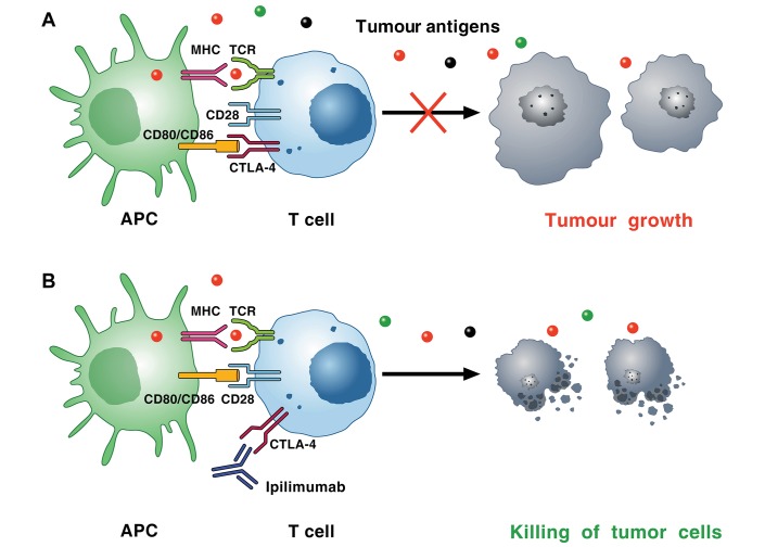
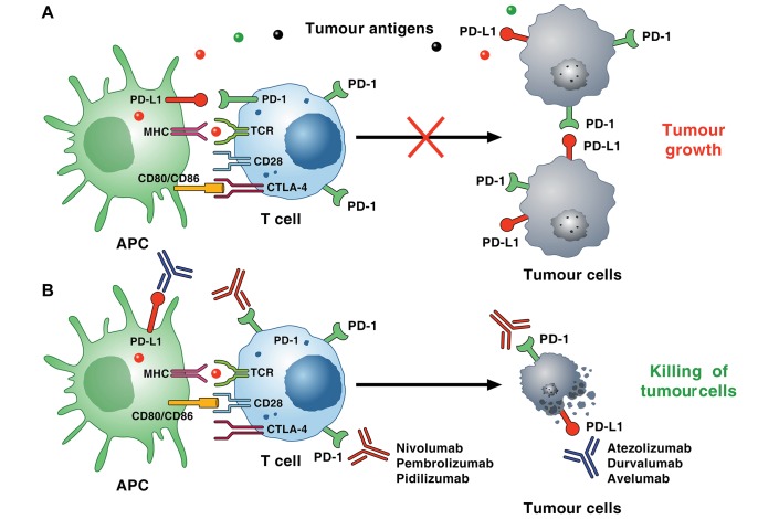
As cancer arises and progresses, malignant cells accumulate genetic alterations resulting in the expression of several neoantigens.45 The innate and adaptive immune responses initially prevent tumour outgrowth. However, cancer cells can escape the immune response (immunosurveillance) by selection of non-immunogenic tumour cells (immunoediting) or suppression of immune responses.46 47 For decades, immunologists and oncologists have attempted to stimulate antitumour immune responses to fight cancer. These initial attempts displayed marginal success for a number of reasons. In particular, several inhibitory pathways, such as CTLA-4, PD-1 and PD-L1, profoundly dampen the antitumour functions of T lymphocytes.48 These inhibitory pathways play pivotal roles in the maintenance of peripheral tolerance and the prevention of autoimmune diseases.33 49 Tumours exploit these and many other inhibitory pathways to escape T cell-mediated tumour-specific immunity (figures 1 and 2). The pioneering work of Allison and coworkers led to the discovery that activation of these checkpoint inhibitors is a fundamental tool by which tumour cells evade the immune system.8 50 Blockade of these immune checkpoints with specific mAbs (eg, anti-CTLA-4, anti-PD-1, anti-PD-L1) has recently revolutionised the entire branch of immunotherapy of a wide spectrum of tumours.51 52
Figure 1.
Mechanism of CTLA-4-induced immunosuppression. (A) Cancer cells synthesise and release neoantigens (dots of different colours) that are captured by APCs. These cells present peptides in the context of MHC I molecules/TCRs on the surface of CD8+ cytotoxic T cells within lymph nodes. APCs can also present peptides bound to MHC II molecules to CD4+ T helper cells. T cell activation on TCR signalling requires costimulatory signals transmitted via CD28, which is activated by binding to CD80, and/or CD86, on the surface of APCs. Activated T cells upregulate CTLA-4, which competes with CD28 for binding to CD80 and/or CD86. The interaction of CTLA-4 with CD80 or CD86 results in inhibitory signalling promoting tumour growth. The immunosuppressive activity of CTLA-4 is mediated by downregulation of Th cells and enhancement of Treg cells. (B) CTLA-4 blockade by ipilimumab results in a broad enhancement of immune responses against neoantigen expressing tumour cells, which results in killing of tumour cells. APC, antigen presenting cell; CTLA-4, cytotoxic T lymphocyte-associated antigen 4; TCR, T cell receptor; Th cells, helper CD4+ T cells; Treg, regulatory T cell.
Figure 2.
Mechanism of PD-1/PD-L1 pathway-induced immunosuppression within the tumour microenvironment. (A) Tumour neoantigens (dots of different colours) released by cancer cells are captured by APCs. These cells present peptides in the context of MHC molecules/TCRs on the surface of CD8+ cytotoxic T cells. PD-1 is induced on T cells on activation through the TCR and through several cytokines. Tumour cells and other cells in the tumour microenvironment (eg, endothelial cells, mast cells) can express high levels of PD-L1 and/or PD-L2 that binds to PD-1 on T cells, resulting in inhibitory checkpoint signalling that decreases cytotoxicity and leads to T cell exhaustion. Recent evidence suggests that murine and human cancer cell subpopulations can express PD-1 and promote tumour growth. (B) PD-1 blocking antibodies (nivolumab, pembrolizumab, pidilizumab and so on) inhibit the interaction of PD-1 with both PD-L1 and PD-L2, resulting in enhanced T cell cytotoxicity, TAM activity, increased cytokine production, and ultimately killing of tumour cells. PD-L1+ tumour cells can also induce T cell apoptosis, anergy, functional exhaustion and interleukin-10 production. Anti-PD-L1 antibodies (atezolizumab, durvalumab, avelumab) have similar effects, but only inhibit the interaction between PD-L1 and PD-1. PD-1, programmed cell death 1; PD-L1, programmed cell death ligand 1; TAM, tumour-associated macrophage; TCR, T cell receptor.
Anti-CTLA-4: the first generation of checkpoint inhibitors
The development of blocking mAbs against immune checkpoint molecules is based on the role of these molecules as coinhibitory receptors of T lymphocytes. Indeed, the activation of naïve T lymphocytes requires antigen presentation by dendritic cells (DCs) to T cells through the interaction of major histocompatibility complex (MHC) and T cell receptor (TCR) (signal 1). The process of T cell activation is strengthened by costimulatory signals.53 Receptors delivering coinhibitory signals (eg, CTLA-4, PD-1) function as immune checkpoints and play a role in maintaining tolerance and in preventing autoimmunity.49 54 55 The pathways involving either CD80 (also known as B7.1) or CD86 (also known as B7.2), plus either CD28 or CTLA-4, are crucial in T cell activation and tolerance (figure 1).
CTLA-4, expressed almost exclusively on T cells, modulates the amplitude of early stages of T cell activation.56 57 CTLA-4 competes with CD28 for binding to B7.1 and/or B7.2.58 CD28 and CTLA-4 share identical ligands, CD80 and/or CD86.59–62 Overexpression of CTLA-4 on activated T cells dampens their activation competing CD28 in binding CD80 and/or CD86. The crucial role of CTLA-4 was demonstrated by the lethal immune hyperactivation phenotype of Ctla-4 knockout mice.54 55 The immunosuppressive activity of CTLA-4 is mediated by downmodulation of helper CD4+ T cell and enhancement of regulatory T cell (Treg) activity.63 64
The discovery of these immunoregulating CTLA-4 functions as a negative regulator of immune responses led to a radical shift in cancer immunotherapy: removal of inhibitory signals that block antitumour T cell responses rather than direct activation of the immune system.50 Indeed, mice bearing immunogenic tumours and treated with an anti-CTLA-4 antibody showed an efficient antitumour response.65 This seminal observation led to the development of a fully human mAb anti-CTLA-4 (ipilimumab), which was the prototype mAb to demonstrate a survival benefit for patients with metastatic melanoma,66 and it was approved by the Food and Drug Administration in 2010 and the European Medicines Agency in 2011.
Anti-PD-1 pathway: the second generation of checkpoint inhibitors
The immune system has developed several coinhibitory pathways to maintain T cell tolerance and to prevent autoimmunity.26 67 The pathway consisting of PD-1 (also called CD279) and its ligands, PD-L1 (B7-H1 or CD274) and PD-L2 (B7-DC or CD276), is another important target to stimulate antitumour immune responses. Several mAbs targeting PD-1 and PD-L1 have been developed based on the role of these checkpoint molecules as coinhibitory receptors of T cell activation (figure 2 and table 1). mAbs against PD-1 and/or PD-L1 restore antitumour immune responses and have shown favourable clinical responses across various cancers.68
Table 1.
Immune checkpoint inhibitors under preclinical and clinical development
| Target | Agent | Antibody | Manufacturer |
| CTLA-4 | Ipilimumab | Human IgG1 | Bristol-Myers Squibb |
| PD-1 | Nivolumab | Human IgG4 | Bristol-Myers Squibb |
| Pembrolizumab | Humanised IgG4 | Merck | |
| MEDI0680 | Humanised | MedImmune | |
| REGN2810 | Human IgG4 | Regeneron/Sanofi | |
| PDR001 | Humanised IgG4 | Novartis | |
| BGB-A317 | Humanised | BeiGene | |
| Pidilizumab | Humanised IgG1 | Medivation/CureTech | |
| AMP-224 | PD-L2 IgG2a fusion protein | GSK | |
| AMP-514 | PD-L2 fusion protein | GSK | |
| SHR-1210 | Human IgG4 | Incyte/Jiangsu | |
| JS001 | Humanised | Shanghai Junshi Biosciences | |
| Tsr-042 | Humanised | Tesaro | |
| PD-L1 | Atezolizumab | Humanised IgG1 | Genentech/Roche |
| Durvalumab | Human IgG1 | MedImmune/AstraZeneca | |
| Avelumab | Human IgG1 | Merck Serono/Pfizer | |
| BMS-936559 | Human IgG4 | Bristol-Myers Squibb | |
| LY3300054 | Not available | Eli Lilly | |
| MEDI4736 | Humanised IgG1 | AstraZeneca | |
| KNO35 | Not available | 3D Medicines | |
| PD-L2 | rHIgM12B7 | Mayo Clinic/NCI | |
| TIM-3 | Anti-TIM-3 antibody | ||
| LAG-3 | Dual anti-LAG-3/anti-PD-1 antibody | ||
| TIGIT | Anti-TIGIT antibody | ||
| BTLA | Anti-BTLA antibody | ||
| VISTA | Anti-VISTA antibody |
BTLA, B and T lymphocyte attenuator; CTLA-4, cytotoxic T lymphocyte-associated antigen 4; LAG-3, lymphocyte-activated gene-3; PD-1, programmed cell death 1; PD-L1, programmed cell death ligand 1; TIGIT, T cell immunoreceptor with Ig and ITIM domains; TIM-3, T cell immunoglobulin and mucin-containing protein 3; VISTA, V-domain Ig suppressor of T cell activation.
PD-1 is induced on T cells on activation through the TCR and cytokines.69 PD-1 is expressed at low levels on T cells in the thymus, activated natural killer (NK) cells, B cells, monocytes, tumour-associated macrophage (TAM), immature Langerhans cells and cardiomyocytes.35 69–71 During T cell activation, PD-1 is translocated to TCR microclusters.72 Engagement of PD-1 by PD-L1 inhibits the activation of TCR proximal kinases.73 PD-1 ligation inhibits T cell–APC contacts and thereby contributes to the cessation of T cell effector functions. The role of PD-1 in peripheral tolerance was demonstrated by the development of lupus-like glomerulonephritis and arthritis,49 as well as in dilated cardiomyopathy in PD-1-deficient mice.33
PD-L1 and PD-L2, the ligands of PD-1, display different expression.69 74 PD-L1 is constitutively expressed at low levels on both professional APCs and non-professional APCs, as well as on non-haematopoietic cells (ie, endothelial cells, pancreatic islet cells, testes and eye).75 PD-L1 is also expressed by cardiomyocytes.36 70 PD-L1 pathway suppresses effector T cells, maintains self-tolerance and promotes the resolution of inflammation.
The expression of PD-L1 and, to a lesser extent, PD-L2 in several tumours11 75 76 stimulated the exploitation of the PD-1–PD-L1 pathway in cancer immunotherapy. In fact, PD-L1 delivers antiapoptotic signals to cancer cells and prevents immune-mediated cancer cell killing.77 Cancer cells dampen the host immune response through the upregulation of PD-L1 and PD-L2 in tumour microenvironment and their ligation to PD-1 expressed by tumour-specific CD8+ T cells. PD-L1 and PD-L2 can be upregulated by cancer cells through several cytokines (interferon (IFN), tumour necrosis factor-α (TNF-α) and vascular endothelial growth factor (VEGF)). PD-L1 expression is also modulated by epigenetic mechanisms through microRNAs.78
In tumour microenvironment, tumour neoantigens released by dying cancer cells activate T cells that overexpress PD-1. Recent evidence indicates that mouse and human TAM express PD-1.71 TAM PD-1 expression increases with increasing disease stage in human tumours and dampens macrophage phagocytosis of tumour cells. These events result in the activation of PD-1–PD-L1/PD-L2 inhibitory mechanism(s) leading to selective inhibition of tumour-specific T cells and TAM. Therefore, targeting of the PD-1/PD-L1 checkpoint pathway with mAbs results in the expansion of tumour-infiltrating CD8+ T cells, which recognise tumour antigens.71 79 CD8+ T cells at the invasive tumour front progressively increase during immunotherapy and correlate with a reduction in tumour size.12 These findings suggest that anti-PD-1 antibody increases CD8+ memory T cell and TAM functions in the tumour microenvironment.71 80 In addition, they also suggest that, although anti-PD-1/PD-L1 inhibitors are administered systematically, their mechanism of action is presumably locally active in cancer tissues.
The new generation of checkpoint inhibitors
Immunotherapy targeting CTLA-4 (ipilimumab), PD-1 (nivolumab and pembrolizumab) and PD-L1 (atezolizumab, avelumab, durvalumab) has revolutionised the management of several tumours.12 81–85 Checkpoint inhibitors provide clinical benefits for a subset of patients with a wide range of solid (melanoma, non-small cell lung cancer (NSCLC), renal carcinoma, ovarian cancer)9 85 86 and haematological tumours (Hodgkin’s lymphoma, primary lymphoma, chronic lymphocytic leukaemia, multiple myeloma).19–23
Despite these promising results, dramatic responses are confined to a minority of patients.28 87 88 This is likely due to the complex network of immunosuppressive pathways in tumour microenvironment, which are unlikely overcome by the blockage of a single signalling checkpoint molecule. In fact, combined anti-CTLA-4 and anti-PD-1 blockade further enhances antitumour activity and patient survival.66 83 89 90 In addition, combination of four different types of immunotherapies eradicated several experimental tumours viewed as intractable.91 A new generation of checkpoint inhibitors, beyond CTLA-4, PD-1 and PD-L1, are under preclinical and clinical development for safer and more effective treatment of human cancers (table 1).
The T cell immunoglobulin and mucin-containing protein 3 (TIM-3), expressed on Treg cells, monocytes/macrophages and APCs, regulate their functions.92 93 Anti-TIM-3 inhibits tumour growth.92 The lymphocyte-activated gene-3 (LAG-3, CD223) is expressed by CD4+ and CD8+ T cells, Treg and Tr1 cells.94–96 LAG-3 blockade synergises with PD-1 inhibition to induce tumour regression.97 98
The T cell immunoreceptor with Ig and ITIM domains (TIGIT) is a novel member of the immunoglobulin superfamily expressed on activated T cells, Tregs, NK and natural killer t cells (NKT) cells.99 100 Similar to PD-1, TIM-3 and LAG-3, TIGIT is upregulated on T cells in cancer.101 TIGIT and PD-1 coblockade inhibits tumour growth in mice.101 B and T lymphocyte attenuator (BTLA), structurally related to CTLA-4 and PD-1, is expressed on B cells, αβ and γδ T cells and DCs.102 103 BTLA blockade induces tumour regression in mice.104
The V-domain Ig suppressor of T cell activation (VISTA), also known as PD-1 homologue, is expressed on neutrophils, monocytes/macrophages, DCs, Myeloiddendritic cells (MDSDs) and Treg cells, and at lower levels on CD4+ and CD8+ T cells.105 VISTA blockade, combined with a peptide vaccine, induces tumour regression in a melanoma model.106
These novel checkpoint inhibitors, alone or in combination, restore antitumour immunological responses in preclinical models. It is likely that immunotherapeutic approaches of human cancers may enlist the combined use of several checkpoint inhibitors.91 The assessment of adverse events, including cardiac toxicity, of these novel checkpoint regulators, alone and in combination, will be of fundamental clinical relevance.
IRAEs associated with checkpoint inhibitors
Due to the pivotal role played by immune checkpoints in the maintenance of self-tolerance, their therapeutic blockade can alter immunological tolerance,87 and give rise to autoimmune or inflammatory side effects, termed ‘immune-related adverse events’.31 107–109 IRAEs associated with the use of ipilimumab were already evident in phase I studies, but now their incidence and severity are well-recognised.66 110 IRAEs are common, usually reversible and not severe in most patients.29 However, endocrinopathies (6%–8% of patients)111 are associated with a high risk of irreversible toxicity. They are caused by the immune infiltration into the thyroid or pituitary glands, resulting in thyroiditis or hypophysitis, respectively.112
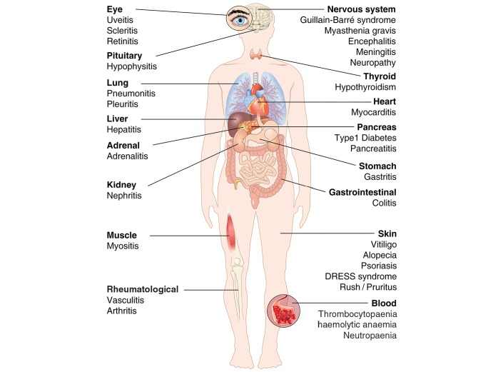
PD-1 and PD-L1 blocking agents display different adverse effect profiles compared with ipilimumab.107 The most common adverse events are mild fatigue, rash, pruritus and diarrhoea.87 The incidence of IRAEs seems to be lower with anti-PD-1 therapy than with ipilimumab.9 66 In general, IRAEs caused by ICIs resemble the autoimmune manifestations observed in PD-1-deficient mice.33 49 113 Even though adverse events with combined checkpoint inhibitors are reported as well-tolerated,114 115 combination of ipilimumab plus nivolumab requires discontinuation of therapy in nearly 40% of patients.89 90 116 IRAEs can affect nearly every organ in association with checkpoint inhibitors (figure 3). Knowledge of the early-onset and late-onset toxic effects associated with checkpoint inhibitors, as well as effective algorithms for the identification and management of these effects, will be fundamental to optimise the safety and efficacy of these immunotherapies.117 Finally, the long-term impact of immune checkpoint blockers on quality of life must be evaluated in future studies.
Figure 3.
Some of the immune-related adverse effects (IRAEs) associated with checkpoints inhibitors in patients with cancer. DRESS, drug rash with eosinophilia and systemic symptoms.
Cardiac toxicity in PD-1-deficient and CTLA-4-deficient animals
PD-L1 is expressed in human34 and murine heart.35 Freeman and colleagues concluded that PD-L1 may regulate potentially autoreactive lymphocytes at effector sites, thus playing a role in limiting activities of T cells in the heart, where PD-L1 is highly expressed. Nishimura and coworkers demonstrated that disruption of the gene encoding for PD-1 in mice caused dilated cardiomyopathy.33 They concluded that PD-1 may be an important receptor contributing to autoimmune diseases.
Several mouse models of T cell-dependent myocarditis exist. In a model of CD8+ T cell myocarditis, IFN-γ induced the overexpression of PD-L1 on endothelial cells.38 Genetic deletion of PD-L1/PD-L2, as well as treatment with anti-PD-L1, transformed transient myocarditis into lethal disease. Deletion of PD-L1 in murphy Roths Large (MRL) mice (genetically predisposed to autoimmunity) resulted in lethal autoimmune myocarditis.39 Similarly, PD-1 deficiency in MRL mice causes a fatal myocarditis,40 reminiscent of CTLA-4-deficient mice.55 In two models of T cell-dependent myocarditis, PD-1 protected against inflammation and myocyte damage.41
Recently, the role of PD-1/PD-L1 has been explored in models of cardiac ischaemia-reperfusion injury and myocardial infarction. Isolated ischaemic-reperfused rat hearts showed increased expression of PD-1 and PD-L1 in cardiomyocytes.70 Interestingly, PD-1 and PD-L1 were not coexpressed on the same myocytes. Furthermore, myocardial infarction in BALB/c mice increased the percentage of PD-1+ and PD-L1+ cardiac cells. These experimental studies suggest that PD-1/PD-L1 and CTLA-4 play important roles in limiting T cell-mediated autoimmune myocarditis.
Cardiac toxicity of checkpoint inhibitors in patients with cancer
With few recent exceptions,118–121 the vast majority of papers on the toxicities of checkpoint inhibitors have underestimated or even neglected cardiac toxicity.87 114 115 117 In a multicentre retrospective study on 752 patients with melanoma treated with ipilimumab, one case of myocardial fibrosis was reported.122 One case report revealed left ventricular dysfunction123 and one case of Takotsubo cardiomyopathy after treatment with ipilimumab.44 Interestingly, a late-onset ipilimumab-induced pericarditis was reported in a patient with melanoma.124 In a multicentre study, six cases of cardiotoxicity after ipilimumab were identified.30 Two out of six cases were fatal despite intensive treatment. In the same series, one case of myocarditis after ipilimumab plus nivolumab and one case of cardiac arrest after pembrolizumab were described. A case of cardiac arrest was reported in a clinical trial of ipilimumab in melanoma110 and a fatal case of myocardial infarction in a patient with NSCLC treated with pembrolizumab.16 Similar case reports have confirmed these findings outside the clinical trial setting.125 126 Autoimmune myocarditis with variable severity has been described.127 128 In a multicentre, phase II, non-controlled study on 26 patients with advanced Merkel cell carcinoma treated with pembrolizumab, adverse events occurred in 77% of patients and one case of myocarditis was reported after the first dose of pembrolizumab.15 Thus, a number of cardiotoxic events (myocarditis, HF, heart block, myocardial fibrosis and cardiomyopathy) were documented in these groups of patients.
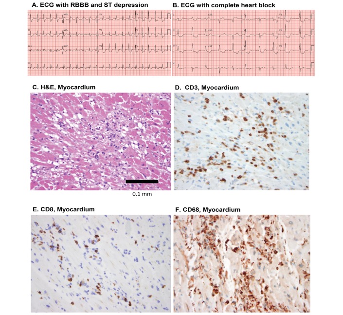
Recently, two cases of fulminant myocarditis and myositis associated with combination of ipilimumab plus nivolumab were carefully described.36 Despite intensive treatment, these two cases were fatal after receiving the first doses of checkpoint inhibitors. Both patients with melanoma had hypertension, but did not display other cardiac risk factors. Histological analysis demonstrated lymphocytes (CD4+ and CD8+ T cells) and macrophages infiltrating the myocardium, the cardiac sinus and the atrioventricular nodes. PD-L1 was highly expressed on injured cardiomyocytes and on infiltrating CD8+ T cells. Figure 4 (reproduced with permission from Johnson et al36) illustrates the ECGs and the histological findings of the heart of patient 2 described by Johnson and collaborators. Importantly, the overexpression of PD-L1 in the injured myocardium in the two patients described by Johnson and collaborators is consistent with the constitutive expression of PD-L1 in human heart34 35 and its upregulation in T cell-mediated myocarditis in mice.38 Recently, PD-1 and PD-L1 were detected on rat cardiomyocytes and overexpressed in the ischaemic-reperfused heart.70 An analysis of T cells infiltrating the myocardium, skeletal muscle and tumour revealed clonality of TCR. The authors suggested that common antigens present in these tissues could be recognised by clonal lymphocytes. Moreover, overexpression of IFN-γ, granzyme B and TNF-α, presumably produced by activated T cells, might contribute to cardiac damage.
Figure 4.
ECG and histological findings of the heart in a 63-year-old man with metastatic melanoma who developed fulminant lymphocytic myocarditis following initial doses of nivolumab and ipilimumab and who developed complete heart block.36 Despite intense treatment (intravenous methylprednisolone 1 g/kg daily for 4 days plus infliximab 5 mg/kg), fatal complete heart block occurred. Initial right bundle branch block (RBBB) and ST depression (A) progressed rapidly to complete heart block and cardiac arrest (B). Autopsy showed lymphocytic infiltration in myocardium (C) comprised CD3+ T cells (D), many of which were CD8+ lymphocytes (E) and CD68+ macrophages (F) (adapted with permission from Johnson et al36).
The authors also assessed the frequency of myocarditis in the safety databases of Bristol-Myers Squibb Corporate to verify the occurrence of events in patients treated with nivolumab, ipilimumab or both. Among 20 594 patients treated with these checkpoint inhibitors, 18 drug-related severe adverse events of myocarditis were reported (0.09%). Combination therapy with both drugs was associated with more severe and frequent myocarditis than those who received nivolumab alone (0.27% vs 0.06%).36 Myocarditis, diagnosed at a median of 17 days after the first treatment, suggests the occurrence of early cardiotoxicity.
In conclusion, although combined immune checkpoint inhibition has produced durable antitumour responses in a percentage of patients with different tumours, IRAEs required discontinuation in nearly 40% of patients.89 90 Most of these events are manageable with a high dose of glucocorticoids, although severe and even fatal events have occurred in rare instances. Better characterisation of the real incidence of cardiovascular toxicity of checkpoint blockers, alone and in combination, even if uncommon, is a major priority.
It is important to note that extensive cardiac monitoring, including the assessment of troponin, a sensitive and specific marker of cardiotoxicity,129 130 is not routinely performed in most immunotherapy trials. Therefore, the real incidence of early and late cardiotoxicity associated with immune checkpoint blockade is largely unknown.
Management of myocarditis associated with ICIs
Oncologists and cardiologists should be aware of early onset of myocarditis, in patients treated with ICIs alone and in combination. Our understanding of the pathophysiology of myocarditis comes largely from animal studies.131 In the 2000s it was demonstrated that deletion of CTLA-4 and PD-1 axis can cause autoimmune myocarditis.33 37 Myocarditis is an insidious disease with a wide spectrum of clinical presentation reflecting the different aetiologies and the variability of local or diffuse involvement. In addition, patients with systemic autoimmune disorders (eg, rheumatoid arthritis, lupus erythematosus, psoriatic arthritis, systemic sclerosis, vasculitis, polymyositis) can have subclinical myocarditis. In the absence of specific studies, clinical experience should guide the use of checkpoint inhibitors in patients with cancer with pre-existing autoimmune diseases.132
Similarly, at present there is urgent need of validated guidelines for treatment of myocarditis associated with checkpoint inhibitors. Wang and coworkers118, based on their extensive personal experience, have proposed an interesting algorithm for management of myocarditis in patients treated with checkpoint inhibitors. Their algorithm represents an excellent basis for an urgently needed consensus guideline for management of different forms of immune-mediated myocarditis.
Concluding remarks
Checkpoint blockade has introduced clinical benefits by inducing regression of advanced metastatic tumours, improving patient survival and inducing durable effects in a percentage of patients with a broad spectrum of cancer types. Although IRAEs associated with monoclonal anti-CTLA-4 and anti-PD-1/PD-L1 antibodies are common and usually reversible, increasing reports of severe cardiac toxicity introduce important questions relevant for future oncology trials and clinical practice.
Patients with autoimmune disorders are usually excluded from clinical trials with checkpoint inhibitors. Patients with a wide spectrum of autoimmune disorders presumably represent 20–50 million people in the USA alone.119 In addition, approximately 14% of patients with lung cancer have a concurrent diagnosis of autoimmune disease.133 These findings indicate that clinical and subclinical autoimmune disorders are an important consideration before initiation of checkpoint inhibitor therapy. In 12 patients treated with ipilimumab, worsening or exacerbation of pre-existing autoimmune diseases was observed in 50% of cases.134 135 Recently, Johnson and collaborators reported their experience with two groups of patients with pre-existing autoimmune disease and melanoma treated with ipilimumab or anti-PD-1.36 132 Although 20%–30% of these patients experienced an autoimmune flare, the authors concluded that treatment with either ipilimumab or anti-PD-1 is feasible for patients with certain types of pre-existing autoimmunity.119 Given the heterogeneity of autoimmune disorders and the wide spectrum of their severity, specific guidelines regarding exclusion criteria and treatment are urgently needed.
Cardiac parameters and levels of troponins are not routinely evaluated in most oncology trials. Therefore, the true incidence of cardiac toxicity associated with checkpoint inhibitors may be higher in the real-world population.
Combination therapies such as combined checkpoint inhibitors, or sequential therapies with conventional chemotherapy plus checkpoint inhibitors, or checkpoint inhibitors plus antiangiogenic agents are increasingly being used.136 137 Cardiovascular monitoring is necessary to assess the occurrence of early and late cardiac toxicity associated with these newer cancer immunotherapies. The use of ICIs is expected to increase within the next years for treatment of new tumour types and presumably also for other immune-mediated disorders such as HIV138 and infectious diseases.139 Therefore, prospective cardiovascular evaluation appears necessary to detect potential cardiotoxicity in these disorders.
All cases of cardiotoxicity associated with checkpoint inhibitors reported so far occurred immediately after the infusion or during the first year of therapy.15 30 36 44 Prospective studies should assess whether late-onset chronic cardiotoxicity can occur after completion of therapy.
Interestingly, interindividual differences in intestinal microbiota are a source of the heterogeneity in immunotherapeutic efficacy and toxicity of ICIs in cancer.7 140 141 It will be important to investigate whether gut microbiota can also influence cardiac toxicity of ICIs.
Constitutive expression of PD-1 and PD-L1 occurs in human and murine myocytes.34 35 70 In addition, overexpression of PD-L1 on the surface of injured myocytes has been demonstrated in patients with fulminant myocarditis treated with checkpoint inhibitors.36 The latter observations open the possibility that, in certain clinical conditions (eg, myocardial ischaemia), cytokines and chemokines produced by infiltrating immune cells can upregulate PD-1/PD-L1 pathway in human myocardium. Additional in vitro and in vivo research is urgently needed to understand the immunological and molecular mechanisms underlying the development of these cardiac toxicities.
Today, oncologists and cardiologists work together mainly to detect and manage cardiotoxicity of antineoplastic treatments. Cardiologists are not always involved in the initial anticancer treatment planning or in the assessment of early cardiac dysfunction. With a growing number of patients treated with different types of ICIs, a collaboration of oncologists, cardiologists and immunologists is now necessary for a better characterisation of the mechanisms of cardiotoxicity of this novel class of anticancer drugs and for a comprehensive identification and management of patients at risk for cardiac adverse events.
Acknowledgments
The authors apologise to the many authors who have contributed importantly to this field and whose work has not been cited due to space restrictions. The authors thank Fabrizio Fiorbianco for preparing the figures.
Footnotes
Contributors: Gilda Varricchi, Gianni Marone and Carlo Gabriele Tocchetti drafted the first version of the manuscript. All authors revised the manuscript and approved the final version of the manuscript. Gilda Varricchi and Carlo Gabriele Tocchetti are responsible for the overall content as guarantors.
Funding: This work was supported in part by grants from Regione Campania CISI-Lab, CRÈME Project and TIMING Project, and Ricerca di Ateneo.
Competing interests: CGT received travel support from Alere.
Provenance and peer review: Not commissioned; internally peer reviewed.
References
- 1.Bloom MW, Hamo CE, Cardinale D, et al. Cancer therapy-related cardiac dysfunction and heart failure: part 1: definitions, pathophysiology, risk factors, and imaging. Circ Heart Fail 2016;9:e002661 10.1161/CIRCHEARTFAILURE.115.002661 [DOI] [PMC free article] [PubMed] [Google Scholar]
- 2.Hamo CE, Bloom MW, Cardinale D, et al. Cancer therapy-related cardiac dysfunction and heart failure: part 2: prevention, treatment, guidelines, and future directions. Circ Heart Fail 2016;9:e002843 10.1161/CIRCHEARTFAILURE.115.002843 [DOI] [PMC free article] [PubMed] [Google Scholar]
- 3.Yeh ET, Bickford CL. Cardiovascular complications of cancer therapy: incidence, pathogenesis, diagnosis, and management. J Am Coll Cardiol 2009;53:2231–47. 10.1016/j.jacc.2009.02.050 [DOI] [PubMed] [Google Scholar]
- 4.Tocchetti CG, Gallucci G, Coppola C, et al. The emerging issue of cardiac dysfunction induced by antineoplastic angiogenesis inhibitors. Eur J Heart Fail 2013;15:482–9. 10.1093/eurjhf/hft008 [DOI] [PubMed] [Google Scholar]
- 5.Suter TM, Ewer MS. Cancer drugs and the heart: importance and management. Eur Heart J 2013;34:1102–11. 10.1093/eurheartj/ehs181 [DOI] [PubMed] [Google Scholar]
- 6.Moslehi JJ. Cardiovascular toxic effects of targeted cancer therapies. N Engl J Med 2016;375:1457–67. 10.1056/NEJMra1100265 [DOI] [PubMed] [Google Scholar]
- 7.Pitt JM, Vétizou M, Daillère R, et al. Resistance mechanisms to immune-checkpoint blockade in cancer: tumor-intrinsic and -extrinsic factors. Immunity 2016;44:1255–69. 10.1016/j.immuni.2016.06.001 [DOI] [PubMed] [Google Scholar]
- 8.Hurst JH. Cancer immunotherapy innovator James Allison receives the 2015 Lasker~DeBakey clinical medical research award. J Clin Invest 2015;125:3732–6. 10.1172/JCI84236 [DOI] [PMC free article] [PubMed] [Google Scholar]
- 9.Topalian SL, Hodi FS, Brahmer JR, et al. Safety, activity, and immune correlates of anti-PD-1 antibody in cancer. N Engl J Med 2012;366:2443–54. 10.1056/NEJMoa1200690 [DOI] [PMC free article] [PubMed] [Google Scholar]
- 10.Powles T, Eder JP, Fine GD, et al. MPDL3280A (anti-PD-L1) treatment leads to clinical activity in metastatic bladder cancer. Nature 2014;515:558–62. 10.1038/nature13904 [DOI] [PubMed] [Google Scholar]
- 11.Herbst RS, Soria JC, Kowanetz M, et al. Predictive correlates of response to the anti-PD-L1 antibody MPDL3280A in cancer patients. Nature 2014;515:563–7. 10.1038/nature14011 [DOI] [PMC free article] [PubMed] [Google Scholar]
- 12.Tumeh PC, Harview CL, Yearley JH, et al. PD-1 blockade induces responses by inhibiting adaptive immune resistance. Nature 2014;515:568–71. 10.1038/nature13954 [DOI] [PMC free article] [PubMed] [Google Scholar]
- 13.Brahmer J, Reckamp KL, Baas P, et al. Nivolumab versus docetaxel in advanced squamous-cell non-small-cell lung cancer. N Engl J Med 2015;373:123–35. 10.1056/NEJMoa1504627 [DOI] [PMC free article] [PubMed] [Google Scholar]
- 14.Borghaei H, Paz-Ares L, Horn L, et al. Nivolumab versus docetaxel in advanced nonsquamous non-small-cell lung cancer. N Engl J Med 2015;373:1627–39. 10.1056/NEJMoa1507643 [DOI] [PMC free article] [PubMed] [Google Scholar]
- 15.Nghiem PT, Bhatia S, Lipson EJ, et al. PD-1 Blockade with pembrolizumab in advanced merkel-cell Carcinoma. N Engl J Med 2016;374:2542–52. 10.1056/NEJMoa1603702 [DOI] [PMC free article] [PubMed] [Google Scholar]
- 16.Herbst RS, Baas P, Kim DW, et al. Pembrolizumab versus docetaxel for previously treated, PD-L1-positive, advanced non-small-cell lung cancer (KEYNOTE-010): a randomised controlled trial. Lancet 2016;387:1540–50. 10.1016/S0140-6736(15)01281-7 [DOI] [PubMed] [Google Scholar]
- 17.Fehrenbacher L, Spira A, Ballinger M, et al. Atezolizumab versus docetaxel for patients with previously treated non-small-cell lung cancer (POPLAR): a multicentre, open-label, phase 2 randomised controlled trial. Lancet 2016;387:1837–46. 10.1016/S0140-6736(16)00587-0 [DOI] [PubMed] [Google Scholar]
- 18.Bellmunt J, de Wit R, Vaughn DJ, et al. Pembrolizumab as second-line therapy for advanced urothelial Carcinoma. N Engl J Med 2017;376:1015–26. 10.1056/NEJMoa1613683 [DOI] [PMC free article] [PubMed] [Google Scholar]
- 19.Lesokhin AM, Ansell SM, Armand P, et al. Nivolumab in patients with relapsed or refractory hematologic malignancy: preliminary results of a phase Ib study. J Clin Oncol 2016;34:2698–704. 10.1200/JCO.2015.65.9789 [DOI] [PMC free article] [PubMed] [Google Scholar]
- 20.Ding W, LaPlant BR, Call TG, et al. Pembrolizumab in patients with CLL and richter transformation or with relapsed CLL. Blood 2017;129:3419–27. 10.1182/blood-2017-02-765685 [DOI] [PMC free article] [PubMed] [Google Scholar]
- 21.Badros A, Hyjek E, Ma N, et al. Pembrolizumab, pomalidomide, and low-dose dexamethasone for relapsed/refractory multiple myeloma. Blood 2017;130:1189–97. 10.1182/blood-2017-03-775122 [DOI] [PubMed] [Google Scholar]
- 22.Nayak L, Iwamoto FM, LaCasce A, et al. PD-1 blockade with nivolumab in relapsed/refractory primary central nervous system and testicular lymphoma. Blood 2017;129:3071–3. 10.1182/blood-2017-01-764209 [DOI] [PMC free article] [PubMed] [Google Scholar]
- 23.Westin JR, Chu F, Zhang M, et al. Safety and activity of PD1 blockade by pidilizumab in combination with rituximab in patients with relapsed follicular lymphoma: a single group, open-label, phase 2 trial. Lancet Oncol 2014;15:69–77. 10.1016/S1470-2045(13)70551-5 [DOI] [PMC free article] [PubMed] [Google Scholar]
- 24.Nguyen LT, Ohashi PS. Clinical blockade of PD1 and LAG3-potential mechanisms of action. Nat Rev Immunol 2015;15:45–56. 10.1038/nri3790 [DOI] [PubMed] [Google Scholar]
- 25.Sharma P, Allison JP. The future of immune checkpoint therapy. Science 2015;348:56–61. 10.1126/science.aaa8172 [DOI] [PubMed] [Google Scholar]
- 26.Le Mercier I, Lines JL, Noelle RJ. Beyond CTLA-4 and PD-1, the generation Z of negative checkpoint regulators. Front Immunol 2015;6:418 10.3389/fimmu.2015.00418 [DOI] [PMC free article] [PubMed] [Google Scholar]
- 27.Chen L, Han X. Anti-PD-1/PD-L1 therapy of human cancer: past, present, and future. J Clin Invest 2015;125:3384–91. 10.1172/JCI80011 [DOI] [PMC free article] [PubMed] [Google Scholar]
- 28.Goodman A, Patel SP, Kurzrock R. PD-1-PD-L1 immune-checkpoint blockade in B-cell lymphomas. Nat Rev Clin Oncol 2017;14:203–20. 10.1038/nrclinonc.2016.168 [DOI] [PubMed] [Google Scholar]
- 29.Lacouture ME, Wolchok JD, Yosipovitch G, et al. Ipilimumab in patients with cancer and the management of dermatologic adverse events. J Am Acad Dermatol 2014;71:161–9. 10.1016/j.jaad.2014.02.035 [DOI] [PubMed] [Google Scholar]
- 30.Heinzerling L, Ott PA, Hodi FS, et al. Cardiotoxicity associated with CTLA4 and PD1 blocking immunotherapy. J Immunother Cancer 2016;4:50 10.1186/s40425-016-0152-y [DOI] [PMC free article] [PubMed] [Google Scholar]
- 31.Spain L, Walls G, Julve M, et al. Neurotoxicity from immune-checkpoint inhibition in the treatment of melanoma: a single centre experience and review of the literature. Ann Oncol 2017;28:377–85. 10.1093/annonc/mdw558 [DOI] [PubMed] [Google Scholar]
- 32.Hofmann L, Forschner A, Loquai C, et al. Cutaneous, gastrointestinal, hepatic, endocrine, and renal side-effects of anti-PD-1 therapy. Eur J Cancer 2016;60:190–209. 10.1016/j.ejca.2016.02.025 [DOI] [PubMed] [Google Scholar]
- 33.Nishimura H, Okazaki T, Tanaka Y, et al. Autoimmune dilated cardiomyopathy in PD-1 receptor-deficient mice. Science 2001;291:319–22. 10.1126/science.291.5502.319 [DOI] [PubMed] [Google Scholar]
- 34.Dong H, Zhu G, Tamada K, et al. B7-H1, a third member of the B7 family, co-stimulates T-cell proliferation and interleukin-10 secretion. Nat Med 1999;5:1365–9. 10.1038/70932 [DOI] [PubMed] [Google Scholar]
- 35.Freeman GJ, Long AJ, Iwai Y, et al. Engagement of the PD-1 immunoinhibitory receptor by a novel B7 family member leads to negative regulation of lymphocyte activation. J Exp Med 2000;192:1027–34. 10.1084/jem.192.7.1027 [DOI] [PMC free article] [PubMed] [Google Scholar]
- 36.Johnson DB, Balko JM, Compton ML, et al. Fulminant myocarditis with combination immune checkpoint blockade. N Engl J Med 2016;375:1749–55. 10.1056/NEJMoa1609214 [DOI] [PMC free article] [PubMed] [Google Scholar]
- 37.Okazaki T, Tanaka Y, Nishio R, et al. Autoantibodies against cardiac troponin I are responsible for dilated cardiomyopathy in PD-1-deficient mice. Nat Med 2003;9:1477–83. 10.1038/nm955 [DOI] [PubMed] [Google Scholar]
- 38.Grabie N, Gotsman I, DaCosta R, et al. Endothelial programmed death-1 ligand 1 (PD-L1) regulates CD8+ T-cell mediated injury in the heart. Circulation 2007;116:2062–71. 10.1161/CIRCULATIONAHA.107.709360 [DOI] [PubMed] [Google Scholar]
- 39.Lucas JA, Menke J, Rabacal WA, et al. Programmed death ligand 1 regulates a critical checkpoint for autoimmune myocarditis and pneumonitis in MRL mice. J Immunol 2008;181:2513–21. 10.4049/jimmunol.181.4.2513 [DOI] [PMC free article] [PubMed] [Google Scholar]
- 40.Wang J, Okazaki IM, Yoshida T, et al. PD-1 deficiency results in the development of fatal myocarditis in MRL mice. Int Immunol 2010;22:443–52. 10.1093/intimm/dxq026 [DOI] [PubMed] [Google Scholar]
- 41.Tarrio ML, Grabie N, Bu DX, et al. PD-1 protects against inflammation and myocyte damage in T cell-mediated myocarditis. J Immunol 2012;188:4876–84. 10.4049/jimmunol.1200389 [DOI] [PMC free article] [PubMed] [Google Scholar]
- 42.Semper H, Muehlberg F, Schulz-Menger J, et al. Drug-induced myocarditis after nivolumab treatment in a patient with PDL1- negative squamous cell carcinoma of the lung. Lung Cancer 2016;99:117–9. 10.1016/j.lungcan.2016.06.025 [DOI] [PubMed] [Google Scholar]
- 43.Tadokoro T, Keshino E, Makiyama A, et al. Acute Lymphocytic Myocarditis With Anti-PD-1 Antibody Nivolumab. Circ Heart Fail 2016;9:e003514 10.1161/CIRCHEARTFAILURE.116.003514 [DOI] [PubMed] [Google Scholar]
- 44.Geisler BP, Raad RA, Esaian D, et al. Apical ballooning and cardiomyopathy in a melanoma patient treated with ipilimumab: a case of takotsubo-like syndrome. J Immunother Cancer 2015;3:4 10.1186/s40425-015-0048-2 [DOI] [PMC free article] [PubMed] [Google Scholar]
- 45.Chen DS, Mellman I. Oncology meets immunology: the cancer-immunity cycle. Immunity 2013;39:1–10. 10.1016/j.immuni.2013.07.012 [DOI] [PubMed] [Google Scholar]
- 46.Zitvogel L, Apetoh L, Ghiringhelli F, et al. The anticancer immune response: indispensable for therapeutic success? J Clin Invest 2008;118:1991–2001. 10.1172/JCI35180 [DOI] [PMC free article] [PubMed] [Google Scholar]
- 47.Mohme M, Riethdorf S, Pantel K. Circulating and disseminated tumour cells - mechanisms of immune surveillance and escape. Nat Rev Clin Oncol 2017;14:155–67. 10.1038/nrclinonc.2016.144 [DOI] [PubMed] [Google Scholar]
- 48.Mahoney KM, Freeman GJ, McDermott DF. The next immune-checkpoint inhibitors: PD-1/PD-L1 blockade in melanoma. Clin Ther 2015;37:764–82. 10.1016/j.clinthera.2015.02.018 [DOI] [PMC free article] [PubMed] [Google Scholar]
- 49.Nishimura H, Nose M, Hiai H, et al. Development of lupus-like autoimmune diseases by disruption of the PD-1 gene encoding an ITIM motif-carrying immunoreceptor. Immunity 1999;11:141–51. 10.1016/S1074-7613(00)80089-8 [DOI] [PubMed] [Google Scholar]
- 50.Allison JP, Hurwitz AA, Leach DR. Manipulation of costimulatory signals to enhance antitumor T-cell responses. Curr Opin Immunol 1995;7:682–6. 10.1016/0952-7915(95)80077-8 [DOI] [PubMed] [Google Scholar]
- 51.Sharma P, Allison JP. Immune checkpoint targeting in cancer therapy: toward combination strategies with curative potential. Cell 2015;161:205–14. 10.1016/j.cell.2015.03.030 [DOI] [PMC free article] [PubMed] [Google Scholar]
- 52.Pardoll DM. The blockade of immune checkpoints in cancer immunotherapy. Nat Rev Cancer 2012;12:252–64. 10.1038/nrc3239 [DOI] [PMC free article] [PubMed] [Google Scholar]
- 53.Bretscher PA. A two-step, two-signal model for the primary activation of precursor helper T cells. Proc Natl Acad Sci U S A 1999;96:185–90. 10.1073/pnas.96.1.185 [DOI] [PMC free article] [PubMed] [Google Scholar]
- 54.Tivol EA, Borriello F, Schweitzer AN, et al. Loss of CTLA-4 leads to massive lymphoproliferation and fatal multiorgan tissue destruction, revealing a critical negative regulatory role of CTLA-4. Immunity 1995;3:541–7. 10.1016/1074-7613(95)90125-6 [DOI] [PubMed] [Google Scholar]
- 55.Waterhouse P, Penninger JM, Timms E, et al. Lymphoproliferative disorders with early lethality in mice deficient in Ctla-4. Science 1995;270:985–8. 10.1126/science.270.5238.985 [DOI] [PubMed] [Google Scholar]
- 56.Brunet JF, Denizot F, Luciani MF, et al. A new member of the immunoglobulin superfamily-CTLA-4. Nature 1987;328:267–70. 10.1038/328267a0 [DOI] [PubMed] [Google Scholar]
- 57.Linsley PS, Brady W, Urnes M, et al. CTLA-4 is a second receptor for the B cell activation antigen B7. J Exp Med 1991;174:561–9. 10.1084/jem.174.3.561 [DOI] [PMC free article] [PubMed] [Google Scholar]
- 58.Martin PJ, Ledbetter JA, Morishita Y, et al. A 44 kilodalton cell surface homodimer regulates interleukin 2 production by activated human T lymphocytes. J Immunol 1986;136:3282–7. [PubMed] [Google Scholar]
- 59.Hathcock KS, Laszlo G, Dickler HB, et al. Identification of an alternative CTLA-4 ligand costimulatory for T cell activation. Science 1993;262:905–7. 10.1126/science.7694361 [DOI] [PubMed] [Google Scholar]
- 60.Freeman GJ, Gribben JG, Boussiotis VA, et al. Cloning of B7-2: a CTLA-4 counter-receptor that costimulates human T cell proliferation. Science 1993;262:909–11. 10.1126/science.7694363 [DOI] [PubMed] [Google Scholar]
- 61.Azuma M, Ito D, Yagita H, et al. B70 antigen is a second ligand for CTLA-4 and CD28. Nature 1993;366:76–9. 10.1038/366076a0 [DOI] [PubMed] [Google Scholar]
- 62.Linsley PS, Clark EA, Ledbetter JA. T-cell antigen CD28 mediates adhesion with B cells by interacting with activation antigen B7/BB-1. Proc Natl Acad Sci U S A 1990;87:5031–5. 10.1073/pnas.87.13.5031 [DOI] [PMC free article] [PubMed] [Google Scholar]
- 63.Wing K, Onishi Y, Prieto-Martin P, et al. CTLA-4 control over Foxp3+ regulatory T cell function. Science 2008;322:271–5. 10.1126/science.1160062 [DOI] [PubMed] [Google Scholar]
- 64.Peggs KS, Quezada SA, Chambers CA, et al. Blockade of CTLA-4 on both effector and regulatory T cell compartments contributes to the antitumor activity of anti-CTLA-4 antibodies. J Exp Med 2009;206:1717–25. 10.1084/jem.20082492 [DOI] [PMC free article] [PubMed] [Google Scholar]
- 65.Leach DR, Krummel MF, Allison JP. Enhancement of antitumor immunity by CTLA-4 blockade. Science 1996;271:1734–6. 10.1126/science.271.5256.1734 [DOI] [PubMed] [Google Scholar]
- 66.Hodi FS, O’Day SJ, McDermott DF, et al. Improved survival with ipilimumab in patients with metastatic melanoma. N Engl J Med 2010;363:711–23. 10.1056/NEJMoa1003466 [DOI] [PMC free article] [PubMed] [Google Scholar]
- 67.Ni L, Dong C. New checkpoints in cancer immunotherapy. Immunol Rev 2017;276:52–65. 10.1111/imr.12524 [DOI] [PubMed] [Google Scholar]
- 68.Boussiotis VA. Molecular and biochemical aspects of the PD-1 checkpoint pathway. N Engl J Med 2016;375:1767–78. 10.1056/NEJMra1514296 [DOI] [PMC free article] [PubMed] [Google Scholar]
- 69.Okazaki T, Honjo T. PD-1 and PD-1 ligands: from discovery to clinical application. Int Immunol 2007;19:813–24. 10.1093/intimm/dxm057 [DOI] [PubMed] [Google Scholar]
- 70.Baban B, Liu JY, Qin X, et al. Upregulation of programmed death-1 and Its ligand in cardiac injury models: interaction with GADD153. PLoS One 2015;10:e0124059 10.1371/journal.pone.0124059 [DOI] [PMC free article] [PubMed] [Google Scholar]
- 71.Gordon SR, Maute RL, Dulken BW, et al. PD-1 expression by tumour-associated macrophages inhibits phagocytosis and tumour immunity. Nature 2017;545:495–9. 10.1038/nature22396 [DOI] [PMC free article] [PubMed] [Google Scholar]
- 72.Yokosuka T, Takamatsu M, Kobayashi-Imanishi W, et al. Programmed cell death 1 forms negative costimulatory microclusters that directly inhibit T cell receptor signaling by recruiting phosphatase SHP2. J Exp Med 2012;209:1201–17. 10.1084/jem.20112741 [DOI] [PMC free article] [PubMed] [Google Scholar]
- 73.Sheppard KA, Fitz LJ, Lee JM, et al. PD-1 inhibits T-cell receptor induced phosphorylation of the ZAP70/CD3zeta signalosome and downstream signaling to PKCtheta. FEBS Lett 2004;574:37–41. 10.1016/j.febslet.2004.07.083 [DOI] [PubMed] [Google Scholar]
- 74.Francisco LM, Sage PT, Sharpe AH. The PD-1 pathway in tolerance and autoimmunity. Immunol Rev 2010;236:219–42. 10.1111/j.1600-065X.2010.00923.x [DOI] [PMC free article] [PubMed] [Google Scholar]
- 75.Dong H, Strome SE, Salomao DR, et al. Tumor-associated B7-H1 promotes T-cell apoptosis: a potential mechanism of immune evasion. Nat Med 2002;8:1039–800. 10.1038/nm0902-1039c [DOI] [PubMed] [Google Scholar]
- 76.Latchman Y, Wood CR, Chernova T, et al. PD-L2 is a second ligand for PD-1 and inhibits T cell activation. Nat Immunol 2001;2:261–8. 10.1038/85330 [DOI] [PubMed] [Google Scholar]
- 77.Azuma T, Yao S, Zhu G, et al. B7-H1 is a ubiquitous antiapoptotic receptor on cancer cells. Blood 2008;111:3635–43. 10.1182/blood-2007-11-123141 [DOI] [PMC free article] [PubMed] [Google Scholar]
- 78.Chen J, Jiang CC, Jin L, et al. Regulation of PD-L1: a novel role of pro-survival signalling in cancer. Ann Oncol 2016;27:409–16. 10.1093/annonc/mdv615 [DOI] [PubMed] [Google Scholar]
- 79.Rizvi NA, Hellmann MD, Snyder A, et al. Cancer immunology. Mutational landscape determines sensitivity to PD-1 blockade in non-small cell lung cancer. Science 2015;348:124–8. 10.1126/science.aaa1348 [DOI] [PMC free article] [PubMed] [Google Scholar]
- 80.Ribas A, Shin DS, Zaretsky J, et al. PD-1 blockade expands intratumoral memory T cells. Cancer Immunol Res 2016;4:194–203. 10.1158/2326-6066.CIR-15-0210 [DOI] [PMC free article] [PubMed] [Google Scholar]
- 81.Brahmer JR, Drake CG, Wollner I, et al. Phase I study of single-agent anti-programmed death-1 (MDX-1106) in refractory solid tumors: safety, clinical activity, pharmacodynamics, and immunologic correlates. J Clin Oncol 2010;28:3167–75. 10.1200/JCO.2009.26.7609 [DOI] [PMC free article] [PubMed] [Google Scholar]
- 82.Topalian SL, Sznol M, McDermott DF, et al. Survival, durable tumor remission, and long-term safety in patients with advanced melanoma receiving nivolumab. J Clin Oncol 2014;32:1020–30. 10.1200/JCO.2013.53.0105 [DOI] [PMC free article] [PubMed] [Google Scholar]
- 83.Robert C, Long GV, Brady B, et al. Nivolumab in previously untreated melanoma without BRAF mutation. N Engl J Med 2015;372:320–30. 10.1056/NEJMoa1412082 [DOI] [PubMed] [Google Scholar]
- 84.Hamid O, Robert C, Daud A, et al. Safety and tumor responses with lambrolizumab (anti-PD-1) in melanoma. N Engl J Med 2013;369:134–44. 10.1056/NEJMoa1305133 [DOI] [PMC free article] [PubMed] [Google Scholar]
- 85.Rittmeyer A, Barlesi F, Waterkamp D, et al. Atezolizumab versus docetaxel in patients with previously treated non-small-cell lung cancer (OAK): a phase 3, open-label, multicentre randomised controlled trial. Lancet 2017;389:255–65. 10.1016/S0140-6736(16)32517-X [DOI] [PMC free article] [PubMed] [Google Scholar]
- 86.Hamanishi J, Mandai M, Ikeda T, et al. Safety and antitumor activity of anti-PD-1 antibody, nivolumab, in patients with platinum-resistant ovarian cancer. J Clin Oncol 2015;33:4015–22. 10.1200/JCO.2015.62.3397 [DOI] [PubMed] [Google Scholar]
- 87.Boutros C, Tarhini A, Routier E, et al. Safety profiles of anti-CTLA-4 and anti-PD-1 antibodies alone and in combination. Nat Rev Clin Oncol 2016;13:473–86. 10.1038/nrclinonc.2016.58 [DOI] [PubMed] [Google Scholar]
- 88.Garon EB. Cancer immunotherapy trials not immune from imprecise selection of patients. N Engl J Med 2017;376:2483–5. 10.1056/NEJMe1705692 [DOI] [PubMed] [Google Scholar]
- 89.Larkin J, Chiarion-Sileni V, Gonzalez R, et al. Combined nivolumab and ipilimumab or monotherapy in untreated melanoma. N Engl J Med 2015;373:23–34. 10.1056/NEJMoa1504030 [DOI] [PMC free article] [PubMed] [Google Scholar]
- 90.Postow MA, Chesney J, Pavlick AC, et al. Nivolumab and ipilimumab versus ipilimumab in untreated melanoma. N Engl J Med 2015;372:2006–17. 10.1056/NEJMoa1414428 [DOI] [PMC free article] [PubMed] [Google Scholar]
- 91.Moynihan KD, Opel CF, Szeto GL, et al. Eradication of large established tumors in mice by combination immunotherapy that engages innate and adaptive immune responses. Nat Med 2016;22:1402–10. 10.1038/nm.4200 [DOI] [PMC free article] [PubMed] [Google Scholar]
- 92.Sakuishi K, Ngiow SF, Sullivan JM, et al. TIM3(+)FOXP3(+) regulatory T cells are tissue-specific promoters of T-cell dysfunction in cancer. Oncoimmunology 2013;2:e23849 10.4161/onci.23849 [DOI] [PMC free article] [PubMed] [Google Scholar]
- 93.Yan J, Zhang Y, Zhang JP, et al. Tim-3 expression defines regulatory T cells in human tumors. PLoS One 2013;8:e58006 10.1371/journal.pone.0058006 [DOI] [PMC free article] [PubMed] [Google Scholar]
- 94.Huang CT, Workman CJ, Flies D, et al. Role of LAG-3 in regulatory T cells. Immunity 2004;21:503–13. 10.1016/j.immuni.2004.08.010 [DOI] [PubMed] [Google Scholar]
- 95.Gagliani N, Magnani CF, Huber S, et al. Coexpression of CD49b and LAG-3 identifies human and mouse T regulatory type 1 cells. Nat Med 2013;19:739–46. 10.1038/nm.3179 [DOI] [PubMed] [Google Scholar]
- 96.Okamura T, Fujio K, Shibuya M, et al. CD4+CD25-LAG3+ regulatory T cells controlled by the transcription factor Egr-2. Proc Natl Acad Sci U S A 2009;106:13974–9. 10.1073/pnas.0906872106 [DOI] [PMC free article] [PubMed] [Google Scholar]
- 97.Woo SR, Turnis ME, Goldberg MV, et al. Immune inhibitory molecules LAG-3 and PD-1 synergistically regulate T-cell function to promote tumoral immune escape. Cancer Res 2012;72:917–27. 10.1158/0008-5472.CAN-11-1620 [DOI] [PMC free article] [PubMed] [Google Scholar]
- 98.Matsuzaki J, Gnjatic S, Mhawech-Fauceglia P, et al. Tumor-infiltrating NY-ESO-1-specific CD8+ T cells are negatively regulated by LAG-3 and PD-1 in human ovarian cancer. Proc Natl Acad Sci U S A 2010;107:7875–80. 10.1073/pnas.1003345107 [DOI] [PMC free article] [PubMed] [Google Scholar]
- 99.Yu X, Harden K, Gonzalez LC, et al. The surface protein TIGIT suppresses T cell activation by promoting the generation of mature immunoregulatory dendritic cells. Nat Immunol 2009;10:48–57. 10.1038/ni.1674 [DOI] [PubMed] [Google Scholar]
- 100.Stanietsky N, Simic H, Arapovic J, et al. The interaction of TIGIT with PVR and PVRL2 inhibits human NK cell cytotoxicity. Proc Natl Acad Sci U S A 2009;106:17858–63. 10.1073/pnas.0903474106 [DOI] [PMC free article] [PubMed] [Google Scholar]
- 101.Johnston RJ, Comps-Agrar L, Hackney J, et al. The immunoreceptor TIGIT regulates antitumor and antiviral CD8(+) T cell effector function. Cancer Cell 2014;26:923–37. 10.1016/j.ccell.2014.10.018 [DOI] [PubMed] [Google Scholar]
- 102.Watanabe N, Gavrieli M, Sedy JR, et al. BTLA is a lymphocyte inhibitory receptor with similarities to CTLA-4 and PD-1. Nat Immunol 2003;4:670–9. 10.1038/ni944 [DOI] [PubMed] [Google Scholar]
- 103.Han P, Goularte OD, Rufner K, et al. An inhibitory Ig superfamily protein expressed by lymphocytes and APCs is also an early marker of thymocyte positive selection. J Immunol 2004;172:5931–9. 10.4049/jimmunol.172.10.5931 [DOI] [PubMed] [Google Scholar]
- 104.Lasaro MO, Sazanovich M, Giles-Davis W, et al. Active immunotherapy combined with blockade of a coinhibitory pathway achieves regression of large tumor masses in cancer-prone mice. Mol Ther 2011;19:1727–36. 10.1038/mt.2011.88 [DOI] [PMC free article] [PubMed] [Google Scholar]
- 105.Lines JL, Pantazi E, Mak J, et al. VISTA is an immune checkpoint molecule for human T cells. Cancer Res 2014;74:1924–32. 10.1158/0008-5472.CAN-13-1504 [DOI] [PMC free article] [PubMed] [Google Scholar]
- 106.Le Mercier I, Chen W, Lines JL, et al. VISTA Regulates the development of protective antitumor immunity. Cancer Res 2014;74:1933–44. 10.1158/0008-5472.CAN-13-1506 [DOI] [PMC free article] [PubMed] [Google Scholar]
- 107.Gangadhar TC, Vonderheide RH. Mitigating the toxic effects of anticancer immunotherapy. Nat Rev Clin Oncol 2014;11:91–9. 10.1038/nrclinonc.2013.245 [DOI] [PubMed] [Google Scholar]
- 108.Michot JM, Bigenwald C, Champiat S, et al. Immune-related adverse events with immune checkpoint blockade: a comprehensive review. Eur J Cancer 2016;54:139–48. 10.1016/j.ejca.2015.11.016 [DOI] [PubMed] [Google Scholar]
- 109.Buchbinder EI, Desai A. CTLA-4 and PD-1 Pathways: similarities, differences, and implications of their inhibition. Am J Clin Oncol 2016;39:98–106. 10.1097/COC.0000000000000239 [DOI] [PMC free article] [PubMed] [Google Scholar]
- 110.Robert C, Thomas L, Bondarenko I, et al. Ipilimumab plus dacarbazine for previously untreated metastatic melanoma. N Engl J Med 2011;364:2517–26. 10.1056/NEJMoa1104621 [DOI] [PubMed] [Google Scholar]
- 111.Weber JS, Kähler KC, Hauschild A. Management of immune-related adverse events and kinetics of response with ipilimumab. J Clin Oncol 2012;30:2691–7. 10.1200/JCO.2012.41.6750 [DOI] [PubMed] [Google Scholar]
- 112.Corsello SM, Barnabei A, Marchetti P, et al. Endocrine side effects induced by immune checkpoint inhibitors. J Clin Endocrinol Metab 2013;98:1361–75. 10.1210/jc.2012-4075 [DOI] [PubMed] [Google Scholar]
- 113.Keir ME, Liang SC, Guleria I, et al. Tissue expression of PD-L1 mediates peripheral T cell tolerance. J Exp Med 2006;203:883–95. 10.1084/jem.20051776 [DOI] [PMC free article] [PubMed] [Google Scholar]
- 114.Eigentler TK, Hassel JC, Berking C, et al. Diagnosis, monitoring and management of immune-related adverse drug reactions of anti-PD-1 antibody therapy. Cancer Treat Rev 2016;45:7–18. 10.1016/j.ctrv.2016.02.003 [DOI] [PubMed] [Google Scholar]
- 115.Costa R, Carneiro BA, Agulnik M, et al. Toxicity profile of approved anti-PD-1 monoclonal antibodies in solid tumors: a systematic review and meta-analysis of randomized clinical trials. Oncotarget 2017;8:8910–20. 10.18632/oncotarget.13315 [DOI] [PMC free article] [PubMed] [Google Scholar]
- 116.Wolchok JD, Kluger H, Callahan MK, et al. Nivolumab plus ipilimumab in advanced melanoma. N Engl J Med 2013;369:122–33. 10.1056/NEJMoa1302369 [DOI] [PMC free article] [PubMed] [Google Scholar]
- 117.Naidoo J, Page DB, Li BT, et al. Toxicities of the anti-PD-1 and anti-PD-L1 immune checkpoint antibodies. Ann Oncol 2015;26:2375–91. 10.1093/annonc/mdv383 [DOI] [PMC free article] [PubMed] [Google Scholar]
- 118.Wang DY, Okoye GD, Neilan TG, et al. Cardiovascular toxicities associated with cancer immunotherapies. Curr Cardiol Rep 2017;19:21 10.1007/s11886-017-0835-0 [DOI] [PMC free article] [PubMed] [Google Scholar]
- 119.Johnson DB, Sullivan RJ, Menzies AM. Immune checkpoint inhibitors in challenging populations. Cancer 2017;123:1904–11. 10.1002/cncr.30642 [DOI] [PMC free article] [PubMed] [Google Scholar]
- 120.Jain D, Russell RR, Schwartz RG, et al. Cardiac complications of cancer therapy: pathophysiology, identification, prevention, treatment, and future directions. Curr Cardiol Rep 2017;19:36 10.1007/s11886-017-0846-x [DOI] [PubMed] [Google Scholar]
- 121.Tocchetti CG, Cadeddu C, Di Lisi D, et al. From molecular mechanisms to clinical management of antineoplastic drug-induced cardiovascular Toxicity: a translational overview. Antioxid Redox Signal 2017. (Epub ahead of print: 15 May 2017). 10.1089/ars.2016.6930 [DOI] [PMC free article] [PubMed] [Google Scholar]
- 122.Voskens CJ, Goldinger SM, Loquai C, et al. The price of tumor control: an analysis of rare side effects of anti-CTLA-4 therapy in metastatic melanoma from the ipilimumab network. PLoS One 2013;8:e53745 10.1371/journal.pone.0053745 [DOI] [PMC free article] [PubMed] [Google Scholar]
- 123.Roth ME, Muluneh B, Jensen BC, et al. Left ventricular dysfunction after treatment with ipilimumab for metastatic melanoma. Am J Ther 2016;23:e1925–8. 10.1097/MJT.0000000000000430 [DOI] [PubMed] [Google Scholar]
- 124.Yun S, Vincelette ND, Mansour I, et al. Late onset ipilimumab-induced pericarditis and pericardial effusion: a rare but life threatening complication. Case Rep Oncol Med 2015;2015:1–5. 10.1155/2015/794842 [DOI] [PMC free article] [PubMed] [Google Scholar]
- 125.Läubli H, Balmelli C, Bossard M, et al. Acute heart failure due to autoimmune myocarditis under pembrolizumab treatment for metastatic melanoma. J Immunother Cancer 2015;3:11 10.1186/s40425-015-0057-1 [DOI] [PMC free article] [PubMed] [Google Scholar]
- 126.Tadokoro T, Keshino E, Makiyama A, et al. Acute lymphocytic myocarditis with anti-PD-1 antibody nivolumab. Circ Heart Fail 2016;9:e003514 10.1161/CIRCHEARTFAILURE.116.003514 [DOI] [PubMed] [Google Scholar]
- 127.Gibson R, Delaune J, Szady A, et al. Suspected autoimmune myocarditis and cardiac conduction abnormalities with nivolumab therapy for non-small cell lung cancer. BMJ Case Rep 2016;2016 (Epub ahead of print: 20 Jul 2016). 10.1136/bcr-2016-216228 [DOI] [PMC free article] [PubMed] [Google Scholar]
- 128.Koelzer VH, Rothschild SI, Zihler D, et al. Systemic inflammation in a melanoma patient treated with immune checkpoint inhibitors-an autopsy study. J Immunother Cancer 2016;4:13 10.1186/s40425-016-0117-1 [DOI] [PMC free article] [PubMed] [Google Scholar]
- 129.Cardinale D, Sandri MT, Colombo A, et al. Prognostic value of troponin I in cardiac risk stratification of cancer patients undergoing high-dose chemotherapy. Circulation 2004;109:2749–54. 10.1161/01.CIR.0000130926.51766.CC [DOI] [PubMed] [Google Scholar]
- 130.Cardinale D, Cipolla CM. Chemotherapy-induced cardiotoxicity: importance of early detection. Expert Rev Cardiovasc Ther 2016;14:1297–9. 10.1080/14779072.2016.1239528 [DOI] [PubMed] [Google Scholar]
- 131.Cooper LT. Myocarditis. N Engl J Med 2009;360:1526–38. 10.1056/NEJMra0800028 [DOI] [PMC free article] [PubMed] [Google Scholar]
- 132.Menzies AM, Johnson DB, Ramanujam S, et al. Anti-PD-1 therapy in patients with advanced melanoma and preexisting autoimmune disorders or major toxicity with ipilimumab. Ann Oncol 2017;28:368–76. 10.1093/annonc/mdw443 [DOI] [PubMed] [Google Scholar]
- 133.Khan SA, Pruitt SL, Xuan L, et al. Prevalence of autoimmune disease among patients with lung cancer: implications for immunotherapy treatment options. JAMA Oncol 2016;2:1507–8. 10.1001/jamaoncol.2016.2238 [DOI] [PMC free article] [PubMed] [Google Scholar]
- 134.Prashanth P, Grant AM, Atkinson V, et al. Efficacy and toxicity of treatment with the anti-CTLA-4 antibody ipilimumab in patients with metastatic melanoma who have progressed on anti-PD-1 therapy. J Clin Oncol 2015;33(Suppl):abstr 9059. [Google Scholar]
- 135.Douglas BJ, Nikhil IK, Puzanov I, et al. Ipilimumab in metastatic melanoma patients with pre-existing autoimmune disorders. J Clin Oncol 2015;33(Suppl):abstr 9019. [Google Scholar]
- 136.Atkins MB, Larkin J. Immunotherapy combined or sequenced with targeted therapy in the treatment of solid tumors: current perspectives. J Natl Cancer Inst 2016;108:414 10.1093/jnci/djv414 [DOI] [PubMed] [Google Scholar]
- 137.Smyth MJ, Ngiow SF, Ribas A, et al. Combination cancer immunotherapies tailored to the tumour microenvironment. Nat Rev Clin Oncol 2016;13:143–58. 10.1038/nrclinonc.2015.209 [DOI] [PubMed] [Google Scholar]
- 138.Gay CL, Bosch RJ, Ritz J, et al. Clinical trial of the anti-PD-L1 antibody BMS-936559 in HIV-1 infected participants on suppressive antiretroviral therapy. J Infect Dis 2017;215:1725–33. 10.1093/infdis/jix191 [DOI] [PMC free article] [PubMed] [Google Scholar]
- 139.Dyck L, Mills KHG. Immune checkpoints and their inhibition in cancer and infectious diseases. Eur J Immunol 2017;47:765–79. 10.1002/eji.201646875 [DOI] [PubMed] [Google Scholar]
- 140.Vétizou M, Pitt JM, Daillère R, et al. Anticancer immunotherapy by CTLA-4 blockade relies on the gut microbiota. Science 2015;350:1079–84. 10.1126/science.aad1329 [DOI] [PMC free article] [PubMed] [Google Scholar]
- 141.Sivan A, Corrales L, Hubert N, et al. Commensal Bifidobacterium promotes antitumor immunity and facilitates anti-PD-L1 efficacy. Science 2015;350:1084–9. 10.1126/science.aac4255 [DOI] [PMC free article] [PubMed] [Google Scholar]


