Figure 1.
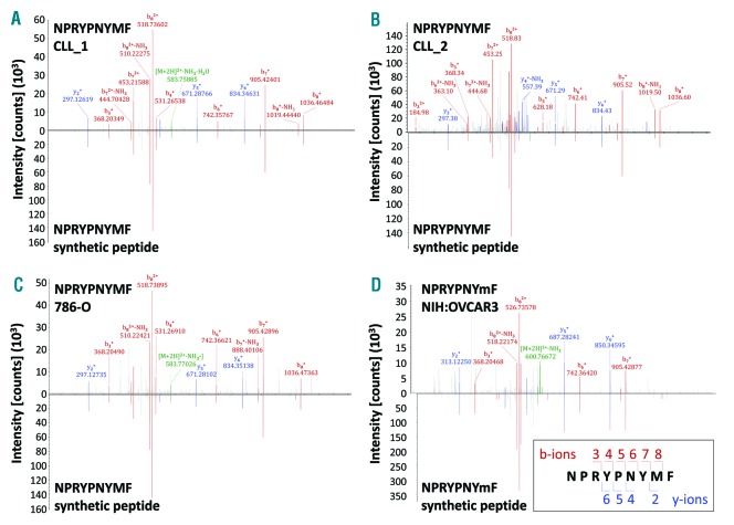
Identification of a novel ROR1-derived peptide naturally presented by tumor cells. Comparison of the mass spectrum of the experimental spectrum (displayed as peaks above the x axis) of the ROR1-derived peptide NPRYPNYMF presented by (A) CLL_1, (B) CLL_2, (C) 786-O, and (D) NIH:OVCAR3 with the synthetic peptide NPRYPNYMF (displayed as peaks below the x axis). The assigned peaks of the b-ions (red) and y-ions (blue) as well as the precursor ions (green) are displayed in color. The masses of b- and y-ion fragments are correlated with the obtained sequence using fragment numbers (right bottom). Analyzing the NIH:OVCAR3 cell line we were able to identify the ROR1 peptide only when applying the parallel reaction monitoring method. Here the peptide was identified with an oxidized methionine, which is indicated by a small ‘m’ (NPRYPNYmF). The spectrum was therefore compared with a spectrum of the synthetic peptide containing also the oxidized methionine. In all other cases data from the data-dependent acquisition method (a top speed collision-induced dissociation fragmentation method) is shown. CLL: chronic lymphocytic leukemia.

